
Tag Archives: Pain Management

Facet Thermal Ablation: Revolutionary Relief for Chronic Pain

Breaking Free from the Chains of Chronic Pain

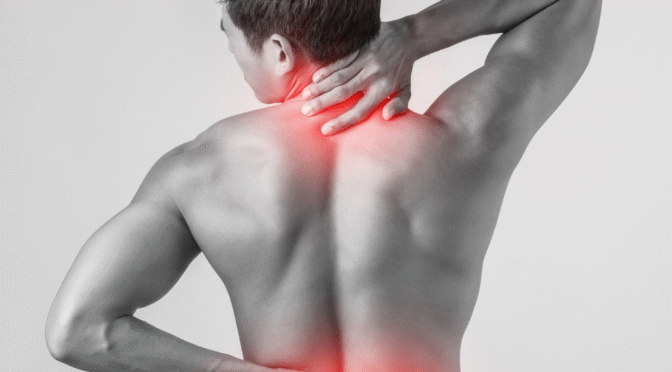
Have you been living with persistent back or neck pain that refuses to respond to traditional treatments? You’re not alone in this struggle. Millions of people worldwide wake up each day to the unwelcome companion of chronic spinal pain, watching as it slowly erodes their quality of life, relationships, and dreams.

Facet thermal ablation represents a revolutionary breakthrough in pain management, offering hope where traditional treatments have failed. This advanced, minimally invasive procedure has transformed the lives of countless patients who had nearly given up on finding lasting relief.

Imagine waking up without that familiar ache radiating through your spine. Picture yourself returning to activities you love – gardening, playing with grandchildren, or simply enjoying a peaceful night’s sleep. This isn’t just wishful thinking; it’s the reality many patients experience after facet joint thermal ablation.

In this comprehensive guide, we’ll explore how this innovative treatment works, who benefits most from the procedure, and why leading pain management specialists consider it a game-changer in chronic back pain treatment. You’ll discover the science behind the success, real patient outcomes, and whether this breakthrough treatment might be the answer you’ve been searching for.


Understanding Facet Joint Pain: The Hidden Culprit Behind Your Suffering

The Anatomy of Agony

Your spine contains small joints called facet joints that connect each vertebra, allowing smooth movement while providing stability. These joints, no larger than your thumbnail, play a crucial role in your spine’s function. When they become inflamed, arthritic, or injured, they can generate intense, persistent pain that radiates throughout your back and neck.

Facet joint pain often manifests as:

  • Deep, aching discomfort that worsens with movement
  • Sharp, stabbing sensations when twisting or bending
  • Morning stiffness that improves throughout the day
  • Radiating pain into shoulders, buttocks, or thighs
  • Muscle spasms and tension

When Traditional Treatments Fall Short

Many patients spend years cycling through conventional treatments – physical therapy, medications, injections – with limited success. The frustration builds as temporary relief gives way to recurring pain, and the cycle begins anew.

Sarah, a 54-year-old teacher, describes her journey: “I tried everything – anti-inflammatories, steroid injections, months of physical therapy. The pain would improve briefly, then return with a vengeance. It was as if I was stuck inside myself.”

This is where radiofrequency ablation for spine conditions becomes a beacon of hope, offering a fundamentally different approach to pain management.


What is Facet Thermal Ablation? The Science of Lasting Relief

Revolutionary Technology Meets Precision Medicine

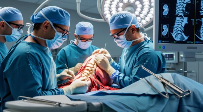
Facet thermal ablation, also known as radiofrequency ablation, represents a quantum leap in pain management technology. This minimally invasive procedure uses precisely controlled thermal energy to interrupt pain signals from problematic facet joints, providing relief that can last months or even years.

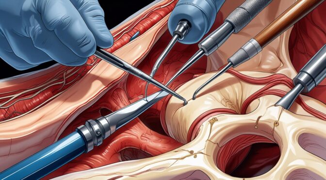
The procedure works by targeting the medial branch nerves – tiny nerve fibers that carry pain signals from facet joints to your brain. By carefully applying radiofrequency energy, specialists can create a controlled lesion on these nerves, effectively “turning off” the pain signals without damaging surrounding tissue.

The Technology Behind the Breakthrough

Modern facet joint thermal ablation employs sophisticated equipment that allows surgeons to:

  • Navigate with millimeter precision using fluoroscopic guidance
  • Monitor temperature in real-time ensuring optimal treatment
  • Customize energy delivery based on individual anatomy
  • Minimize tissue damage while maximizing therapeutic effect

Dr. Rodriguez explains, “What makes this procedure remarkable is its level of precision.” We’re not masking pain with medications – we’re addressing the source directly, giving patients their lives back.”

Beyond Traditional Pain Management

Unlike temporary solutions, chronic back pain treatment through thermal ablation offers several unique advantages:

Immediate Benefits:

  • Minimal recovery time (most patients resume normal activities within a few days)
  • No general anesthesia required
  • Outpatient procedure lasting 30-60 minutes
  • Reduced dependency on pain medications

Long-term Advantages:

  • Pain relief lasting 6 months to 2 years or longer
  • Improved mobility and function
  • Enhanced quality of life
  • Potential to repeat the procedure if needed

The Facet Thermal Ablation Procedure: Your Journey to Pain-Free Living

Pre-Procedure Preparation: Setting the Stage for Success

Your journey begins with comprehensive evaluation and preparation. Leading specialists conduct thorough assessments to ensure facet thermal ablation is the optimal treatment for your specific condition.

Diagnostic Phase:

  • Detailed medical history and physical examination
  • Advanced imaging studies (MRI, CT scans)
  • Diagnostic facet joint injections to confirm pain source
  • Discussion of expectations and potential outcomes

“The diagnostic phase is crucial,” notes Dr. Martinez, a leading interventional pain specialist. He said Our goal is to make sure each patient gets the treatment that best fits their individual needs.”

 
The Procedure Day: Precision in Action

On procedure day, patients arrive at our state-of-the-art facility where comfort and safety are paramount priorities.

Step-by-Step Process:

  1. Patient Positioning: Comfortable positioning on the procedure table with continuous monitoring
  2. Local Anesthesia: Numbing medication applied to ensure patient comfort
  3. Fluoroscopic Guidance: Real-time X-ray imaging guides needle placement with extraordinary precision
  4. Nerve Stimulation: Electrical stimulation confirms correct needle positioning
  5. Thermal Treatment: Controlled radiofrequency energy creates therapeutic lesions
  6. Recovery Monitoring: Brief observation period before discharge

The entire procedure typically takes 30-60 minutes, depending on the number of levels treated.

Post-Procedure Recovery: The Path to Pain Relief

Recovery from radiofrequency ablation for spine conditions is remarkably straightforward for most patients.

Immediate Recovery (First 24-48 Hours):

  • Mild soreness at injection sites (normal and expected)
  • Rest and limited activity recommended
  • Ice application for comfort
  • Gradual return to normal activities

Progressive Improvement (Days to Weeks):

  • Pain relief often begins within the first week
  • Continued improvement over 2-4 weeks
  • Gradual increase in activity levels
  • Follow-up appointments to monitor progress

Remarkable Benefits and Success Rates: The Numbers Tell the Story

Clinical Outcomes That Transform Lives

Research consistently demonstrates the effectiveness of facet joint thermal ablation in providing substantial, long-lasting relief for appropriately selected patients.

Impressive Success Rates:

  • 80-90% of patients experience significant pain reduction
  • Average pain relief duration: 12-24 months
  • Functional improvement: 70-85% of patients report enhanced daily activities
  • Medication reduction: Many patients decrease or eliminate pain medications

Real Patient Transformations

Michael, a 62-year-old contractor, shares his experience: “Before the procedure, I couldn’t work for more than an hour without excruciating pain. Six months after facet thermal ablation, I’m back to full-time work and even started hiking again. It gave me my life back.”

Comprehensive Benefits Beyond Pain Relief

Physical Improvements:

  • Increased range of motion and flexibility
  • Enhanced strength and endurance
  • Better sleep quality
  • Reduced muscle tension and spasms

Emotional and Social Benefits:

  • Improved mood and mental health
  • Renewed participation in family activities
  • Return to hobbies and interests
  • Enhanced overall quality of life

Economic Advantages:

  • Reduced medical expenses over time
  • Decreased need for ongoing treatments
  • Potential return to work or increased productivity
  • Lower long-term healthcare costs

Comparing Treatment Options

Treatment MethodDuration of ReliefInvasivenessRecovery TimeSuccess Rate
Facet Thermal Ablation12-24+ monthsMinimally invasive1-3 days80-90%
Steroid Injections2-6 monthsMinimally invasiveSame day60-70%
Physical TherapyVariableNon-invasiveWeeks40-60%
SurgeryPermanent (if successful)Highly invasive6-12 weeks70-80%

Ideal Candidates: Is Facet Thermal Ablation Right for You?

Perfect Candidates for Success

Facet joint pain relief through thermal ablation works best for patients who meet specific criteria, ensuring optimal outcomes and patient satisfaction.

Primary Indicators:

  • Chronic back or neck pain lasting more than 6 months
  • Pain primarily from facet joints (confirmed through diagnostic injections)
  • Limited response to conservative treatments
  • Desire to reduce dependence on pain medications
  • Good overall health status

Specific Conditions That Respond Well

Optimal Conditions for Treatment:

  • Facet joint arthritis causing chronic pain
  • Degenerative joint disease of the spine
  • Post-traumatic facet joint pain following injury
  • Age-related wear and tear of spinal joints

Medical History Considerations

Your pain management specialist will carefully evaluate your medical history to ensure chronic back pain treatment through thermal ablation is appropriate.

Favorable Factors:

  • Previous positive response to diagnostic facet joint injections
  • Localized pain patterns consistent with facet joint involvement
  • Absence of significant nerve compression
  • Realistic expectations about outcomes

Potential Contraindications:

  • Active infections near treatment sites
  • Certain bleeding disorders
  • Pregnancy
  • Specific cardiac conditions with pacemakers

Age and Activity Level Factors

Facet thermal ablation benefits patients across a wide age range, from active young adults to seniors seeking to maintain independence.

Dr. Thompson observes: “We’ve successfully treated patients from their 30s to their 80s. Age alone isn’t a determining factor – it’s about the specific pain pattern and overall health status.”


Your Next Step Toward Pain-Free Living

The journey from chronic pain to renewed vitality doesn’t have to be a distant dream. Facet thermal ablation has already transformed thousands of lives, offering hope where traditional treatments have fallen short.

If you’re tired of living with persistent back or neck pain, if you’ve tried multiple treatments without lasting success, if you’re ready to reclaim your life from chronic discomfort – this breakthrough treatment might be exactly what you’ve been searching for.

Our team of experienced pain management specialists combines cutting-edge technology with compassionate care, ensuring you receive the personalized treatment you deserve. We understand that every patient’s pain journey is unique, and we’re committed to helping you find the relief that has seemed so elusive.

Take action today. Contact our clinic to schedule your comprehensive consultation and discover whether facet joint thermal ablation could be your path to lasting pain relief. Don’t let another day pass living with pain that could be effectively treated. Schedule Your Consultation | Learn More About Our Services

Is Your Foot Pain Linked to Poor Posture or Nerve Issues?

Understanding Foot Pain: An Overview
Foot pain can be a debilitating condition that affects your quality of life. The foot is a complex
structure composed of bones, joints, ligaments, muscles, and nerves, all working in harmony to
support your body and facilitate movement. When you experience foot pain, it can be challenging to
pinpoint the exact cause due to the intricate nature of the foot’s anatomy. Pain may be localized in
one area, such as the heel, arch, or ball of the foot, or it may be more generalized, affecting the
whole foot. Understanding the potential sources of foot pain is the first step in addressing the issue
effectively.

The causes of foot pain are varied and can include injuries, overuse, and medical conditions such as
arthritis or diabetes. Injuries like sprains, fractures, and tendonitis are common and often result
from accidents or excessive physical activity. Overuse injuries typically occur from repetitive stress
on the foot, such as running or walking long distances without adequate rest. Medical conditions can
lead to inflammation or nerve damage, contributing to foot pain. It’s important to consider all
potential sources when evaluating foot pain, as addressing the root cause is essential for effective
treatment.

Moreover, lifestyle factors and daily habits can significantly influence foot health. Wearing ill-fitting
shoes, standing for extended periods, and neglecting foot care can exacerbate issues and lead to
chronic pain. The foot’s structural integrity is crucial for maintaining balance and proper posture,
which in turn influences overall well-being. By gaining a comprehensive understanding of foot pain
and its origins, you can take proactive steps to alleviate discomfort and prevent future issues.
Recognizing the complexity of foot pain is essential for identifying the underlying causes and
exploring effective treatment options.

The Connection Between Poor Posture and Foot Pain
Poor posture is often overlooked as a contributor to foot pain, yet it plays a significant role in how
your body distributes weight and absorbs shock. When you maintain poor posture, your body’s
alignment is disrupted, leading to uneven pressure on your feet. This imbalance can cause stress on
certain areas of the foot, resulting in pain and discomfort. For example, slouching or leaning forward
can shift your center of gravity, placing additional strain on the balls of your feet. Over time, this
added pressure can lead to conditions such as metatarsalgia or plantar fasciitis.

The connection between posture and foot pain becomes more apparent when considering the
body’s kinetic chain. The kinetic chain is the concept that different parts of the body are
interconnected and affect one another. Poor posture can cause a ripple effect throughout the body,
impacting not just the feet but also the knees, hips, and spine. When one part of the body is
misaligned, it forces other parts to compensate, often leading to discomfort and pain in areas that
may seem unrelated. Understanding this connection is crucial for addressing foot pain holistically
and ensuring long-term relief.

Addressing poor posture involves more than just standing up straight. It requires an awareness of
how your body moves and functions throughout the day. Correcting posture involves strengthening
core muscles, developing body awareness, and making conscious adjustments to your stance and
gait. By improving posture, you can alleviate the undue stress placed on your feet and promote
better overall body mechanics. This not only helps reduce existing foot pain but also prevents future
issues from arising. Taking the time to assess and improve your posture can have a profound impact
on your foot health and overall well-being.

Common Postural Issues That Affect Foot Health
Several common postural issues can directly affect foot health, leading to pain and discomfort. One
such issue is overpronation, where the foot rolls inward excessively while walking or running. This
condition can cause undue stress on the arches and lead to conditions like flat feet, plantar fasciitis,
and shin splints. Overpronation is often exacerbated by poor posture, as it can alter the alignment of
the entire body. Similarly, underpronation, or supination, where the foot rolls outward, can also
result in added pressure on the outer edges of the foot, leading to imbalances and pain.

Another postural issue that impacts foot health is anterior pelvic tilt. This condition occurs when the
pelvis tilts forward, often due to muscle imbalances in the hips and lower back. Anterior pelvic tilt
can cause the lower back to arch excessively, shifting the body’s weight distribution toward the front
of the foot. This added pressure on the forefoot can lead to discomfort and exacerbate existing foot
conditions. Correcting anterior pelvic tilt involves strengthening the core and glute muscles while
stretching the hip flexors and lower back muscles to restore proper alignment.

Forward head posture is another common issue that can indirectly affect foot health. When the
head is positioned forward of the shoulders, it disrupts the body’s alignment, causing compensatory
changes in posture. This forward position can lead to a cascade of adjustments throughout the body,
ultimately affecting the feet. The increased strain on the neck and shoulders can cause tension along
the spine, impacting the lower back and hips, which in turn affects the feet. Addressing forward
head posture involves correcting neck alignment and strengthening the upper back and neck
muscles to support proper posture.

Nerve Issues and Their Impact on Foot Pain
Nerve issues are another significant factor in the development of foot pain. When nerves become
compressed, irritated, or damaged, they can send pain signals to the brain, resulting in discomfort in
the feet. One common nerve-related condition is peripheral neuropathy, which affects the
peripheral nerves responsible for transmitting signals between the central nervous system and the
rest of the body. Peripheral neuropathy can be caused by diabetes, infections, or traumatic injuries,
and it often presents as tingling, numbness, or burning sensations in the feet.

Another nerve-related condition that impacts foot pain is tarsal tunnel syndrome, which is similar to
carpal tunnel syndrome in the wrist. Tarsal tunnel syndrome occurs when the tibial nerve, which
runs along the inside of the ankle, becomes compressed. This compression can lead to pain, tingling,
and numbness in the sole of the foot and the inner ankle. The condition is often caused by repetitive
stress, injury, or anatomical abnormalities such as flat feet. Treating tarsal tunnel syndrome often
involves reducing inflammation, using orthotic supports, and sometimes surgical intervention.

Morton’s neuroma is another nerve issue that can cause foot pain. It involves the thickening of
tissue around one of the nerves leading to the toes, often between the third and fourth toes. This
thickening can compress the nerve, causing sharp, burning pain, and a sensation of a pebble in the
shoe. High-heeled or tight shoes can exacerbate the condition, so wearing appropriate footwear is
essential for relief. Treatment for Morton’s neuroma may include corticosteroid injections, physical
therapy, or surgery in more severe cases. Understanding nerve-related foot pain is crucial for
identifying the appropriate treatment and finding relief.

Symptoms of Nerve-Related Foot Pain
Nerve-related foot pain often presents with a distinct set of symptoms that can help differentiate it
from other types of foot pain. One of the most common symptoms is a burning or tingling sensation,
often described as pins and needles, which can occur in the toes, ball of the foot, or the entire foot.
This sensation may come and go or be constant, depending on the severity and underlying cause of
the nerve issue. Recognizing these symptoms is essential for diagnosis and treatment, as nerve-
related pain requires specific interventions to address the underlying cause.

Numbness is another common symptom of nerve-related foot pain. Affected individuals may
experience a loss of sensation in the feet, which can make it difficult to perceive temperature
changes or feel pressure. This numbness can impact balance and coordination, increasing the risk of
falls and injuries. Additionally, muscle weakness may accompany numbness, further affecting
mobility. These symptoms can be particularly concerning for individuals with conditions like
diabetes, where nerve damage can lead to serious complications if left untreated.

Sharp, shooting pain is also characteristic of nerve-related foot pain. This pain may radiate along the
path of the affected nerve and can be triggered by specific movements or pressure on the foot. In
some cases, the pain may be exacerbated by certain activities, such as walking or standing for
prolonged periods. Identifying the specific symptoms and patterns of nerve-related foot pain is
crucial for determining the appropriate course of treatment. Early intervention can help prevent
further nerve damage and improve the overall quality of life for those affected.

How to Differentiate Between Postural and Nerve-Related Foot Pain
Differentiating between postural and nerve-related foot pain can be challenging, as both can present
with overlapping symptoms. However, understanding the nuances of each type of pain can aid in
accurate diagnosis and treatment. Postural foot pain is often associated with specific activities or

positions, such as standing for long periods or wearing improper footwear. It may be accompanied
by other postural symptoms, such as lower back pain or shoulder discomfort. In contrast, nerve-
related pain is typically characterized by tingling, numbness, or burning sensations that may persist
regardless of activity or position.

A thorough physical examination and medical history are essential for distinguishing between
postural and nerve-related foot pain. A healthcare professional may assess your posture, gait, and
alignment to identify any imbalances contributing to postural pain. They may also perform
neurological tests to evaluate nerve function and identify any areas of nerve compression or
damage. Imaging studies, such as X-rays or MRIs, may be used to rule out other conditions and gain
a better understanding of the underlying issues.

In some cases, electromyography (EMG) or nerve conduction studies may be recommended to
assess nerve health and function. These tests can help identify the specific nerves affected and the
extent of any damage. By combining clinical evaluation with diagnostic tests, healthcare
professionals can accurately differentiate between postural and nerve-related foot pain. This
differentiation is crucial for developing an effective treatment plan tailored to the individual’s needs
and addressing the root cause of the pain.

Exercises to Improve Posture and Alleviate Foot Pain
Incorporating targeted exercises into your routine can significantly improve posture and alleviate
foot pain. Strengthening exercises for the core, hips, and lower back can help correct postural
imbalances and reduce stress on the feet. For example, planks and bridges are effective exercises for
building core stability and promoting proper alignment. These exercises engage the abdominal and
back muscles, supporting the spine and pelvis in a neutral position. Consistent practice can lead to
improved posture and reduced foot pain over time.

Stretching exercises are also crucial for maintaining flexibility and preventing muscle tightness that
can contribute to postural issues. Stretching the calf muscles, hamstrings, and hip flexors can relieve
tension and improve range of motion, allowing for better alignment and weight distribution.
Incorporating yoga or Pilates into your routine can provide a comprehensive approach to stretching
and strengthening, promoting balance and flexibility throughout the body. These practices
emphasize mindfulness and body awareness, helping you maintain correct posture in daily activities.

Foot-specific exercises can further alleviate pain and improve foot health. Simple exercises like toe
curls and heel raises can strengthen the muscles and ligaments in the feet, providing better support
and stability. Additionally, using a foam roller or massage ball can help release tension and improve
circulation in the feet. Regularly performing these exercises can enhance foot function, reduce pain,
and prevent future issues. By adopting a holistic approach to exercise, you can address both postural
and foot-specific concerns, promoting overall well-being and reducing the risk of foot pain.

When to Seek Professional Help for Foot Pain
While self-care measures and exercises can be effective for managing mild foot pain, there are times
when professional help is necessary. If you experience severe pain, swelling, or an inability to bear
weight on the affected foot, it’s important to seek medical attention promptly. These symptoms may
indicate a more serious condition, such as a fracture or severe nerve damage, requiring professional
evaluation and treatment. Early intervention can prevent further complications and support a
quicker recovery.

Persistent or worsening foot pain despite self-care measures also warrants professional evaluation.
A healthcare provider can conduct a thorough assessment to determine the underlying cause of the
pain and develop an appropriate treatment plan. They may recommend physical therapy, orthotic
supports, or medications to address specific issues. In some cases, surgical intervention may be
necessary to correct structural abnormalities or relieve nerve compression. Seeking professional
help ensures that you receive the most effective treatment for your specific condition.

Additionally, if you have underlying medical conditions such as diabetes, arthritis, or neuropathy, it’s
important to regularly consult with a healthcare professional to monitor your foot health. These
conditions can increase the risk of complications, and early detection of any changes in foot health is
crucial for preventing serious issues. Regular check-ups with a podiatrist or specialist can help
manage your condition and maintain optimal foot health. By being proactive and seeking
professional help when needed, you can effectively manage foot pain and improve your overall
quality of life.

Preventative Measures for Maintaining Foot Health
Maintaining foot health requires a proactive approach that includes adopting healthy lifestyle habits
and taking preventative measures. One of the most important steps is wearing appropriate footwear
that provides adequate support and cushioning. Choose shoes with a proper fit, arch support, and a
wide toe box to prevent crowding and pressure on the toes. Avoid high heels and shoes with narrow
toe boxes, as they can exacerbate foot pain and contribute to conditions like bunions and
hammertoes. Investing in quality footwear can significantly reduce the risk of foot pain and promote
overall foot health.

Regular foot care is also essential for preventing issues and maintaining healthy feet. This includes
washing and drying your feet thoroughly, trimming toenails properly, and inspecting your feet
regularly for any signs of changes or abnormalities. Moisturizing your feet can prevent dry, cracked
skin, while exfoliating can remove dead skin cells and promote circulation. If you notice any unusual
symptoms, such as redness, swelling, or persistent pain, it’s important to address them promptly to
prevent more serious issues.

Incorporating foot and ankle exercises into your routine can strengthen the muscles and improve
flexibility, reducing the risk of injuries and strains. Balance exercises, such as standing on one foot or

using a balance board, can enhance stability and coordination, supporting overall foot health.
Maintaining a healthy weight is also crucial, as excess weight can place additional stress on the feet
and lead to pain and discomfort. By adopting these preventative measures, you can support foot
health, reduce the risk of pain, and enjoy a more active and comfortable lifestyle.

Conclusion: Finding the Right Approach to Foot Pain Relief
Foot pain can be a complex and multifaceted issue, with causes ranging from poor posture to nerve-
related conditions. Understanding the interplay between these factors is crucial for finding effective
relief and addressing the root causes of discomfort. By recognizing the impact of posture on foot
health and the potential for nerve issues to contribute to pain, you can take a more informed and
targeted approach to treatment. Whether through exercises, lifestyle changes, or professional
intervention, addressing foot pain requires a comprehensive and individualized strategy.

Taking proactive steps to improve posture, engage in regular exercise, and maintain proper foot care
can significantly reduce the risk of foot pain and enhance overall well-being. Seeking professional
help when necessary ensures that you receive the appropriate diagnosis and treatment for your
specific condition. By prioritizing foot health and being mindful of the factors that contribute to pain,
you can reclaim your stride and enjoy a more active and pain-free lifestyle.

Ultimately, the key to finding relief from foot pain lies in understanding your body’s unique needs
and addressing the underlying causes of discomfort. By adopting a holistic approach that considers
posture, nerve health, and overall lifestyle, you can achieve lasting relief and maintain optimal foot health. Don’t let foot pain hold you back any longer—take charge of your well-being and step
confidently into a future free from discomfort and limitations.

When Pain Lingers: Identifying Chronic vs. Acute Pain

Understanding Pain: The Basics
Pain is an intricate and multifaceted experience that serves as both a protective mechanism and a
signal of potential harm. It is an essential part of the human condition, warning us of danger and
prompting us to take necessary action to prevent further injury. Pain can be as straightforward as
the sharp sting from a paper cut or as complex as the persistent ache of fibromyalgia. While it seems
straightforward, the mechanisms of pain and its implications are far from simple. Understanding the
basics of pain is the first step in recognizing the type of pain you are experiencing and finding the
appropriate course of action.

The sensation of pain is mediated by a complex network of nerves and brain pathways that work
together to alert us to potential damage. When tissue is damaged, sensory receptors called
nociceptors are activated and send signals through the nervous system to the brain. The brain then
interprets these signals as pain, prompting us to react. This reaction can help us avoid further injury
and begin the healing process. However, not all pain is created equal, and understanding the
distinction between different types of pain is crucial for effective management.

Pain is typically categorized into two main types: acute and chronic. While both types serve
important functions, they differ significantly in duration, cause, and treatment approaches. Acute
pain is typically short-lived and directly related to tissue damage or inflammation. It serves as a
warning signal and usually resolves as the underlying cause is treated or heals. Chronic pain, on the
other hand, persists for weeks, months, or even years, often without a clear cause. This type of pain
can become a condition in its own right, affecting a person’s quality of life and requiring specialized
treatment strategies.

What is Acute Pain?
Acute pain is a sudden and severe form of pain that typically results from injury, surgery, or illness. It
is the body’s immediate response to damage, signaling that something is wrong and requires
attention. Unlike chronic pain, acute pain is usually well-defined and localized, making it easier to
diagnose and treat. It can range from mild discomfort to excruciating agony, depending on the
extent of the injury or illness. The primary purpose of acute pain is to alert the individual to potential
harm and facilitate healing by encouraging rest and caution.

The duration of acute pain is typically short, lasting from a few moments to a few weeks, depending
on the severity of the condition. It is often described as sharp, throbbing, or aching, and is usually
accompanied by other symptoms such as swelling, redness, or warmth in the affected area. In many
cases, acute pain resolves once the underlying cause is addressed, such as when a broken bone is set
or an infection is treated with antibiotics.

Acute pain can be effectively managed with a variety of treatments, including medication, physical
therapy, and rest. Over-the-counter pain relievers like ibuprofen or acetaminophen are commonly

used to alleviate mild to moderate acute pain, while stronger prescription medications may be
necessary for more severe cases. In addition to medication, other interventions such as ice or heat
therapy, elevation, and immobilization can help reduce pain and promote healing. By addressing the
root cause of the pain and providing appropriate treatment, acute pain can often be resolved quickly
and effectively.

Characteristics of Chronic Pain
Chronic pain is a persistent and often debilitating form of pain that lasts for an extended period,
typically beyond three to six months. Unlike acute pain, which serves as a warning signal for injury or
illness, chronic pain can persist long after the initial cause has healed, and in some cases, it occurs
without any apparent cause. This type of pain is not just a symptom but can become a condition in
its own right, affecting every aspect of a person’s life, from physical functioning to emotional well-
being.

The nature of chronic pain is complex and multifaceted, with its intensity and characteristics varying
widely among individuals. It may be described as a dull ache, burning sensation, or sharp stabbing
pain, and can fluctuate in severity over time. Chronic pain is often accompanied by other symptoms
such as fatigue, sleep disturbances, and mood changes, which can further complicate its
management. The persistence of pain can lead to increased stress, anxiety, and depression, creating
a vicious cycle that can be difficult to break.

Managing chronic pain requires a comprehensive and multidisciplinary approach that addresses
both the physical and psychological aspects of the condition. Treatment strategies may include
medication, physical therapy, behavioral therapy, and lifestyle modifications. Medications such as
non-steroidal anti-inflammatory drugs (NSAIDs), antidepressants, and anticonvulsants can help
manage pain, while physical therapy can improve mobility and strength. Behavioral therapies, such
as cognitive-behavioral therapy (CBT), can help patients develop coping strategies and address the
emotional impact of chronic pain.

Common Causes of Acute Pain
Acute pain is typically associated with specific, identifiable causes that are often short-lived and
responsive to treatment. One of the most common causes of acute pain is injury, which can occur
from accidents, falls, or direct trauma to the body. Injuries such as fractures, sprains, lacerations,
and burns can result in sudden and intense pain, alerting the individual to seek medical attention
and begin the healing process. In these cases, acute pain serves as a protective mechanism,
encouraging rest and caution to prevent further damage.

Surgical procedures are another common source of acute pain, as they often involve cutting or
manipulating tissues, leading to inflammation and discomfort. Postoperative pain is a normal and
expected part of the recovery process, and it is typically managed with a combination of medications

and therapies to ensure patient comfort and facilitate healing. Effective pain management after
surgery is crucial for promoting mobility, preventing complications, and improving overall outcomes.

In addition to injury and surgery, acute pain can also result from infections or illnesses that cause
inflammation and tissue damage. Conditions such as appendicitis, kidney stones, and infections of
the skin or internal organs can lead to sudden and severe pain as the body responds to the
underlying pathology. In these cases, addressing the root cause of the pain, such as treating the
infection or removing an obstruction, is essential for resolving the acute pain and preventing further
complications.

Common Causes of Chronic Pain
Chronic pain is a complex and often elusive condition that can arise from a variety of underlying
causes. While it may begin as acute pain due to an injury or illness, it can persist long after the initial
cause has healed, sometimes without a clear explanation. One of the most common causes of
chronic pain is musculoskeletal disorders, such as arthritis and back pain. These conditions can lead
to ongoing discomfort and inflammation in the joints, muscles, and connective tissues, significantly
impacting a person’s quality of life.

Nerve damage or neuropathic pain is another frequent cause of chronic pain. This type of pain
occurs when there is damage to the nerves themselves, often resulting from conditions such as
diabetes, shingles, or multiple sclerosis. Neuropathic pain is typically described as burning, tingling,
or shooting, and can be challenging to manage due to its complex nature. In some cases, chronic
pain may arise from central sensitization, a condition in which the central nervous system becomes
hypersensitive to pain signals, amplifying the perception of pain even in the absence of a clear cause.

In addition to physical causes, chronic pain can also be influenced by psychological and emotional
factors. Stress, anxiety, and depression can exacerbate pain perception and contribute to the
persistence of chronic pain. The relationship between mental health and chronic pain is complex and
bidirectional, with each condition potentially influencing the other. As a result, addressing the
psychological aspects of chronic pain is an essential component of effective management, often
involving a combination of therapy, medication, and lifestyle changes.

How to Differentiate Between Acute and Chronic Pain
Differentiating between acute and chronic pain is essential for determining the appropriate course
of treatment and management. One of the key distinctions between the two types of pain is their
duration. Acute pain is typically short-lived, lasting from a few moments to a few weeks, while
chronic pain persists for months or even years. The temporal aspect of pain is often the first clue in
identifying its nature, with acute pain resolving as the underlying condition heals and chronic pain
continuing despite treatment.

The characteristics and underlying causes of the pain can also provide valuable insights into its
classification. Acute pain is often sharp, well-defined, and directly related to an identifiable cause,
such as an injury or illness. In contrast, chronic pain may be more diffuse, with varying intensity and
lacking a clear origin. It is often accompanied by other symptoms such as fatigue, sleep disturbances,
and mood changes, which can further complicate its diagnosis and management. Understanding the
context and presentation of the pain is crucial for distinguishing between acute and chronic pain.

Medical evaluation and diagnostic testing can aid in differentiating between acute and chronic pain
by identifying the underlying cause and assessing the extent of the condition. Imaging studies, blood
tests, and physical examinations can provide valuable information about the source of the pain and
guide the development of a targeted treatment plan. A comprehensive assessment by a healthcare
professional is essential for accurately diagnosing the type of pain and determining the most
effective approach to management.

The Role of Diagnosis in Pain Management
Accurate diagnosis is a critical component of effective pain management, as it provides the
foundation for developing a targeted and individualized treatment plan. Identifying the underlying
cause of the pain allows healthcare providers to address the root issue, rather than merely masking
symptoms. This approach not only improves outcomes but also reduces the risk of complications
and the potential for pain to become chronic. Diagnosis involves a thorough evaluation of the
patient’s medical history, physical examination, and diagnostic testing to determine the nature and
source of the pain.

In cases of acute pain, diagnosis often focuses on identifying the specific injury or illness responsible
for the discomfort. This may involve imaging studies, such as X-rays or MRI scans, to assess the
extent of the damage and guide treatment decisions. For chronic pain, diagnosis can be more
challenging due to the complex and multifaceted nature of the condition. In addition to identifying
any underlying physical causes, healthcare providers may also evaluate psychological and social
factors that could contribute to the persistence of pain.

Once a diagnosis is made, healthcare providers can develop a comprehensive pain management plan
tailored to the individual’s needs. This plan may include a combination of medication, physical
therapy, behavioral therapy, and lifestyle modifications to address the various aspects of the pain.
Regular follow-up and reassessment are essential to ensure the effectiveness of the treatment plan
and make any necessary adjustments. By focusing on accurate diagnosis and individualized care,
patients can achieve better pain control and improved quality of life.

Treatment Options for Acute Pain
The treatment of acute pain focuses on addressing the underlying cause and providing relief from
discomfort. This often involves a combination of medication, physical therapy, and supportive
measures to promote healing and restore function. Over-the-counter pain relievers, such as

ibuprofen or acetaminophen, are commonly used for mild to moderate acute pain, while stronger
prescription medications, such as opioids, may be necessary for more severe cases. It is important to
use these medications as directed to avoid potential side effects and complications.

In addition to medication, non-pharmacological interventions can play a key role in managing acute
pain. Physical therapy and rehabilitation exercises can help improve mobility, strength, and
flexibility, reducing pain and promoting recovery. Techniques such as ice or heat therapy, elevation,
and immobilization can also help alleviate pain and inflammation in the affected area. Supportive
measures, such as rest and adequate nutrition, are essential for facilitating the healing process and
preventing further injury.

Communication with healthcare providers is crucial for effective acute pain management. Patients
should discuss any concerns or side effects they experience with their treatment plan, as well as any
changes in their pain levels. Healthcare providers can offer guidance and adjustments to the
treatment plan as needed to ensure optimal outcomes. By actively participating in their care and
following the recommended treatment plan, patients can achieve effective pain control and a
smooth recovery.

Treatment Options for Chronic Pain
Managing chronic pain requires a comprehensive and multidisciplinary approach that addresses
both the physical and psychological aspects of the condition. Treatment often involves a
combination of medication, physical therapy, behavioral therapy, and lifestyle modifications to
improve pain control and quality of life. Medications such as NSAIDs, antidepressants, and
anticonvulsants can help manage pain and reduce inflammation, while opioids may be used in
certain cases under careful supervision to minimize the risk of dependency.

Physical therapy is a key component of chronic pain management, focusing on improving mobility,
strength, and flexibility. A physical therapist can develop a personalized exercise program tailored to
the individual’s needs and limitations, helping to reduce pain and improve function. Techniques such
as massage, acupuncture, and chiropractic care may also be beneficial for some patients, providing
additional pain relief and relaxation.

Behavioral therapies, such as cognitive-behavioral therapy (CBT), play an important role in
addressing the emotional and psychological impact of chronic pain. These therapies can help
patients develop coping strategies, manage stress, and improve their overall mental health. Lifestyle
modifications, such as regular exercise, a balanced diet, and adequate sleep, are also essential for
managing chronic pain and promoting overall well-being. By taking a holistic and individualized
approach, patients can achieve better pain control and improved quality of life.

When to Seek Professional Help

Knowing when to seek professional help for pain is crucial for effective management and prevention
of complications. While some mild or temporary pain can be managed at home with self-care
measures, certain situations warrant prompt medical attention. If the pain is severe, persistent, or
accompanied by other concerning symptoms, such as fever, swelling, or loss of function, it is
important to seek evaluation by a healthcare provider. Early intervention can prevent the pain from
becoming chronic and improve outcomes.

For acute pain, seeking professional help is often necessary to identify the underlying cause and
receive appropriate treatment. This may involve diagnostic testing, such as imaging studies or blood
tests, to assess the extent of the injury or illness. For chronic pain, a comprehensive evaluation by a
healthcare provider is essential to determine the contributing factors and develop a targeted
treatment plan. This may include referral to specialists, such as pain management or rehabilitation
experts, for further assessment and care.

Regular follow-up with healthcare providers is important for ongoing pain management and
monitoring of treatment effectiveness. Patients should communicate any changes in their pain levels
or response to treatment, as well as any side effects or concerns. By working closely with their
healthcare team, patients can achieve better pain control and improve their overall quality of life.
Seeking professional help when needed is an essential step in managing pain and taking control of
one’s health journey.

Why Hip and Pelvic Pain Often Go Hand in Hand

Understanding Hip and Pelvic Pain

Hip and pelvic pain can be a confounding experience, often leaving individuals feeling trapped in a cycle of discomfort and confusion. The hips and pelvis serve as crucial support structures for the body, playing a vital role in maintaining balance and facilitating movement. When pain arises in these areas, it can significantly disrupt daily activities, making even the simplest tasks challenging. Understanding the anatomy and function of the hips and pelvis is the first step towards unraveling the mystery behind the pain that often links them together.

The hips are ball-and-socket joints where the femur (thigh bone) connects to the pelvis. This configuration allows for a wide range of motion, essential for activities like walking, running, and bending. The pelvis, on the other hand, is a basin-shaped structure composed of several bones including the ilium, ischium, and pubis. It supports the weight of the upper body and connects the spine to the lower limbs. The complex interplay between these structures means that an issue in one can easily affect the other, leading to concurrent pain.

Many people underestimate the interconnectedness of the hips and pelvis, often viewing them as separate entities. However, the reality is that they work in tandem, supported by a network of muscles, ligaments, and tendons. This intricate relationship means that stress or injury in one area can create a ripple effect, causing discomfort in the other. Recognizing this connection is vital for those seeking to understand and address their hip and pelvic pain effectively.

Common Causes of Hip and Pelvic Pain

Hip and pelvic pain can stem from a myriad of causes, some of which are more common than others. One prevalent cause is osteoarthritis, a degenerative joint disease that affects the cartilage cushioning the hip joint. As the cartilage wears down, it leads to pain and stiffness, which can extend to the pelvic region. Osteoarthritis is particularly common in older adults, though it can affect younger individuals as well, especially those with a family history of the condition.

Another common culprit is tendinitis, which occurs when the tendons connecting muscles to bones become inflamed. This inflammation can result from repetitive motions or overuse, often seen in athletes or individuals with physically demanding jobs. Tendinitis in the hip can cause pain that radiates to the pelvis, as the muscles and tendons work to stabilize the joint. Similarly, bursitis, the inflammation of the small fluid-filled sacs that cushion the joints, can lead to similar discomfort.

In addition to these musculoskeletal issues, hip and pelvic pain can also be caused by injuries such as fractures or dislocations. Trauma from falls, accidents, or sports injuries can impact both the hip and pelvic regions, resulting in acute pain. In some cases, underlying medical conditions like hernias, gynecological issues, or even kidney stones can manifest as hip and pelvic pain, making diagnosis and treatment a complex process. Understanding the potential causes is essential for addressing the root of the problem and finding relief.

The Connection Between Hip and Pelvic Pain

The connection between hip and pelvic pain is a topic that often surprises those unfamiliar with the intricacies of human anatomy. At the heart of this relationship is the shared structural and functional roles that bind the hips and pelvis together. Both areas are integral to the support and movement of the lower body, meaning that dysfunction in one can have a direct impact on the other. This interconnectedness is crucial for understanding why pain in one area is frequently accompanied by discomfort in the other.

Muscular imbalances are a common factor that links hip and pelvic pain. When certain muscles around the hips and pelvis become overactive or underactive, it can lead to changes in posture and movement patterns. For instance, tight hip flexors can pull on the pelvis, creating an anterior tilt that strains the lower back and pelvic region. Conversely, weak gluteal muscles can lead to instability in the hip joint, causing compensatory movements that stress the pelvis.

Additionally, the nerves that supply the hips and pelvis can contribute to their shared pain experience. The sciatic nerve, for example, runs from the lower back through the hips and down each leg. Compression or irritation of this nerve can result in sciatica, a condition characterized by pain that radiates from the lower back to the hips and pelvis. This nerve-related pain highlights the interconnected nature of these regions and underscores the importance of addressing both areas when seeking relief.

Symptoms to Watch For

Recognizing the symptoms of hip and pelvic pain is a critical step towards effective diagnosis and treatment. One of the most common symptoms is a persistent aching or stiffness in the hip joint or pelvic region. This discomfort may worsen with physical activity, prolonged sitting, or standing, and can feel particularly intense in the morning or after periods of rest. Paying attention to the pattern and intensity of the pain can provide valuable insights into its underlying cause.

In addition to general discomfort, individuals may also experience more specific symptoms such as swelling, warmth, or redness around the affected area. These signs suggest inflammation, which is often associated with conditions like tendinitis or bursitis. In some cases, the pain may be accompanied by a reduced range of motion, making it difficult to perform everyday activities like bending, walking, or climbing stairs. Such limitations can significantly impact one’s quality of life, highlighting the need for timely intervention.

Another important symptom to watch for is referred pain, which occurs when discomfort is felt in a location distant from its source. For instance, pain originating in the hip may be perceived in the groin, thigh, or even the knee. Similarly, pelvic pain can manifest as lower back discomfort. Understanding these atypical presentations is essential for accurate diagnosis and ensuring that treatment targets the true source of the pain rather than just its symptoms.

Diagnosing Hip and Pelvic Pain

Accurate diagnosis is paramount when it comes to addressing hip and pelvic pain, as it enables tailored treatment plans that target the specific underlying cause. The diagnostic process typically begins with a comprehensive medical history and physical examination. During this initial assessment, healthcare providers may inquire about the onset, duration, and nature of the pain, as well as any previous injuries or medical conditions that could be contributing factors.

Imaging studies are often employed to gain a clearer picture of the structures involved. X-rays can reveal changes in bone architecture, such as fractures or arthritis-related degeneration. For a more detailed view, magnetic resonance imaging (MRI) or computed tomography (CT) scans may be utilized to assess the soft tissues, including muscles, tendons, and ligaments. These advanced imaging techniques are invaluable in identifying conditions like tendinitis, bursitis, or hernias that may not be visible on standard X-rays.

In some cases, diagnostic injections may be used to pinpoint the source of pain. By injecting a local anesthetic into specific areas, doctors can determine if temporary pain relief occurs, thereby confirming the involvement of that structure in the patient’s symptoms. This targeted approach helps distinguish between multiple potential sources of pain and ensures that treatment strategies are appropriately directed. Ultimately, a thorough diagnostic process is essential for developing a treatment plan that effectively addresses the root cause of hip and pelvic pain.

Treatment Options for Relief

Once the cause of hip and pelvic pain has been identified, a range of treatment options can be explored to provide relief and restore function. One of the most common approaches is physical therapy, which focuses on strengthening the muscles around the hip and pelvis to improve stability and reduce strain. Customized exercise programs tailored to the individual’s needs can enhance flexibility, correct muscular imbalances, and promote better posture and movement patterns.

In addition to physical therapy, medication may be prescribed to manage pain and inflammation. Nonsteroidal anti-inflammatory drugs (NSAIDs) such as ibuprofen or naproxen are often recommended for their ability to reduce swelling and alleviate discomfort. For more severe pain, doctors may consider corticosteroid injections to deliver targeted relief directly to the affected area. These injections can provide significant pain reduction, allowing individuals to participate more effectively in rehabilitation exercises.

In cases where conservative treatments are ineffective, surgical intervention may be necessary. Surgical options vary depending on the specific condition, ranging from minimally invasive procedures to more extensive surgeries. For example, arthroscopic surgery can address issues like labral tears or impingement in the hip joint, while hip replacement surgery may be considered for severe arthritis. The decision to pursue surgery is typically made after a thorough evaluation of the patient’s condition and consideration of potential risks and benefits.

Preventative Measures to Avoid Pain

Prevention plays a vital role in minimizing the risk of developing hip and pelvic pain, and adopting certain lifestyle changes can be highly effective. One of the most important preventative measures is maintaining a healthy weight, as excess body weight places additional stress on the hips and pelvis, increasing the likelihood of joint degeneration and injury. A balanced diet and regular exercise can help manage weight and promote overall musculoskeletal health.

Engaging in regular physical activity is another key factor in preventing hip and pelvic pain. Activities that promote strength, flexibility, and cardiovascular fitness can enhance joint stability and reduce the risk of injury. Low-impact exercises such as swimming, cycling, or walking are particularly beneficial, as they provide cardiovascular benefits without placing excessive strain on the joints. Incorporating strength training exercises that target the core, hips, and lower extremities can further bolster stability and support.

Proper body mechanics and ergonomic considerations are also crucial for preventing pain. Paying attention to posture during daily activities, such as sitting, standing, or lifting, can reduce stress on the hips and pelvis. Using ergonomic furniture and equipment, taking frequent breaks during prolonged activities, and practicing good lifting techniques can all contribute to minimizing the risk of strain or injury. By adopting these preventative measures, individuals can significantly reduce their risk of developing hip and pelvic pain and promote long-term joint health.

The Role of Physical Therapy

Physical therapy is a cornerstone in the treatment and prevention of hip and pelvic pain, offering a non-invasive approach that can yield significant benefits. A physical therapist works closely with patients to develop individualized exercise programs that target specific weaknesses and imbalances. These programs are designed to improve flexibility, strength, and stability, ultimately enhancing joint function and reducing pain.

One of the primary goals of physical therapy is to increase the range of motion in the hip and pelvic joints. Through a combination of stretching exercises and manual therapy techniques, therapists can help release tight muscles and improve joint mobility. This increased flexibility not only alleviates existing discomfort but also helps prevent future pain by promoting more efficient movement patterns.

Another critical aspect of physical therapy is education. Therapists provide valuable guidance on body mechanics, posture, and ergonomic adjustments that can be applied to daily activities. By teaching patients how to move safely and efficiently, therapists empower them to take an active role in managing their condition and preventing recurrence. This educational component, combined with targeted exercises, makes physical therapy an essential tool in addressing hip and pelvic pain effectively.

When to Seek Medical Attention

While many cases of hip and pelvic pain can be managed with conservative measures, there are certain situations where seeking medical attention becomes imperative. If the pain is severe, persistent, or accompanied by other concerning symptoms, such as fever, unexplained weight loss, or changes in bowel or bladder function, it is crucial to consult a healthcare professional. These symptoms may indicate an underlying condition that requires prompt evaluation and treatment.

Additionally, if pain is the result of a traumatic injury, such as a fall or accident, immediate medical attention is necessary to rule out fractures or dislocations. Delaying treatment in such cases can lead to complications and prolonged recovery. Similarly, if conservative treatments like rest, ice, or over-the-counter medications fail to provide relief after a reasonable period, it is advisable to seek further evaluation to determine the cause and explore alternative treatment options.

Ultimately, listening to your body and seeking timely medical attention can prevent minor issues from escalating into more significant problems. Early intervention not only facilitates more effective treatment but also improves the chances of a full recovery. By prioritizing health and being proactive in seeking care when needed, individuals can better manage their hip and pelvic pain and maintain an active, fulfilling lifestyle.

Conclusion: Managing Hip and Pelvic Pain Effectively

Managing hip and pelvic pain requires a multifaceted approach that addresses both the underlying causes and the symptoms. By understanding the intricate relationship between these two areas, individuals can take proactive steps to alleviate discomfort and improve their quality of life. Whether through lifestyle modifications, physical therapy, or medical interventions, there are numerous strategies available to manage and prevent pain effectively.

Educating oneself about the anatomy and function of the hips and pelvis is a crucial first step in this journey. Recognizing the common causes and symptoms of pain allows for more informed discussions with healthcare providers and empowers individuals to make decisions that align with their health goals. With the right knowledge and resources, it is possible to navigate the complexities of hip and pelvic pain successfully.

Ultimately, the key to managing hip and pelvic pain lies in a combination of proactive prevention, timely intervention, and a commitment to maintaining overall health and well-being. By embracing a holistic approach and working closely with healthcare professionals, individuals can reclaim their comfort and mobility, paving the way for a more active and fulfilling lifestyle. Through understanding and action, the puzzle of hip and pelvic pain can be unraveled, leading to lasting relief and improved quality of life.

Top Non-Surgical Treatments for Joint Pain Relief

Understanding Joint Pain: Causes and Symptoms

Joint pain is a common complaint that can stem from a variety of causes, each presenting its own set of symptoms and challenges. At its core, joint pain arises from inflammation or damage within the joints, leading to discomfort and restricted movement. The causes can range from acute injuries, such as sprains and fractures, to chronic conditions like arthritis and bursitis. Arthritis, in particular, is a prevalent cause of joint pain, affecting millions worldwide with its various forms, including osteoarthritis and rheumatoid arthritis. Osteoarthritis results from wear and tear of the cartilage, while rheumatoid arthritis is an autoimmune disorder causing joint inflammation.

Symptoms of joint pain can vary significantly depending on the underlying cause. Commonly, individuals experience aching, stiffness, and a limited range of motion. Swelling and redness may also accompany these symptoms, indicating inflammation. In cases of acute injuries, the pain might be sharp and sudden, whereas chronic conditions often lead to persistent, dull aches. Regardless of the cause, the impact on daily life can be profound, limiting physical activities and diminishing overall quality of life. Recognizing the symptoms early and understanding their implications is crucial for effective management and treatment.

Proper diagnosis is essential in addressing joint pain effectively. Healthcare professionals often rely on a combination of physical examinations, medical history reviews, and imaging tests, such as X-rays or MRIs, to pinpoint the exact cause of the discomfort. Understanding the root cause is the first step towards choosing the most appropriate treatment plan. While surgical options are available, non-surgical treatments are often preferred for their lower risk and less invasive nature. These treatments aim to alleviate symptoms, enhance joint function, and improve overall quality of life without resorting to surgery.

Overview of Non-Surgical Treatment Options

Non-surgical treatments for joint pain are diverse, offering a range of options tailored to address different causes and severities of pain. These treatments prioritize symptom relief and improved joint function while minimizing risks associated with surgical interventions. Among the most common non-surgical treatments are physical therapy, medications, and various forms of injections. Each approach has its unique benefits and may be used alone or in combination to achieve optimal results. By focusing on non-invasive methods, individuals can often manage their joint pain effectively and maintain an active lifestyle without undergoing surgery.

Physical therapy is a cornerstone of non-surgical joint pain management, offering personalized exercise programs designed to strengthen muscles and improve joint flexibility. This approach not only alleviates pain but also addresses potential imbalances that may contribute to joint issues. Medications play a significant role as well, with options ranging from over-the-counter pain relievers to prescription medications aimed at reducing inflammation. Non-steroidal anti-inflammatory drugs (NSAIDs) and analgesics are commonly used to manage mild to moderate pain, while stronger medications may be prescribed for more severe cases.

In addition to physical therapy and medications, injections such as corticosteroids and hyaluronic acid provide targeted relief for certain types of joint pain. These treatments are particularly beneficial for individuals with arthritis or other chronic inflammatory conditions, offering temporary relief and allowing for improved joint function. Alternative therapies, including acupuncture and chiropractic care, have gained popularity as well, providing holistic approaches to pain management. By exploring these non-surgical treatment options, individuals can make informed decisions and choose the best path to regain mobility and enhance their quality of life.

Physical Therapy: Benefits and Techniques

Physical therapy is a highly effective non-surgical treatment for joint pain, offering numerous benefits that extend beyond pain relief. One of the primary advantages of physical therapy is its ability to improve joint mobility and function. Through targeted exercises and stretches, physical therapists help patients regain strength in the surrounding muscles, which can alleviate pressure on the joints. This approach not only reduces pain but also enhances overall joint stability, preventing further injury or deterioration. Moreover, physical therapy is tailored to each individual’s specific needs, ensuring a personalized treatment plan that addresses the root causes of their joint pain.

A variety of techniques are employed in physical therapy to address joint pain. Manual therapy, for example, involves hands-on techniques such as massage and joint mobilization, which can help reduce stiffness and improve circulation. Additionally, therapeutic exercises are a core component of physical therapy, focusing on strengthening and stretching the muscles around the affected joint. These exercises are designed to be safe and effective, promoting healing while preventing further strain. Physical therapists may also incorporate modalities such as heat or cold therapy, ultrasound, and electrical stimulation to complement the treatment and enhance pain relief.

Education plays a crucial role in physical therapy, empowering patients with the knowledge and skills needed to manage their joint pain independently. Physical therapists often provide guidance on proper body mechanics and posture, helping patients avoid movements that could exacerbate their condition. By learning how to perform daily activities safely and effectively, individuals can reduce their risk of future joint problems and maintain their progress over time. As a result, physical therapy not only addresses current joint pain but also equips patients with the tools needed for long-term joint health and well-being.

The Role of Medications in Joint Pain Management

Medications are a vital component of non-surgical joint pain management, offering relief from symptoms and improving overall quality of life. These medications work by targeting the underlying causes of pain, such as inflammation or nerve sensitivity, providing much-needed respite from discomfort. Over-the-counter options, such as acetaminophen and non-steroidal anti-inflammatory drugs (NSAIDs), are commonly used for mild to moderate pain. These medications are easily accessible and provide effective relief for many individuals, allowing them to continue with their daily activities with minimal disruption.

For those with more severe joint pain, prescription medications may be necessary. These can include stronger NSAIDs, corticosteroids, and disease-modifying antirheumatic drugs (DMARDs), which are specifically used for inflammatory conditions such as rheumatoid arthritis. Corticosteroids, for instance, are potent anti-inflammatory agents that can be taken orally or injected directly into the joint for targeted relief. DMARDs, on the other hand, work by modifying the immune system to slow disease progression and reduce joint damage. While these medications offer significant benefits, they may also come with potential side effects, making it essential for patients to work closely with their healthcare provider to find the most suitable option.

Opioids are another class of medications that may be prescribed for severe joint pain, particularly when other treatments have proven ineffective. However, due to their potential for addiction and other adverse effects, they are typically considered a last resort and used only for short-term management. It’s essential for individuals taking any medication for joint pain to follow their healthcare provider’s instructions carefully and to report any side effects or concerns promptly. By understanding the role of medications and using them appropriately, patients can effectively manage their joint pain and improve their overall quality of life.

Injections for Joint Pain Relief: Corticosteroids and Hyaluronic Acid

Injections offer a targeted approach to joint pain relief, providing direct treatment to the affected area. Corticosteroid injections are among the most commonly used, delivering powerful anti-inflammatory medication directly into the joint. This localized treatment can significantly reduce swelling and pain, particularly for individuals with arthritis or other inflammatory joint conditions. The effects of corticosteroid injections can vary, providing relief for several weeks to months, depending on the individual’s condition and response to treatment. While generally safe, repeated use of corticosteroid injections may lead to side effects, such as joint damage or weakening of surrounding tissues, making it important to use them judiciously.

Hyaluronic acid injections are another option for joint pain relief, often used for individuals with osteoarthritis. This treatment involves injecting a synthetic form of hyaluronic acid, a natural component of joint fluid, into the affected joint. By enhancing lubrication and cushioning, hyaluronic acid injections can improve joint function and reduce pain. Unlike corticosteroids, hyaluronic acid does not address inflammation directly but instead focuses on improving the joint’s mechanical properties. This makes it a suitable option for those seeking long-term improvement in joint health. The benefits of hyaluronic acid injections can last several months, providing sustained relief for many patients.

Both corticosteroid and hyaluronic acid injections offer distinct advantages and can be integral components of a comprehensive joint pain management plan. When considering these options, it’s essential for patients to discuss their specific needs and medical history with their healthcare provider. By understanding the potential benefits and limitations of each treatment, individuals can make informed decisions about their care and achieve the best possible outcomes for their joint health.

Alternative Therapies: Acupuncture and Chiropractic Care

Alternative therapies have gained popularity as complementary treatments for joint pain, offering holistic approaches that address the body as a whole. Acupuncture, a traditional Chinese medicine practice, involves the insertion of thin needles into specific points on the body to stimulate energy flow and promote healing. This technique is believed to enhance the body’s natural pain-relief mechanisms, such as the release of endorphins, providing relief for various types of joint pain. Studies have shown that acupuncture can be particularly effective for conditions like osteoarthritis and rheumatoid arthritis, reducing pain and improving function without the need for medication.

Chiropractic care is another alternative therapy that focuses on the alignment and function of the musculoskeletal system. Chiropractors use manual adjustments and manipulations to correct misalignments in the spine and other joints, which can contribute to pain and dysfunction. By restoring proper alignment, chiropractic care aims to improve joint mobility, reduce inflammation, and alleviate pain. This approach is often used to address back, neck, and joint pain, making it a valuable option for those seeking non-invasive treatments.

Both acupuncture and chiropractic care offer unique benefits and can be used alongside traditional treatments to enhance joint pain management. It’s important for individuals considering these therapies to consult with qualified practitioners who are experienced in treating joint pain. By exploring alternative therapies, patients can discover new ways to manage their symptoms, improve their overall well-being, and achieve a more balanced approach to joint health.

Lifestyle Modifications for Joint Health

Lifestyle modifications play a crucial role in managing joint pain and promoting long-term joint health. Simple changes in daily habits can significantly impact joint function and reduce the risk of further damage. One of the most effective lifestyle modifications is maintaining a healthy weight, as excess body weight places additional stress on the joints, particularly those in the lower body. By achieving and maintaining a healthy weight, individuals can alleviate pressure on their joints, reduce pain, and improve mobility.

Regular physical activity is another essential component of joint health. Engaging in low-impact exercises, such as swimming, cycling, or walking, can help strengthen the muscles surrounding the joints, enhance flexibility, and promote overall fitness. Exercise also stimulates the production of synovial fluid, which lubricates the joints and reduces friction. It’s important for individuals to choose activities that are gentle on the joints and to start slowly, gradually increasing intensity as their fitness improves. Consulting with a healthcare professional or physical therapist can provide guidance on appropriate exercises and help prevent injury.

In addition to weight management and exercise, adopting a balanced diet rich in nutrients that support joint health is vital. Foods high in antioxidants, omega-3 fatty acids, and vitamins such as vitamin C and D can help reduce inflammation and promote joint function. Incorporating these dietary changes, along with other lifestyle modifications, can empower individuals to take control of their joint health and improve their overall quality of life.

Dietary Supplements: What Works for Joint Pain?

Dietary supplements have become increasingly popular as a means of supporting joint health and alleviating pain. Among the most well-known supplements for joint pain are glucosamine and chondroitin, which are often used together to enhance their effects. These supplements are believed to support cartilage health and reduce inflammation, offering relief for individuals with osteoarthritis. While research on their effectiveness is mixed, many people report improvements in pain and function, making them a viable option for those seeking non-pharmaceutical interventions.

Omega-3 fatty acids, commonly found in fish oil supplements, are another option for managing joint pain. These essential fatty acids have potent anti-inflammatory properties, which can help reduce joint swelling and discomfort. Studies have shown that omega-3 supplements may be particularly beneficial for individuals with rheumatoid arthritis, as they can decrease morning stiffness and improve overall joint function. Including omega-3-rich foods, such as fatty fish, flaxseeds, and walnuts, in the diet can also provide these benefits naturally.

Other supplements, such as turmeric, ginger, and boswellia, have gained attention for their anti-inflammatory and pain-relieving properties. Curcumin, the active compound in turmeric, is known for its ability to inhibit inflammatory pathways, offering potential relief for various types of joint pain. Ginger and boswellia are also believed to reduce inflammation and improve joint function. As with any supplement, it’s essential for individuals to consult with a healthcare provider before starting a new regimen to ensure safety and effectiveness.

When to Consider Surgical Options

While non-surgical treatments are often effective in managing joint pain, there are instances where surgical intervention may be necessary. Surgery is typically considered when conservative treatments have failed to provide adequate relief, and the individual’s quality of life is significantly impacted. Conditions such as severe osteoarthritis, advanced joint degeneration, or traumatic injuries may require surgical solutions to restore function and alleviate pain. It’s important for patients to thoroughly discuss their options with a healthcare professional, considering the potential risks and benefits of surgery.

Joint replacement surgery, such as total knee or hip replacement, is one of the most common surgical options for individuals with end-stage arthritis. These procedures involve replacing the damaged joint surfaces with prosthetic components, which can significantly reduce pain and improve mobility. While joint replacement surgery has a high success rate, it also requires a significant recovery period and rehabilitation to achieve optimal results. Other surgical options, such as arthroscopy or osteotomy, may be appropriate for less severe cases, offering minimally invasive solutions with shorter recovery times.

Ultimately, the decision to pursue surgery should be based on a comprehensive evaluation of the individual’s condition, lifestyle, and treatment goals. By understanding when surgical intervention may be necessary and working closely with a healthcare team, patients can make informed decisions that align with their needs and aspirations for joint health and overall well-being.

Conclusion: Choosing the Right Treatment for You

Navigating the myriad of non-surgical treatments for joint pain can be overwhelming, but understanding the options available is key to finding the right solution. Each individual’s journey with joint pain is unique, and what works for one person may not be suitable for another. It’s essential to consider factors such as the underlying cause of the pain, the severity of symptoms, and personal preferences when choosing a treatment plan. Consulting with healthcare professionals can provide valuable insights and guidance, helping individuals make informed decisions about their care.

Non-surgical treatments, such as physical therapy, medications, injections, and alternative therapies, offer effective solutions for managing joint pain and improving quality of life. By exploring these options and integrating lifestyle modifications, individuals can take proactive steps towards achieving long-term joint health. Supplements and dietary changes can further support these efforts, providing additional benefits for those seeking holistic approaches to pain management.

Ultimately, the goal is to empower individuals to reclaim their mobility and enjoy a vibrant, active life without the limitations imposed by joint pain. By taking charge of their health and exploring the diverse range of non-surgical treatments available, individuals can find relief and thrive with confidence and ease. Remember, the journey to joint health is personal, and with the right support and resources, it is entirely achievable.

Nerve Pain vs. Muscle Pain: What’s the Difference?

Understanding Pain: An Overview

Pain, in its essence, is a complex and multidimensional experience that serves as a crucial signal from our bodies, alerting us to potential harm or injury. It is an essential component of our sensory system, designed to protect us by prompting a response to avoid further damage. This sensory phenomenon is not solely an unpleasant sensation; it’s a critical mechanism that can indicate underlying health issues. Pain can manifest in various forms, and understanding its nature is key to addressing it effectively.

The human body is equipped with a sophisticated network of nerves and receptors that detect pain and transmit signals to the brain. Once these signals reach the brain, they are processed, resulting in the sensation of pain. However, not all pain is alike. It can be acute or chronic, localized or widespread, and it can vary greatly in intensity. This diversity in pain experiences underscores the importance of identifying the type of pain one is experiencing, as each type requires a different approach for effective management.

Moreover, the perception of pain is highly subjective, influenced by factors such as emotional state, previous experiences, and cultural background. This subjectivity can complicate diagnosis and treatment, as what is excruciating for one person might be tolerable for another. Therefore, understanding the specific characteristics and causes of different types of pain, such as nerve pain and muscle pain, is paramount for both healthcare professionals and individuals seeking relief from their discomfort.

What is Nerve Pain?

Nerve pain, also known as neuropathic pain, arises from damage or dysfunction within the nervous system. Unlike other types of pain, which often result from direct physical injury or inflammation, nerve pain is typically associated with the malfunctioning of the nerves themselves. This malfunction can occur due to a range of factors, including injury, infection, or chronic conditions such as diabetes. The result is a unique and often debilitating form of pain that can significantly impact an individual’s quality of life.

A hallmark of nerve pain is its distinctive and often unpredictable nature. Patients frequently describe it as a burning, shooting, or stabbing sensation, which can be accompanied by tingling or numbness. These sensations can occur spontaneously or be triggered by stimuli that would not normally provoke pain, such as light touch or changes in temperature. This characteristic, known as allodynia, is a key indicator of neuropathic pain and differentiates it from other types of pain.

Nerve pain can be persistent and challenging to manage, often requiring a multifaceted approach to treatment. Conventional pain relief methods, such as over-the-counter painkillers, are often ineffective against neuropathic pain, necessitating the use of medications specifically designed to target nerve dysfunction. These may include anticonvulsants, antidepressants, or topical treatments. In some cases, more advanced interventions, such as nerve blocks or electrical stimulation, may be necessary to provide relief.

What is Muscle Pain?

Muscle pain, or myalgia, is a common condition that most individuals experience at some point in their lives. It is characterized by discomfort or soreness in the muscles, often resulting from overuse, tension, or minor injuries. Unlike nerve pain, which originates from the nervous system, muscle pain is typically related to physical exertion or strain on the muscular system. This type of pain is usually acute and resolves with rest and appropriate care.

One of the primary causes of muscle pain is physical activity, particularly when muscles are pushed beyond their usual limits. This can lead to microscopic damage to muscle fibers, resulting in inflammation and soreness. However, muscle pain can also be caused by other factors, such as poor posture, stress, or underlying medical conditions like fibromyalgia or myositis. Identifying the cause of muscle pain is crucial for effective treatment and prevention of future episodes.

Muscle pain is often described as a dull, aching sensation that can be localized or widespread. It may be accompanied by stiffness or weakness, making it difficult to perform daily activities. Fortunately, muscle pain is generally responsive to self-care measures, such as rest, application of heat or cold, and gentle stretching. In cases where muscle pain is persistent or severe, medical intervention may be required to address any underlying issues or to provide more targeted relief.

Key Differences Between Nerve Pain and Muscle Pain

While both nerve pain and muscle pain can be debilitating, they have distinct characteristics that set them apart. Understanding these differences is essential for accurate diagnosis and effective treatment. One of the primary distinctions lies in the origin of the pain. Nerve pain stems from issues within the nervous system, whereas muscle pain is related to the muscular system and often results from physical strain or injury.

In terms of sensation, nerve pain is often described as sharp, burning, or shooting, with accompanying symptoms such as tingling or numbness. These sensations can occur without any apparent cause or be triggered by non-painful stimuli. In contrast, muscle pain is typically a dull, aching sensation that is directly related to physical activity or tension. It is often localized to specific areas and is associated with stiffness or weakness in the affected muscles.

The treatment approaches for nerve pain and muscle pain also differ significantly. Nerve pain may require medications that specifically target nerve dysfunction, such as anticonvulsants or antidepressants, while muscle pain is usually managed with rest, heat or cold therapy, and over-the-counter pain relievers. Understanding these differences is crucial for developing an effective treatment plan that addresses the underlying cause of the pain and provides lasting relief.

Common Causes of Nerve Pain

Nerve pain can arise from a variety of causes, many of which involve damage or dysfunction of the nervous system. One of the most common causes is injury or trauma to the nerves, which can occur as a result of accidents, surgeries, or even certain medical procedures. When nerves are damaged, they may send incorrect signals to the brain, resulting in the perception of pain.

Chronic conditions such as diabetes are another leading cause of nerve pain. High blood sugar levels can lead to diabetic neuropathy, a condition characterized by damage to the nerves, particularly in the extremities. This can result in pain, tingling, or numbness in the hands and feet, significantly affecting an individual’s quality of life. Other conditions, such as multiple sclerosis or shingles, can also cause nerve pain by affecting the nervous system.

Infections and inflammation can also contribute to the development of nerve pain. Viral infections, such as the herpes zoster virus, can lead to postherpetic neuralgia, a painful condition that persists after the resolution of shingles. Additionally, autoimmune disorders that cause inflammation of the nerves, such as lupus or Guillain-Barré syndrome, can result in severe nerve pain. Identifying the underlying cause of nerve pain is essential for effective management and relief.

Common Causes of Muscle Pain

Muscle pain is often the result of physical exertion or strain, but it can also be caused by other factors. Overuse or injury to the muscles is one of the most common causes, particularly in individuals who engage in strenuous physical activities or sports. This can lead to muscle strains or sprains, which cause inflammation and pain in the affected muscles.

Poor posture and repetitive movements can also contribute to muscle pain, particularly in individuals who spend long hours sitting at a desk or performing manual labor. These activities can lead to muscle imbalances and tension, resulting in discomfort and pain. Stress and anxiety can also manifest as muscle pain, as tension and tightness in the muscles often accompany emotional stress.

Certain medical conditions can also cause muscle pain. Fibromyalgia, for example, is a chronic disorder characterized by widespread muscle pain and tenderness. Myositis, an inflammatory condition affecting the muscles, can also result in persistent pain and weakness. In such cases, addressing the underlying condition is crucial for alleviating muscle pain and improving overall well-being.

Symptoms of Nerve Pain vs. Muscle Pain

The symptoms of nerve pain and muscle pain can differ significantly, making it crucial to recognize these differences for accurate diagnosis and treatment. Nerve pain is often described as a sharp, burning, or shooting sensation that may be accompanied by tingling or numbness. These symptoms can occur spontaneously or be triggered by light touch or changes in temperature, known as allodynia.

In contrast, muscle pain is typically a dull, aching sensation that is directly related to physical exertion or tension. It is often localized to specific areas and may be accompanied by stiffness or weakness. Muscle pain can also lead to limited range of motion or difficulty performing daily activities, especially if the affected muscles are involved in essential movements.

Understanding these symptoms is key to distinguishing between nerve pain and muscle pain, as each type requires a different approach to treatment. Nerve pain may necessitate medications that target nerve dysfunction, while muscle pain is often managed with rest, heat or cold therapy, and over-the-counter pain relief. Recognizing these symptoms can also help individuals identify when to seek medical attention for persistent or severe pain.

Diagnosis: How to Differentiate Between Nerve and Muscle Pain

Diagnosing the source of pain is a critical step in developing an effective treatment plan. Differentiating between nerve pain and muscle pain often requires a thorough evaluation by a healthcare professional, who will consider the patient’s medical history, symptoms, and any potential underlying conditions. This process typically involves a combination of physical examination, diagnostic tests, and imaging studies.

During the physical examination, the healthcare provider will assess the patient’s range of motion, muscle strength, and reflexes. They may also perform specific tests to evaluate the sensitivity of the nerves and identify any areas of tenderness or weakness. These assessments can provide valuable insights into whether the pain is originating from the nerves or the muscles.

Diagnostic tests, such as electromyography (EMG) or nerve conduction studies, may be used to evaluate the function of the nerves and identify any abnormalities. Imaging studies, such as MRI or CT scans, can also be helpful in identifying structural issues that may be contributing to the pain. By gathering this information, healthcare providers can accurately diagnose the source of the pain and develop a targeted treatment plan that addresses the underlying cause.

Treatment Options for Nerve Pain and Muscle Pain

Effective treatment of pain requires a comprehensive approach that addresses the underlying cause and provides relief from symptoms. For nerve pain, treatment may involve medications that specifically target nerve dysfunction, such as anticonvulsants or antidepressants. These medications work by altering the way the nervous system processes pain signals, providing relief from the debilitating symptoms of neuropathic pain.

Topical treatments, such as capsaicin cream or lidocaine patches, can also be effective in managing nerve pain by numbing the affected area and reducing pain signals. In some cases, more advanced interventions, such as nerve blocks or electrical stimulation, may be necessary to provide relief. These treatments work by interrupting the transmission of pain signals to the brain, offering significant relief for individuals with chronic nerve pain.

For muscle pain, treatment typically focuses on relieving inflammation and promoting healing. Rest and gentle stretching are often recommended to prevent further strain on the affected muscles. Heat or cold therapy can also be effective in reducing inflammation and providing pain relief. Over-the-counter pain relievers, such as ibuprofen or acetaminophen, can help alleviate pain and reduce swelling.

In cases where muscle pain is persistent or severe, physical therapy may be recommended to strengthen the muscles and improve flexibility. This can help prevent future episodes of pain and improve overall function. Additionally, addressing any underlying conditions, such as fibromyalgia or myositis, is crucial for managing muscle pain and improving quality of life.

When to Seek Medical Attention

While many cases of nerve pain and muscle pain can be managed with self-care measures and over-the-counter treatments, there are instances when medical attention is necessary. It is important to seek medical evaluation if the pain is severe, persistent, or accompanied by other concerning symptoms, such as fever, sudden weight loss, or loss of bladder or bowel control. These symptoms may indicate a more serious underlying condition that requires prompt intervention.

If the pain is interfering with daily activities or significantly impacting quality of life, it is also advisable to consult a healthcare professional. They can conduct a thorough evaluation, identify the source of the pain, and develop a treatment plan tailored to the individual’s needs. Early intervention can prevent the progression of pain and improve outcomes. In cases where pain is caused by an underlying medical condition, such as diabetes or fibromyalgia, ongoing medical management is essential for controlling symptoms and preventing complications. Regular follow-up with a healthcare provider can help ensure that the treatment plan remains effective and that any changes in the condition are promptly addressed. Seeking medical attention when needed is an important step in managing pain and maintaining overall health and well-being

Extremity Pain: When Should You See a Doctor?

Understanding Extremity Pain: Types and Causes

Extremity pain refers to discomfort or pain felt in the limbs, encompassing arms, legs, hands, and feet. This pain can manifest in various forms, ranging from sharp, shooting pains to dull, persistent aches. Understanding the types of extremity pain is crucial for identifying its underlying causes. Broadly, extremity pain can be categorized into acute and chronic pain. Acute pain is sudden and often results from an injury, while chronic pain persists over time and might be indicative of an underlying medical condition. Recognizing these distinctions can help in determining the appropriate course of action for relief and treatment.

The causes of extremity pain are as diverse as the symptoms themselves. Injuries such as sprains, fractures, or dislocations are common culprits, often resulting from accidents, sports activities, or falls. Moreover, overuse injuries from repetitive motions, such as typing or playing musical instruments, can also lead to significant discomfort. Aside from physical injuries, extremity pain may arise from medical conditions such as arthritis, which causes inflammation in the joints, or peripheral neuropathy, where nerve damage leads to tingling, numbness, or pain in the extremities. Identifying the root cause is essential for effective management and treatment.

In addition to physical and medical causes, lifestyle factors can contribute to extremity pain. Poor posture, especially in those who work long hours at a desk, can lead to strain and pain in the arms and shoulders. Similarly, wearing improper footwear can cause stress on the feet, leading to discomfort and potential long-term damage. Awareness of these contributing factors is vital in both preventing and addressing extremity pain. By understanding the various types and causes of this pain, individuals can take proactive measures to protect their health and well-being.

Common Symptoms Associated with Extremity Pain

When experiencing extremity pain, it’s important to recognize the accompanying symptoms that may provide clues about the underlying issue. Common symptoms include swelling, redness, and warmth in the affected area, often indicating inflammation or infection. These symptoms may be accompanied by a limited range of motion, making it difficult to move the affected limb comfortably. In cases of nerve-related pain, individuals might experience tingling or a pins-and-needles sensation, which can be both uncomfortable and alarming.

Muscle weakness is another symptom frequently associated with extremity pain. This weakness can result from nerve damage, muscle strain, or conditions affecting muscle function. It may manifest as difficulty in gripping objects, climbing stairs, or even standing for extended periods. Additionally, muscle cramps or spasms can occur, causing sudden, intense pain that interrupts daily activities. Recognizing these symptoms and their patterns can help in assessing the severity of the pain and determining whether medical intervention is necessary.

In some cases, extremity pain may be accompanied by systemic symptoms such as fatigue, fever, or unexplained weight loss. These symptoms are warning signs that the pain could be part of a more complex medical condition requiring prompt attention. For instance, autoimmune diseases like lupus or rheumatoid arthritis often present with joint pain and systemic symptoms, necessitating a comprehensive evaluation by a healthcare professional. Understanding these associated symptoms is crucial for identifying when extremity pain is more than just a minor inconvenience.

When to Seek Medical Attention for Extremity Pain

Determining when to seek medical attention for extremity pain can be challenging, but certain red flags should prompt a visit to the doctor. If the pain is severe, persistent, or worsening over time, it’s essential to consult a healthcare professional. Additionally, if the pain results from an injury that limits mobility or causes visible deformity, immediate medical attention is necessary. Ignoring these signs can lead to complications or prolonged recovery, underscoring the importance of timely intervention.

Another critical factor in deciding when to seek medical care is the presence of accompanying symptoms. If extremity pain is accompanied by fever, unexplained swelling, or signs of infection such as redness and warmth, it’s crucial to see a doctor. These symptoms could indicate an underlying infection requiring prompt treatment. Similarly, if there is a sudden loss of sensation or strength in the limb, it may signal nerve damage or a neurological condition that warrants further investigation.

Chronic extremity pain that interferes with daily life and activities is another reason to seek medical advice. Conditions such as arthritis or neuropathy require management strategies to improve quality of life and prevent further deterioration. By consulting a healthcare professional, individuals can obtain a proper diagnosis and develop a tailored treatment plan. Seeking medical attention at the right time can make a significant difference in managing extremity pain effectively and preventing long-term consequences.

Potential Diagnoses for Extremity Pain

The potential diagnoses for extremity pain are varied, reflecting the complex nature of this symptom. One common diagnosis is arthritis, a condition characterized by inflammation of the joints, leading to pain, stiffness, and reduced mobility. Osteoarthritis, the most prevalent form, results from the wear and tear of cartilage, while rheumatoid arthritis is an autoimmune disease that attacks joint linings. Both conditions require specific treatment approaches to manage symptoms and maintain joint function.

Another potential diagnosis for extremity pain is peripheral neuropathy, a disorder affecting the peripheral nerves, which connect the brain and spinal cord to the rest of the body. This condition can cause pain, tingling, and numbness in the extremities, often resulting from diabetes, infections, or exposure to toxins. Early diagnosis and treatment are crucial to prevent irreversible nerve damage and improve quality of life.

In some cases, extremity pain may be linked to vascular issues such as deep vein thrombosis (DVT), where a blood clot forms in a deep vein, typically in the leg. DVT presents with pain, swelling, and warmth in the affected leg and requires immediate medical attention to prevent complications like pulmonary embolism. Other potential diagnoses include tendonitis, bursitis, and carpal tunnel syndrome, each with distinct symptoms and treatment protocols. Accurate diagnosis is essential for effective management and relief from extremity pain.

Diagnostic Tests and Procedures for Extremity Pain

To accurately diagnose the cause of extremity pain, healthcare providers rely on a variety of diagnostic tests and procedures. Initial assessments typically include a thorough medical history and physical examination, where the doctor will evaluate the affected area for signs of swelling, tenderness, or deformity. The patient’s description of symptoms, including their onset, duration, and intensity, provides valuable insights into potential underlying conditions.

Imaging studies are often employed to gain a clearer understanding of the structures involved in the pain. X-rays can reveal fractures, dislocations, or changes in bone structure, while MRI scans offer detailed images of soft tissues, including muscles, tendons, and ligaments. In cases where nerve involvement is suspected, electromyography (EMG) and nerve conduction studies may be conducted to assess nerve function and identify any abnormalities.

Laboratory tests can also play a crucial role in diagnosing extremity pain. Blood tests may be ordered to check for markers of inflammation, infection, or autoimmune diseases. For instance, elevated levels of rheumatoid factor or anti-cyclic citrullinated peptide antibodies can indicate rheumatoid arthritis. In some cases, joint aspiration, where fluid is removed from the joint space for analysis, may be performed to detect infections or gout. These diagnostic tools help pinpoint the cause of extremity pain, guiding appropriate treatment strategies.

Treatment Options for Extremity Pain

Effective treatment of extremity pain depends on the underlying cause and severity of the symptoms. For acute pain resulting from injuries, initial treatment often involves the R.I.C.E. method: rest, ice, compression, and elevation. This approach helps reduce swelling and alleviate pain. Over-the-counter pain relievers, such as ibuprofen or acetaminophen, can also provide temporary relief. If the pain persists or worsens, medical intervention may be necessary to address the specific injury.

In cases of chronic extremity pain, a more comprehensive treatment plan is required. For conditions like arthritis, treatment may include medications such as nonsteroidal anti-inflammatory drugs (NSAIDs), corticosteroids, or disease-modifying antirheumatic drugs (DMARDs). Physical therapy is often recommended to improve joint function and strength, while lifestyle modifications, such as weight management and exercise, play a crucial role in managing symptoms.

For nerve-related pain, treatment options may include medications like anticonvulsants or antidepressants, which help modulate nerve signals and reduce pain. In some cases, nerve blocks or corticosteroid injections may be administered for more targeted relief. Additionally, alternative therapies such as acupuncture, massage, or chiropractic care can complement traditional treatments, providing further relief and improving overall well-being. A personalized treatment plan, developed in consultation with healthcare professionals, is essential for managing extremity pain effectively.

Home Remedies and Self-Care Strategies

In addition to medical treatments, several home remedies and self-care strategies can help alleviate extremity pain and improve overall comfort. Applying heat or cold packs to the affected area can provide relief; heat helps relax tense muscles and improve circulation, while cold reduces inflammation and numbs sharp pain. Alternating between heat and cold therapy can be particularly effective for certain conditions.

Engaging in regular, low-impact exercise is another beneficial strategy for managing extremity pain, especially in chronic conditions like arthritis. Activities such as swimming, cycling, or yoga can help maintain joint flexibility, strengthen muscles, and enhance overall physical fitness without putting undue stress on the joints. It’s important to consult a healthcare provider before starting an exercise regimen to ensure it aligns with individual needs and limitations.

Maintaining a healthy lifestyle through proper nutrition and stress management can also positively impact extremity pain. A balanced diet rich in anti-inflammatory foods, such as fruits, vegetables, and omega-3 fatty acids, supports overall health and reduces inflammation. Stress reduction techniques like meditation, deep breathing exercises, or mindfulness can help alleviate pain by promoting relaxation and improving mental well-being. By incorporating these self-care strategies into daily routines, individuals can enhance their ability to manage extremity pain effectively.

Preventive Measures to Avoid Extremity Pain

Preventing extremity pain involves adopting healthy habits and making lifestyle adjustments to minimize risk factors. One key preventive measure is maintaining proper posture, whether sitting, standing, or moving. Ergonomic workstations, supportive footwear, and mindful body mechanics can help reduce strain on the limbs and prevent injuries. Regular breaks from repetitive tasks and exercises to strengthen core muscles are also beneficial for maintaining good posture.

Engaging in regular physical activity is another crucial preventive measure. Exercise enhances muscle strength, flexibility, and balance, reducing the likelihood of injuries and chronic pain. It’s important to choose activities that are appropriate for individual fitness levels and to warm up properly before exercising to prevent strains or sprains. Staying active not only prevents extremity pain but also contributes to overall health and well-being.

Additionally, managing underlying health conditions is vital for preventing extremity pain. For example, individuals with diabetes should work closely with healthcare providers to control blood sugar levels, reducing the risk of peripheral neuropathy. Similarly, those with a history of arthritis or other chronic conditions should adhere to prescribed treatment plans and attend regular check-ups. By addressing these factors proactively, individuals can significantly reduce the risk of developing extremity pain.

The Importance of Early Intervention

Early intervention is critical in managing extremity pain effectively and preventing further complications. When symptoms first appear, promptly addressing them can lead to quicker recovery and less impact on daily activities. Delaying treatment may result in the progression of underlying conditions or the development of secondary issues, such as muscle atrophy or joint deformities. Seeking medical advice early ensures that appropriate diagnostic tests and treatments are initiated in a timely manner.

Moreover, early intervention can prevent chronic pain from becoming a long-term issue. Acute pain, if not addressed, can transition into chronic pain, which is often more challenging to treat and manage. By identifying and treating the root cause early, individuals can avoid the cycle of chronic pain, improving their quality of life and maintaining their ability to engage in daily activities without limitations.

Education and awareness play a vital role in early intervention. Understanding the signs and symptoms of extremity pain and knowing when to seek medical attention empowers individuals to take charge of their health. Healthcare providers can offer guidance on preventive measures, treatment options, and self-care strategies, enabling patients to make informed decisions about their care. Early intervention is a proactive step towards better health and well-being, emphasizing the importance of addressing extremity pain promptly.

Conclusion: Taking Charge of Your Health

In conclusion, extremity pain is a common issue that can significantly impact one’s quality of life. By understanding the types, causes, and symptoms of extremity pain, individuals can better assess their situation and determine when to seek medical attention. Prompt diagnosis and treatment are crucial in managing pain effectively and preventing further complications. With a comprehensive approach that includes medical treatments, home remedies, and preventive measures, individuals can take charge of their health and minimize the impact of extremity pain on their daily lives.

Empowering yourself with knowledge about extremity pain and maintaining open communication with healthcare providers are essential steps in managing this condition. By staying informed and proactive, you can make informed decisions about your health and well-being. Remember, your health is your most valuable asset, and taking charge of it ensures that you can lead a fulfilling and active life, free from the limitations of extremity pain. Don’t let pain hold you back; take the necessary steps to protect and enhance your health today.

Understanding Laminectomy: Benefits, Risks, and Recovery Insights

What is Laminectomy?

Laminectomy is a surgical procedure that involves the removal of a portion of the vertebra known as the lamina. The lamina is part of the vertebral arch, a bony structure that protects the back side of the spinal cord. By removing this section, surgeons can relieve pressure on the spinal cord and nerves, which can be caused by a variety of factors such as herniated discs, spinal stenosis, or bone spurs. This procedure is often recommended when conservative treatments like medication or physical therapy fail to alleviate symptoms.

The primary goal of a laminectomy is to create more space within the spinal canal, allowing for the decompression of nerves and the spinal cord. This decompression can significantly reduce symptoms such as pain, numbness, and weakness that often accompany nerve compression in the spine. Laminectomy can be performed on different parts of the spine, including the cervical, thoracic, and lumbar regions, depending on where the pressure is occurring.

While the concept of removing a part of the vertebral bone might sound daunting, laminectomy is a well-established procedure with a long history in spinal surgery. Advances in surgical techniques and technology have made it a safe and effective option for many patients. Understanding the specifics of how laminectomy works can alleviate fears and misconceptions about the procedure, paving the way for more informed decision-making regarding spinal health.

Indications for Laminectomy

Laminectomy is typically indicated for individuals experiencing significant nerve compression in the spine, which can manifest as chronic back pain, sciatica, or neurological symptoms. One of the most common conditions leading to a laminectomy is spinal stenosis, a narrowing of the spinal canal that increases pressure on the nerves. This condition often results from age-related changes in the spine, such as the thickening of ligaments, bone spurs, or bulging discs.

Another indication for laminectomy is the presence of herniated discs, where the inner gel of a disc protrudes through its outer layer, pressing on nearby nerves. While many cases of herniated discs can be managed with non-surgical interventions, surgery becomes necessary when symptoms persist or worsen, significantly impacting the patient’s quality of life. Other indications include tumors, cysts, or injuries that compromise the spinal canal’s integrity.

Deciding on a laminectomy involves a thorough evaluation by a healthcare professional, including a detailed medical history, physical examination, and imaging studies such as MRI or CT scans. These assessments help determine the precise cause and location of the nerve compression, ensuring that surgery is the most appropriate and effective treatment option. Patients should engage in open discussions with their medical team to fully understand the reasons for recommending laminectomy and to explore any alternative treatments available.

Benefits of Laminectomy

One of the primary benefits of undergoing a laminectomy is the relief from chronic pain that has been resistant to other forms of treatment. By alleviating pressure on the spinal cord and nerves, patients often experience significant reductions in pain, allowing them to return to daily activities they may have previously avoided. This pain relief can lead to improved mobility and overall well-being, as patients can engage in physical activities without the fear of exacerbating their symptoms.

Laminectomy can also contribute to the restoration of neurological function. Nerve compression can lead to symptoms such as tingling, numbness, and muscle weakness, which can severely impact a person’s ability to perform routine tasks. By decompressing the affected nerves, laminectomy can help restore normal sensation and muscle strength, improving a patient’s ability to function independently and enhancing their quality of life.

Beyond physical benefits, laminectomy can have positive psychological effects. Chronic pain and disability can lead to feelings of frustration, depression, and anxiety. Successful surgery often results in a renewed sense of hope and motivation, as patients regain control over their bodies and lives. While the decision to undergo surgery is never taken lightly, the potential benefits of a laminectomy can be life-changing for those who have exhausted other treatment avenues.

Potential Risks and Complications

As with any surgical procedure, laminectomy carries potential risks and complications that patients should be aware of before making a decision. One of the primary concerns is the risk of infection, which can occur at the surgical site or within deeper tissues. Surgeons take extensive precautions to minimize this risk, including the use of sterile techniques and, in some cases, prescribing antibiotics before and after the procedure.

Bleeding and blood clots are other potential complications associated with laminectomy. While significant bleeding during the procedure is uncommon, it can occur, leading to the need for a blood transfusion or additional interventions. Blood clots, particularly in the legs, can also develop post-surgery, posing a risk of pulmonary embolism if they travel to the lungs. Patients are often encouraged to move around soon after surgery to promote circulation and reduce the likelihood of clot formation.

Nerve damage is a rare but serious complication of laminectomy. Although the procedure is intended to relieve pressure on the nerves, there is a small risk of accidental damage during surgery, which can result in new or worsening symptoms. Additionally, some patients may experience issues related to anesthesia, such as allergic reactions or respiratory problems. Understanding these risks and discussing them with the surgical team can help patients make informed decisions and prepare for potential outcomes.

The Laminectomy Procedure Explained

The laminectomy procedure is typically performed under general anesthesia, ensuring that the patient is unconscious and free of pain during the surgery. The patient is positioned face-down on the operating table, allowing the surgeon access to the affected area of the spine. Once the patient is properly positioned, the surgical team prepares the site by cleaning it with antiseptic solutions to prevent infection.

The surgeon makes an incision over the affected vertebrae, carefully moving aside muscles and tissues to expose the spine. Using specialized instruments, the surgeon removes the lamina, creating more space within the spinal canal. Depending on the specific condition being treated, additional procedures may be performed, such as removing herniated disc material or excising bone spurs. In some cases, a spinal fusion may be recommended to stabilize the spine if significant bone is removed.

Once the decompression is complete, the surgeon closes the incision with sutures or staples and covers it with a sterile dressing. The entire procedure can take several hours, depending on the complexity and extent of the surgery. After the operation, patients are moved to a recovery room, where they are monitored as they awaken from anesthesia. Understanding each step of the laminectomy procedure can help alleviate concerns and prepare patients for what to expect on the day of surgery.

Recovery Process After Laminectomy

Recovery from a laminectomy can vary widely among patients, depending on factors such as age, overall health, and the complexity of the surgery. Initially, patients may experience some discomfort and swelling at the surgical site, which is a normal part of the healing process. Pain management is a crucial aspect of recovery, with medications prescribed to help control discomfort in the days following the surgery.

Patients are typically encouraged to begin moving soon after the procedure to promote healing and reduce the risk of complications such as blood clots. Gentle walking and physical therapy exercises can help improve circulation and prevent stiffness. While it may be tempting to rest extensively, early mobilization is essential for a smooth recovery and can expedite the return to normal activities.

The timeline for returning to work and other activities varies, with some patients able to resume light duties within a few weeks, while others may require a more extended period of recovery. Regular follow-up appointments with the surgeon are important to monitor progress and address any concerns that may arise. By adhering to post-operative instructions and actively participating in the recovery process, patients can optimize their outcomes and achieve the best possible results from their laminectomy.

Pain Management and Rehabilitation

Effective pain management is a cornerstone of successful recovery after laminectomy. In the immediate post-operative period, patients are likely to be prescribed a combination of medications to manage pain and inflammation. These may include opioids for severe pain, nonsteroidal anti-inflammatory drugs (NSAIDs), and muscle relaxants. It’s essential for patients to follow their prescribed medication regimen and communicate with their healthcare provider about any side effects or concerns.

Rehabilitation plays a critical role in restoring function and strength after laminectomy. Physical therapy often begins shortly after surgery and is tailored to the individual needs of the patient. A physical therapist will guide patients through exercises designed to improve flexibility, strength, and stability in the spine. As recovery progresses, the intensity and complexity of exercises are gradually increased to match the patient’s capabilities.

In addition to professional rehabilitation, patients are encouraged to incorporate gentle activities into their routines, such as walking, swimming, or yoga, as tolerated. These activities can enhance cardiovascular health, improve mood, and accelerate the healing process. By actively engaging in both formal rehabilitation and personal activities, patients can facilitate a smoother recovery and reduce the risk of future spinal issues.

Lifestyle Changes Post-Surgery

Adopting healthy lifestyle changes after laminectomy can significantly impact long-term outcomes and prevent recurrence of symptoms. One of the most important changes is maintaining a healthy weight, as excess body weight can place additional stress on the spine and hinder recovery. Patients should focus on a balanced diet rich in nutrients, including protein, vitamins, and minerals, to support tissue healing and overall health.

Incorporating regular exercise into daily life is another crucial lifestyle change. While the type and intensity of exercise should be tailored to individual abilities and recovery progress, staying active can help maintain spinal flexibility and strength, reducing the likelihood of future issues. Patients should consult with their healthcare providers or physical therapists to develop a safe and effective exercise plan that meets their needs.

Smoking cessation is particularly important for individuals recovering from laminectomy, as smoking can impair blood flow and delay healing. Quitting smoking not only benefits spinal health but also improves overall cardiovascular and respiratory health. By embracing these lifestyle changes, patients can enhance their recovery, support spinal health, and improve their quality of life long after surgery.

Frequently Asked Questions About Laminectomy

Patients considering laminectomy often have numerous questions about the procedure, recovery, and long-term outcomes. One common question is how long the effects of the surgery will last. While laminectomy can provide significant relief from symptoms, its longevity varies depending on the underlying condition and adherence to post-surgery recommendations. Some patients experience long-lasting relief, while others may require additional treatments in the future.

Another frequently asked question is whether laminectomy will limit physical activities. Post-surgery, patients are encouraged to gradually resume activities, with modifications as necessary. While some high-impact sports or activities may need to be adjusted, most patients can return to a full range of activities with appropriate precautions and conditioning. Consulting with healthcare providers about specific activities can ensure a safe and effective return to an active lifestyle.

Patients may also wonder about the potential for surgery to be unsuccessful. While laminectomy is generally effective in relieving symptoms, there is a possibility that some patients may not experience the desired outcome. Factors such as the extent of nerve damage or the presence of other spinal issues can influence results. Open communication with the surgical team and realistic expectations are essential components of the decision-making process.

Conclusion and Next Steps

Understanding laminectomy, from its indications and benefits to potential risks and recovery insights, is crucial for anyone considering this surgical option. By comprehensively exploring this procedure, patients can make informed decisions about their spinal health and confidently navigate their treatment journey. Laminectomy offers a promising path to relief for many individuals suffering from chronic back pain and neurological symptoms, enabling them to reclaim their quality of life.

For those considering laminectomy, the next steps involve a thorough discussion with healthcare providers, including surgeons and rehabilitation specialists, to evaluate the appropriateness of the procedure based on individual circumstances. Patients should feel empowered to ask questions, seek second opinions, and explore all available treatment options to ensure the best possible outcome.

Ultimately, laminectomy can be a transformative experience, providing significant relief and improved function for those struggling with debilitating spinal conditions. By approaching the decision with knowledge and preparation, patients can embark on the path to recovery with confidence, supported by a comprehensive understanding of what to expect before, during, and after surgery.

How to Tell If Your Neck Pain Is a Sign of Something Serious

Understanding Neck Pain: Common Causes

Neck pain is a widespread issue that affects millions of people globally, and its origins can be as varied as its sufferers. One of the most prevalent causes of neck discomfort is poor posture, often a result of our modern, sedentary lifestyles. Whether it’s hunching over a computer screen or spending long hours looking down at mobile devices, these habits strain the muscles and ligaments in the neck, leading to pain and stiffness. Additionally, sleeping in an awkward position or using an unsupportive pillow can also contribute to waking up with a sore neck.

Another common cause of neck pain is muscle strain or tension. Everyday activities, such as prolonged driving, carrying heavy bags on one shoulder, or sudden movements during sports, can cause the muscles in the neck to become overstretched or torn. This type of injury often leads to acute pain that can last for several days. Moreover, stress and anxiety can cause muscle tension in the neck and shoulders, exacerbating discomfort.

In some cases, neck pain is associated with underlying medical conditions. Arthritis, for example, can result in chronic neck pain due to inflammation of the joints in the cervical spine. Herniated discs, where the inner gel-like core of a spinal disc pushes out through a tear in the outer shell, can also press on nearby nerves, causing pain that radiates into the arm. While these causes are more severe, they are less common than the everyday strains and sprains most people experience.

When to Worry: Signs of Serious Neck Pain

While many instances of neck pain are benign and resolve on their own, certain symptoms should prompt further investigation. One critical sign to watch out for is pain that persists beyond a few days or becomes progressively worse. If your neck pain doesn’t improve with rest or over-the-counter medications, it might indicate an underlying condition that requires medical attention.

Another red flag is neck pain accompanied by neurological symptoms. If you experience numbness, tingling, or weakness in your arms or hands, it could suggest nerve compression or damage. This is often seen in conditions like cervical radiculopathy, where a nerve root in the neck is pinched. These symptoms should not be ignored, as prompt treatment can prevent permanent nerve damage.

Severe, sudden neck pain following an accident or injury is also a cause for concern. Trauma to the head or neck can result in fractures, dislocations, or even spinal cord injuries. In such cases, it’s crucial to seek immediate medical care to evaluate the extent of the injury and initiate appropriate treatment. Remember, it’s better to err on the side of caution and consult a healthcare professional if you have any doubts about the severity of your neck pain.

The Connection Between Neck Pain and Other Health Issues

Neck pain can sometimes be an indicator of more systemic health issues. For instance, certain infections can cause neck pain. Meningitis, an infection of the protective membranes covering the brain and spinal cord, often presents with a stiff neck, headache, and fever. This condition requires urgent medical treatment, as it can be life-threatening if not addressed promptly.

Heart-related problems can also manifest as neck pain. Angina, which is chest pain caused by reduced blood flow to the heart, can radiate to the neck, jaw, or shoulders. This type of pain is often accompanied by other symptoms like shortness of breath, sweating, or nausea. If you suspect your neck pain is related to a cardiac issue, it’s crucial to seek immediate medical attention.

Autoimmune diseases, such as rheumatoid arthritis or lupus, can cause chronic inflammation in various parts of the body, including the neck. These conditions often require long-term management with medications to control symptoms and prevent joint damage. Understanding the potential link between neck pain and other health issues is vital for accurate diagnosis and effective treatment.

How to Differentiate Between Minor and Severe Neck Pain

Differentiating between minor and severe neck pain can be challenging, but paying attention to specific characteristics can provide valuable clues. Minor neck pain typically resolves within a few days to a week with rest, gentle stretching, and over-the-counter pain relievers. It generally doesn’t interfere with daily activities and tends to improve with changes in posture or ergonomics.

In contrast, severe neck pain often persists for weeks or longer and may not respond to standard home remedies. It may be accompanied by other symptoms, such as headaches, dizziness, or difficulty swallowing, which can indicate a more serious underlying condition. Monitoring the duration and intensity of your pain, as well as any associated symptoms, can help determine whether professional evaluation is necessary.

Another key factor is the onset of pain. Sudden, intense pain following an accident or injury is more concerning than gradual discomfort that develops over time. Additionally, pain that radiates into the arms or causes weakness should be evaluated by a healthcare provider, as it may suggest nerve involvement. When in doubt, it’s always best to err on the side of caution and consult a medical professional for an accurate diagnosis.

Diagnostic Tests for Neck Pain: What to Expect

When neck pain doesn’t resolve on its own or is accompanied by concerning symptoms, your healthcare provider may recommend diagnostic tests to identify the underlying cause. One of the most common initial tests is an X-ray, which can reveal structural abnormalities in the spine, such as fractures or degenerative changes.

If nerve involvement is suspected, your doctor might order an MRI (magnetic resonance imaging) to get a detailed view of the soft tissues, including the spinal cord and nerve roots. This non-invasive test provides comprehensive images that can help identify herniated discs, tumors, or infections that might be causing your symptoms.

In some cases, a CT (computed tomography) scan or myelogram may be necessary to provide additional information about the bones and nerves in the neck. These tests can help pinpoint the cause of your pain and guide appropriate treatment. While the prospect of undergoing diagnostic tests can be daunting, they are valuable tools in understanding and managing your neck pain.

Treatment Options for Neck Pain: When to Seek Help

Treatment for neck pain varies depending on the underlying cause and severity of symptoms. For minor, acute pain, conservative measures such as rest, ice or heat application, and over-the-counter pain relievers are often sufficient. Gentle stretching and strengthening exercises can also help alleviate discomfort and prevent future episodes.

However, if your neck pain persists despite these measures or is accompanied by other concerning symptoms, it may be time to seek professional help. Physical therapy is a common treatment option that can improve range of motion, strengthen neck muscles, and reduce pain. A physical therapist can also provide guidance on posture and ergonomics to prevent future issues.

In more severe cases, interventions such as corticosteroid injections, nerve blocks, or even surgery may be necessary to address the underlying cause of your pain. It’s crucial to work closely with your healthcare provider to develop an individualized treatment plan that addresses both your symptoms and the root cause of your neck pain.

Lifestyle Changes to Prevent Neck Pain

Preventing neck pain often involves making lifestyle changes that reduce strain and promote overall neck health. One of the most effective strategies is improving posture, both at work and during leisure activities. Ensure your computer monitor is at eye level, use a chair with proper lumbar support, and take regular breaks to stretch and move around.

Incorporating regular exercise into your routine can also help prevent neck pain by strengthening the muscles that support your spine. Focus on exercises that improve flexibility and core strength, such as yoga or pilates, which can help maintain proper alignment and reduce the risk of injury.

Managing stress is another important aspect of preventing neck pain, as tension and anxiety can contribute to muscle tightness. Techniques such as deep breathing, meditation, or massage therapy can help reduce stress levels and promote relaxation. By adopting these healthy habits, you can significantly reduce the likelihood of experiencing neck pain.

The Importance of Posture in Neck Health

Good posture is fundamental to maintaining neck health and preventing pain. When your posture is aligned, the muscles and ligaments in your neck can function optimally, reducing the risk of strain and injury. Conversely, poor posture places excessive stress on your neck and shoulders, leading to discomfort and potential long-term issues.

To improve your posture, be mindful of your body position throughout the day. Keep your shoulders back, your head level, and avoid slouching or leaning forward. When sitting, ensure your feet are flat on the floor and your back is supported. Simple adjustments like these can have a significant impact on reducing neck pain and promoting overall well-being.

In addition to maintaining good posture while sitting, it’s important to be mindful of your posture during other activities, such as driving or using a smartphone. Use hands-free devices when possible, and hold your phone at eye level to avoid straining your neck. By making these small changes, you can protect your neck from unnecessary stress and discomfort.

When to Consult a Specialist: Chiropractors vs. Medical Doctors

Deciding whether to consult a chiropractor or a medical doctor for neck pain can depend on the nature and severity of your symptoms. Chiropractors specialize in the diagnosis and treatment of musculoskeletal disorders, often using manual adjustments to improve alignment and relieve pain. If your neck pain is related to posture or muscle tension, a chiropractor may provide effective relief.

However, if your neck pain is accompanied by neurological symptoms, such as numbness or weakness, or if it persists despite conservative treatment, consulting a medical doctor is advisable. A primary care physician or a specialist, such as a neurologist or orthopedist, can perform a comprehensive evaluation and recommend appropriate diagnostic tests and treatments.

Ultimately, the decision to see a chiropractor or medical doctor should be based on your specific symptoms and needs. In some cases, a combination of both chiropractic care and medical treatment may be beneficial. Collaborating with a healthcare team that understands your condition and goals can lead to the most effective management of your neck pain.

Conclusion: Taking Action for Your Neck Health

Understanding the potential causes and warning signs of neck pain is crucial for taking proactive steps to protect your health. By recognizing when neck pain may indicate a more serious issue, you can seek timely medical attention and avoid potential complications. Implementing lifestyle changes, such as improving posture and managing stress, can also play a significant role in preventing neck pain.

Whether you choose to consult a chiropractor, a medical doctor, or a combination of specialists, it’s important to advocate for your health and communicate openly about your symptoms and concerns. With the right support and treatment, most cases of neck pain can be effectively managed, allowing you to return to your normal activities and enjoy a pain-free life.

Don’t let neck pain disrupt your daily routine or diminish your quality of life. By staying informed and taking action, you can address neck pain head-on and maintain your overall well-being. Remember, your neck is an essential part of your body’s support system, and taking care of it is an investment in your health and future.

Understanding the Root Causes of Chronic Back Pain

What is Chronic Back Pain?

Chronic back pain is a persistent or recurring discomfort in the back that lasts for 12 weeks or more. Unlike acute back pain, which typically resolves with treatment, chronic back pain persists despite medical intervention. It can be a debilitating condition, leading to decreased quality of life, reduced mobility, and significant emotional distress. Chronic back pain does not discriminate; it affects people of all ages, backgrounds, and lifestyles, though certain populations may be more at risk due to occupational or genetic factors.

The nature of chronic back pain is complex, involving both physical and psychological components. It often manifests as a dull ache, sharp pain, or even a burning sensation that can radiate to other areas such as the legs or hips. This pain can vary in intensity, sometimes flaring up to become almost unbearable, while at other times, it may seem to fade into the background, only to return later. Understanding the patterns and triggers of your pain is crucial for effective management and treatment.

Recognizing chronic back pain as a multifaceted condition is the first step toward managing it effectively. It requires a comprehensive approach that addresses not just the physical symptoms but also the mental and emotional toll it can take. Effective management begins with understanding the underlying causes, which range from lifestyle factors to medical conditions, and involves a combination of treatments tailored to the individual needs of the sufferer.

Common Symptoms and Diagnosis

Common symptoms of chronic back pain include persistent pain in the lower, middle, or upper back that does not subside over time. This pain can lead to stiffness, reduced flexibility, and difficulty performing everyday activities. Sufferers may also experience muscle spasms, shooting pain down the legs, or numbness in the extremities. These symptoms can significantly affect one’s ability to work, engage in physical activities, and even perform simple tasks like sitting or standing for extended periods.

Accurate diagnosis of chronic back pain is essential for effective treatment. It typically begins with a thorough medical history and physical examination conducted by a healthcare professional. During this process, the doctor will assess the location, intensity, and duration of the pain, as well as any accompanying symptoms. They may also inquire about past injuries, lifestyle habits, and family history to identify potential contributing factors. Diagnostic imaging tests such as X-rays, MRI, or CT scans may be ordered to get a clearer picture of the spine and surrounding structures.

In some cases, further tests may be required to rule out specific conditions such as herniated discs, spinal stenosis, or degenerative disc disease. Blood tests may also be conducted to identify any underlying infections or inflammatory conditions. An accurate diagnosis is crucial as it forms the basis for developing a personalized treatment plan aimed at managing symptoms and addressing the root causes of the pain.

The Anatomy of the Back: Understanding the Structure

The human back is a complex structure composed of bones, muscles, ligaments, nerves, and other tissues that work together to support the body’s framework and facilitate movement. At the heart of this structure is the spine, a column of vertebrae that runs from the base of the skull to the pelvis. The spine is divided into three main sections: the cervical (neck), thoracic (upper back), and lumbar (lower back) regions. Each section plays a crucial role in maintaining posture and allowing flexibility and movement.

Intervertebral discs lie between each vertebra, acting as shock absorbers that cushion the bones during motion. These discs have a tough outer layer and a gel-like center, providing both support and flexibility to the spine. Ligaments and tendons connect the bones to each other and to muscles, enabling coordinated movement. The muscles in the back, including the erector spinae, latissimus dorsi, and trapezius, among others, contribute to the strength and stability necessary for a wide range of activities.

Nerves branching from the spinal cord pass through spaces between the vertebrae, transmitting signals between the brain and the rest of the body. Any disruption or damage to these structures can lead to pain, reduced mobility, and other symptoms associated with chronic back pain. Understanding the intricate anatomy of the back is essential for identifying the sources of pain and devising effective treatment strategies.

Lifestyle Factors Contributing to Chronic Back Pain

Several lifestyle factors can contribute to the development and persistence of chronic back pain. One of the most significant is sedentary behavior, which is increasingly common in modern society. Prolonged periods of sitting, particularly with poor posture, can put undue stress on the spine and supporting muscles, leading to pain and discomfort. Regular physical activity is crucial for maintaining spinal health, as it strengthens the muscles that support the back and enhances flexibility.

Obesity is another lifestyle factor that can exacerbate back pain. Excess body weight, particularly around the abdomen, increases the strain on the lower back, leading to discomfort and potential injury. A balanced diet and regular exercise are important for managing weight and reducing the risk of chronic back pain. Smoking is also a risk factor, as it reduces blood flow to the spine and can contribute to the degeneration of spinal discs.

Stress and poor sleep can further compound the problem. High stress levels can lead to muscle tension and exacerbate pain, while inadequate sleep can hinder the body’s ability to repair and heal itself. Adopting a healthy lifestyle that includes regular exercise, a nutritious diet, and stress management techniques can go a long way in preventing and alleviating chronic back pain.

Medical Conditions Linked to Chronic Back Pain

Several medical conditions are closely linked to chronic back pain. One of the most common is degenerative disc disease, a condition where the intervertebral discs lose hydration and elasticity over time, leading to pain and reduced mobility. It often occurs as a result of aging or injury and can be exacerbated by lifestyle factors such as smoking or obesity. The pain associated with degenerative disc disease can be chronic and may radiate to other areas of the body.

Another condition frequently associated with chronic back pain is spinal stenosis, which involves the narrowing of the spinal canal, placing pressure on the spinal cord and nerves. This can lead to symptoms such as numbness, tingling, or weakness in the legs. Herniated discs, where the inner gel of a disc protrudes through a tear in its outer layer, can also cause chronic back pain by compressing nearby nerves.

Arthritis, particularly osteoarthritis and rheumatoid arthritis, can affect the spine and lead to chronic back pain. These conditions cause inflammation of the joints, leading to pain, stiffness, and reduced range of motion. Fibromyalgia, a disorder characterized by widespread musculoskeletal pain, fatigue, and tenderness, can also contribute to chronic back pain. Identifying and addressing these underlying medical conditions is crucial for effective pain management and treatment.

The Role of Posture and Ergonomics

Posture plays a critical role in the onset and progression of chronic back pain. Poor posture, whether sitting, standing, or lying down, can place additional stress on the spine and supporting muscles, leading to discomfort and pain. Slouching, for example, can cause the spine to curve unnaturally, placing strain on the back muscles and ligaments. Over time, this can lead to chronic pain and other musculoskeletal issues.

Ergonomics, the study of people’s efficiency in their working environment, is closely related to posture. An ergonomic workspace is designed to support proper posture and reduce the risk of injury. This includes adjusting the height of chairs and desks, ensuring computer monitors are at eye level, and using supportive seating. Simple changes in the work environment can significantly reduce the strain on the back and help prevent chronic pain.

Being mindful of posture and making ergonomic adjustments can have a profound impact on back health. Regular breaks to stretch and move around during prolonged periods of sitting can also help alleviate tension and reduce the risk of developing chronic back pain. By prioritizing good posture and ergonomics, individuals can take proactive steps to protect their spine and maintain a healthy back.

Psychological Factors and Chronic Pain

The relationship between psychological factors and chronic back pain is complex and significant. Mental health issues such as depression, anxiety, and stress can exacerbate the perception of pain and contribute to its persistence. Chronic pain itself can lead to emotional distress, creating a vicious cycle that is difficult to break. Understanding the psychological components of pain is crucial for effective management and treatment.

Cognitive-behavioral therapy (CBT) is one approach that has been shown to be effective in managing chronic back pain. CBT helps individuals identify and change negative thought patterns and behaviors that may be contributing to their pain. By addressing the psychological aspects of pain, individuals can develop coping strategies that reduce the impact of pain on their daily lives.

Mindfulness and relaxation techniques can also play a role in managing chronic back pain. Practices such as meditation, deep breathing, and progressive muscle relaxation can help reduce stress and promote a sense of calm, which can alleviate pain. By incorporating psychological interventions into a comprehensive treatment plan, individuals can improve their overall well-being and reduce the impact of chronic back pain.

Treatment Options: From Medication to Therapy

There are numerous treatment options available for managing chronic back pain, ranging from medication to therapy. Over-the-counter pain relievers such as acetaminophen and nonsteroidal anti-inflammatory drugs (NSAIDs) are often the first line of treatment. These medications can help reduce pain and inflammation, providing temporary relief. In some cases, prescription medications such as muscle relaxants or opioids may be necessary, though these should be used with caution due to the risk of side effects and dependency.

Physical therapy is a highly effective treatment option for chronic back pain. A physical therapist can design a customized exercise program that strengthens the muscles supporting the spine, improves flexibility, and reduces pain. Techniques such as manual therapy, ultrasound, and electrical stimulation may also be used to enhance treatment outcomes. Consistent physical therapy can lead to significant improvements in pain and function.

Complementary therapies such as acupuncture, chiropractic care, and massage therapy can also provide relief for chronic back pain. These approaches focus on restoring balance to the body and promoting natural healing. In some cases, surgical intervention may be necessary, particularly if there is a structural issue that cannot be addressed through conservative treatments. A multidisciplinary approach that combines various treatments is often the most effective way to manage chronic back pain.

Prevention Strategies for Chronic Back Pain

Preventing chronic back pain involves adopting a proactive approach to spinal health. Regular exercise is one of the most effective ways to maintain a healthy back. Engaging in activities that strengthen the core muscles, improve flexibility, and promote cardiovascular fitness can help prevent injury and reduce the risk of chronic pain. Low-impact exercises such as swimming, walking, and yoga are particularly beneficial for back health.

Maintaining a healthy weight is also crucial for preventing chronic back pain. Excess weight can strain the spine and lead to pain and discomfort. A balanced diet that includes plenty of fruits, vegetables, lean proteins, and whole grains can support a healthy weight and provide the nutrients necessary for strong bones and muscles. Staying hydrated and avoiding smoking are additional lifestyle choices that can support back health.

Proper posture and ergonomics are important preventive measures. Being mindful of posture, particularly during activities such as sitting, lifting, and sleeping, can reduce the risk of chronic back pain. Ergonomic adjustments to the workspace, such as using supportive seating and adjusting the height of desks and monitors, can also help prevent strain on the spine. By taking these preventive steps, individuals can reduce their risk of developing chronic back pain and maintain a healthy, active lifestyle.

Conclusion: Taking Control of Your Back Health

Understanding the root causes of chronic back pain is an essential step in managing and preventing this debilitating condition. By recognizing the various factors that contribute to back pain, individuals can take proactive steps to address their symptoms and improve their overall well-being. From lifestyle changes and ergonomic adjustments to medical treatments and psychological interventions, there are numerous strategies available to manage chronic back pain effectively.

Taking control of your back health involves a comprehensive approach that addresses both the physical and psychological aspects of pain. Regular exercise, a balanced diet, and stress management techniques can support a healthy spine and reduce the risk of chronic pain. Seeking professional guidance from healthcare providers, physical therapists, and mental health professionals can provide valuable support and expertise in managing pain.

Empowering yourself with knowledge about chronic back pain can lead to a path of relief and recovery. By understanding the complexities of this condition and exploring various treatment options, individuals can take charge of their health and work towards a pain-free life. Remember, the journey to managing chronic back pain is personal and ongoing, but with determination and the right strategies, it is possible to achieve lasting relief and improved quality of life.

Understanding Foraminotomy: A Comprehensive Guide to Relief from Nerve Compression

What is Foraminotomy?

Foraminotomy is a surgical procedure designed to relieve nerve compression in the spine, specifically targeting the foramen. The foramen is a small opening between the bones in the spine through which nerve roots exit. When these openings become narrowed, either due to bone spurs, herniated discs, or other spinal conditions, it can compress the nerves, leading to pain, numbness, and weakness. Foraminotomy involves enlarging this passageway to alleviate the pressure on the affected nerves.

This minimally invasive surgery is often performed using advanced techniques that require only small incisions, reducing recovery time and minimizing risks compared to traditional open surgery. Surgeons typically use specialized instruments and imaging technology to guide the procedure with precision. The primary goal is to remove the source of compression while preserving as much of the natural anatomy as possible. This approach not only leads to pain relief but also helps maintain spinal stability.

Foraminotomy can be an effective solution for individuals who have not found relief through conservative treatments such as physical therapy, medication, or spinal injections. It offers a targeted approach to resolving nerve compression issues while minimizing collateral damage to surrounding tissues. Understanding the specifics of this procedure is crucial for patients considering it as a potential pathway to alleviate debilitating symptoms.

The Anatomy of Nerve Compression

Nerve compression occurs when excessive pressure is placed on a nerve by surrounding tissues, such as bones, cartilage, muscles, or tendons. In the context of spinal health, this pressure often arises from changes in the structure of the spine, including the narrowing of the foraminal canals. This narrowing, medically known as foraminal stenosis, can result from various degenerative conditions, impacting the normal functioning of spinal nerves.

The spinal column is a complex structure made up of vertebrae, intervertebral discs, ligaments, and nerve roots. The nerve roots exit the spinal column through the foramina, serving as conduits for nerve signals traveling between the spinal cord and the rest of the body. When these foramina are constricted, the nerve roots can become compressed, leading to a range of symptoms, from localized pain to radiating discomfort that follows the path of the nerve.

Understanding the anatomy involved in nerve compression is essential for recognizing the potential benefits of foraminotomy. By addressing the specific areas where compression occurs, foraminotomy aims to restore the normal function of the affected nerves. This targeted approach not only alleviates symptoms but also prevents further deterioration of the spinal structure, promoting long-term spinal health.

Common Causes of Nerve Compression

Several factors can contribute to the development of foraminal stenosis and subsequent nerve compression. One of the most common causes is degenerative disc disease, a condition wherein the intervertebral discs lose their cushioning ability over time. As the discs degenerate, they can bulge or herniate into the foraminal space, exerting pressure on the nerve roots and causing pain.

Another prevalent cause of foraminal narrowing is the formation of bone spurs, or osteophytes. These bony projections often develop as a natural response to degeneration or arthritis, as the body attempts to stabilize the spine. However, the growth of these spurs can encroach upon the foramina, leading to compression of the nerves. Other conditions, such as arthritis or scoliosis, can also contribute to foraminal stenosis by altering the structural integrity of the spine.

Traumatic injuries, such as fractures or dislocations, can further exacerbate nerve compression by altering the alignment of the spinal column. Additionally, lifestyle factors, including poor posture, obesity, and lack of physical activity, can increase the stress on the spine, potentially leading to or aggravating foraminal stenosis. Understanding these causes is vital for prevention and for determining when surgical intervention like foraminotomy may be necessary.

Symptoms Indicating the Need for Foraminotomy

Recognizing the symptoms of nerve compression is critical for determining the appropriate course of treatment. Common indicators that may suggest the need for foraminotomy include persistent pain in the back or neck that radiates into the arms or legs. This pain is often accompanied by numbness, tingling, or weakness in the extremities, which can significantly impact daily activities and overall quality of life.

Patients may also experience a loss of motor function or coordination, making it difficult to perform tasks that require fine motor skills. In severe cases, nerve compression can lead to muscle atrophy or changes in reflexes, further highlighting the need for medical evaluation. The intensity and location of these symptoms can vary depending on the specific nerves affected and the degree of compression.

It is essential for individuals experiencing these symptoms to seek medical consultation. A thorough evaluation, which may include imaging studies such as MRI or CT scans, can help identify the exact cause and location of nerve compression. This diagnostic process is crucial for determining whether foraminotomy is a viable option and ensuring the best possible outcome for the patient.

The Foraminotomy Procedure: What to Expect

Undergoing a foraminotomy involves several stages, beginning with a preoperative assessment to evaluate the patient’s overall health and suitability for surgery. This assessment typically includes a review of medical history, physical examination, and imaging studies to confirm the diagnosis of nerve compression. Once deemed a suitable candidate, the patient is scheduled for the procedure, which is usually performed on an outpatient basis.

During the operation, the surgeon makes a small incision near the affected area of the spine. Using specialized instruments and real-time imaging guidance, the surgeon carefully removes the bone or tissue causing the compression. The goal is to enlarge the foramina and relieve pressure on the nerve roots without compromising the stability of the spine. This precision minimizes trauma to the surrounding tissues and promotes faster healing.

Post-operatively, patients are monitored for a brief period before being discharged. Recovery instructions are provided, including guidelines for pain management, activity restrictions, and follow-up appointments. While the surgery itself is relatively quick, typically lasting one to two hours, the preparation and recovery processes are equally important in achieving successful outcomes.

Benefits of Foraminotomy for Nerve Compression Relief

Foraminotomy offers several significant benefits for individuals suffering from nerve compression. One of the primary advantages is the relief of pain and other debilitating symptoms, allowing patients to resume their normal activities with reduced discomfort. By directly addressing the source of compression, foraminotomy provides targeted relief that can significantly improve the patient’s quality of life.

The minimally invasive nature of the procedure means that patients often experience shorter recovery times and less postoperative pain compared to traditional open surgeries. The use of small incisions reduces the risk of infection, scarring, and other complications, making foraminotomy a safer option for many patients. Additionally, the preservation of spinal stability is a critical benefit, as it helps maintain the overall function and alignment of the spine.

Beyond physical improvements, foraminotomy can also have positive psychological effects. The reduction of chronic pain and restoration of mobility can lead to improved mental health and well-being. Patients often report increased energy levels and a more active lifestyle following successful foraminotomy, highlighting the comprehensive benefits of this surgical intervention.

Risks and Complications Associated with Foraminotomy

As with any surgical procedure, foraminotomy carries certain risks and potential complications. While the surgery is generally considered safe, it is important for patients to be aware of these risks to make informed decisions. Common complications include infection, bleeding, and adverse reactions to anesthesia, though these are relatively rare due to the minimally invasive nature of the procedure.

Nerve damage is another potential risk, albeit infrequent. Given the proximity of the surgical site to the spinal nerves, there is a small chance of injury during the procedure, which could lead to worsening of symptoms or new neurological deficits. Surgeons take great care to minimize this risk by using advanced imaging techniques and specialized instruments to guide their movements with precision.

Other complications may include cerebrospinal fluid leaks or recurrence of symptoms if the underlying cause is not fully addressed. It is crucial for patients to discuss these risks with their healthcare provider, weighing them against the potential benefits of the surgery. A thorough understanding of the procedure and its associated risks allows patients to approach foraminotomy with confidence and realistic expectations.

Recovery and Rehabilitation After Foraminotomy

The recovery process following foraminotomy is a critical component of achieving successful outcomes. Patients are typically encouraged to engage in a rehabilitation program that includes physical therapy and exercises designed to strengthen the spine and improve flexibility. These activities are tailored to the individual’s needs and progress, gradually increasing in intensity as the patient heals.

Pain management is an integral part of the recovery phase, with medications prescribed to alleviate discomfort and inflammation. Patients are advised to adhere to activity restrictions to prevent strain on the surgical site. This may include avoiding heavy lifting, twisting movements, and prolonged periods of sitting or standing during the initial weeks of recovery.

Regular follow-up appointments with the healthcare provider are essential to monitor the healing process and address any concerns that may arise. These visits provide an opportunity to assess the effectiveness of the surgery, adjust rehabilitation plans, and ensure that the patient is on track for a full recovery. With proper care and adherence to postoperative instructions, most patients can expect to return to their normal activities within a few months.

Alternative Treatments for Nerve Compression

While foraminotomy is an effective solution for many individuals with nerve compression, it is not the only treatment option available. Conservative approaches, such as physical therapy, chiropractic care, and pain management techniques, are often recommended as initial interventions. These methods aim to alleviate symptoms and improve function without the need for surgery.

For some patients, spinal injections, including epidural steroid injections, can provide temporary relief by reducing inflammation and swelling around the affected nerve. These injections are typically used in conjunction with other treatments to enhance their effectiveness and provide longer-lasting benefits. Additionally, lifestyle modifications, such as weight management, ergonomic adjustments, and exercise programs, can play a significant role in managing symptoms and preventing further deterioration.

In cases where conservative treatments fail to provide adequate relief, other surgical options may be considered. Procedures such as laminectomy, discectomy, or spinal fusion may be appropriate depending on the specific cause and location of the nerve compression. It is important for patients to explore all available options and work closely with their healthcare providers to determine the most suitable approach for their condition.

Conclusion: Is Foraminotomy Right for You?

Deciding whether foraminotomy is the right choice involves careful consideration of various factors, including the severity of symptoms, the underlying cause of nerve compression, and the patient’s overall health. For individuals who have not achieved relief through conservative treatments, foraminotomy offers a promising solution with its minimally invasive approach and targeted effectiveness.

Potential candidates should engage in open discussions with their healthcare providers, weighing the benefits and risks associated with the procedure. Understanding the specifics of the surgery, including the recovery process and long-term outcomes, is crucial for making informed decisions. Additionally, exploring alternative treatments and considering second opinions can provide a comprehensive view of available options.

Ultimately, the decision to undergo foraminotomy should be based on a thorough evaluation of individual needs and goals. With the right approach and support, foraminotomy can pave the way towards a pain-free future, restoring mobility and enhancing quality of life for those affected by nerve compression.

Common Causes of Shoulder Pain and How to Treat Them

Understanding Shoulder Anatomy

The shoulder is one of the most complex and flexible joints in the human body, allowing for a wide range of motion. This remarkable flexibility is due to its unique structure, comprising bones, muscles, tendons, and ligaments that work in harmony. The primary bones involved in the shoulder joint are the humerus (the upper arm bone), the scapula (shoulder blade), and the clavicle (collarbone). These bones form two main joints: the glenohumeral joint, which is the ball-and-socket joint allowing arm rotation, and the acromioclavicular joint, where the clavicle meets the scapula.

The rotator cuff, consisting of four key muscles and their tendons, envelops the shoulder joint, providing stability and facilitating movement. These muscles include the supraspinatus, infraspinatus, teres minor, and subscapularis. Each plays a crucial role in shoulder dynamics, enabling actions such as lifting, rotating, and reaching. Additionally, the bursa, a small sac filled with lubricating fluid, reduces friction and allows smooth gliding of the shoulder components during movement.

Understanding this intricate anatomy is essential as it lays the foundation for identifying the root causes of shoulder pain. The interdependence of muscles, tendons, and bones makes the shoulder susceptible to various injuries and conditions. When any of these components are compromised, it can lead to discomfort and hinder functionality, prompting the need for effective diagnosis and targeted treatment to restore health and mobility.

Common Causes of Shoulder Pain

Shoulder pain can arise from a multitude of sources, often stemming from overuse, injury, or underlying medical conditions. Identifying the specific cause is crucial for devising an appropriate treatment plan. Among the most prevalent causes are rotator cuff injuries, which account for a significant portion of shoulder-related complaints. These injuries can range from inflammation of the tendons to complete tears and are often the result of repetitive motions or acute trauma.

Another common culprit is shoulder impingement syndrome, which occurs when the rotator cuff tendons are compressed during shoulder movements. This condition is frequently seen in athletes and individuals who engage in activities that involve overhead motions. Frozen shoulder, or adhesive capsulitis, is another painful disorder characterized by stiffness and limited range of motion. It typically develops gradually and can severely impact daily activities.

Arthritis, particularly osteoarthritis and rheumatoid arthritis, also contributes significantly to shoulder pain. These conditions lead to the degeneration of cartilage, resulting in pain, swelling, and reduced mobility. Tendonitis and bursitis, involving inflammation of the tendons and bursa, respectively, are additional contributors to shoulder discomfort. Understanding these common causes is the first step toward addressing shoulder pain effectively.

Rotator Cuff Injuries

Rotator cuff injuries are among the most frequently diagnosed conditions affecting the shoulder, with symptoms ranging from mild discomfort to severe pain and restricted movement. The rotator cuff is essential for stabilizing the shoulder and facilitating various arm movements. Injuries can occur due to acute trauma, such as a fall, or through chronic wear and tear, often seen in individuals who perform repetitive overhead activities.

Tears in the rotator cuff can be partial or complete, with symptoms including pain during rest or activity, weakness in the shoulder, and difficulty performing tasks that require lifting or rotating the arm. Diagnosis typically involves a combination of physical examinations, imaging tests like MRI or ultrasound, and an assessment of medical history. Early diagnosis is crucial to prevent further damage and to start an effective treatment regimen.

Treatment for rotator cuff injuries ranges from conservative approaches such as rest, ice application, and physical therapy to more invasive procedures like corticosteroid injections or surgery. Physical therapy focuses on strengthening the shoulder muscles, improving flexibility, and restoring function. In cases where conservative treatments fail to alleviate symptoms, surgical intervention may be necessary to repair torn tendons and restore shoulder stability.

Shoulder Impingement Syndrome

Shoulder impingement syndrome is a common cause of shoulder pain, particularly in those who frequently engage in activities requiring repetitive overhead motions, such as swimming, tennis, or painting. This condition occurs when the shoulder’s rotator cuff tendons and bursa become compressed between the acromion (part of the scapula) and the humeral head during arm elevation. This compression leads to inflammation and irritation, manifesting as pain and reduced range of motion.

Symptoms of shoulder impingement include a persistent ache in the shoulder, pain when lifting the arm, and discomfort during activities that involve overhead movement. If left untreated, impingement can lead to more severe conditions, such as rotator cuff tears. Diagnosis is typically achieved through a combination of physical examinations, patient history, and imaging tests, such as X-rays or MRIs, to rule out other potential causes of the symptoms.

The primary goal of treatment is to reduce inflammation and restore normal function. Conservative treatments include rest, ice application, anti-inflammatory medications, and physical therapy to strengthen the shoulder muscles and improve flexibility. In cases where symptoms persist despite conservative measures, surgical intervention may be necessary to remove the impinging structures and create more space for the rotator cuff tendons to move freely.

Frozen Shoulder (Adhesive Capsulitis)

Frozen shoulder, or adhesive capsulitis, is a condition characterized by pain and stiffness in the shoulder joint, leading to a significant reduction in range of motion. This condition develops gradually and can last from several months to years, typically progressing through three stages: the freezing stage, the frozen stage, and the thawing stage. Each stage presents distinct symptoms and challenges, impacting the individual’s ability to perform daily activities.

The exact cause of frozen shoulder remains unclear, but it is often associated with prolonged immobilization, such as after surgery or injury, and certain medical conditions like diabetes. During the freezing stage, individuals experience increasing pain, particularly at night, and a gradual loss of shoulder movement. As the condition progresses into the frozen stage, pain may diminish, but stiffness persists, making it difficult to move the shoulder. The thawing stage is characterized by a gradual return of movement and decreased pain.

Treatment focuses on pain management and restoring mobility. Non-surgical options include physical therapy to improve range of motion, pain relief through medications, and, in some cases, corticosteroid injections to reduce inflammation. In severe cases, surgical intervention might be necessary to release the tight joint capsule. Early intervention and adherence to a rehabilitation program are crucial for recovery and preventing long-term disability.

Arthritis and Shoulder Pain

Arthritis is a prevalent cause of shoulder pain, with osteoarthritis and rheumatoid arthritis being the most common types affecting the shoulder joint. Osteoarthritis involves the degeneration of cartilage within the joint, leading to pain, stiffness, and reduced mobility. This type of arthritis is often age-related, but it can also occur due to previous injuries or repetitive stress on the shoulder.

Rheumatoid arthritis is an autoimmune disorder that causes inflammation of the joint lining, resulting in pain, swelling, and joint deformity. Unlike osteoarthritis, rheumatoid arthritis can affect individuals at any age and typically involves multiple joints. Symptoms include persistent pain, swelling, and a reduced range of motion, which can severely impact daily activities and quality of life.

Diagnosis of arthritis involves a combination of physical examinations, patient history, and imaging tests such as X-rays or MRI. Blood tests may also be conducted to identify markers of rheumatoid arthritis. Treatment aims to manage symptoms and slow disease progression. Conservative measures include pain management with medications, physical therapy to maintain joint function, and lifestyle modifications. In advanced cases, surgical options such as joint replacement may be considered to restore function and alleviate pain.

Tendonitis and Bursitis in the Shoulder

Tendonitis and bursitis are inflammatory conditions that commonly affect the shoulder, leading to pain and discomfort. Tendonitis involves inflammation of the tendons, often resulting from overuse or repetitive movements, while bursitis is the inflammation of the bursa, the small fluid-filled sacs that cushion the shoulder joint. Both conditions share similar symptoms, including pain, swelling, and reduced range of motion.

Tendonitis in the shoulder often occurs in individuals who engage in repetitive activities, such as athletes or manual laborers. The condition can cause sharp pain during movement and tenderness around the affected area. Bursitis, on the other hand, may present as a dull ache, particularly when moving the shoulder or applying pressure to the area. These conditions can significantly impact daily activities and lead to chronic pain if left untreated.

Treatment for tendonitis and bursitis focuses on reducing inflammation and restoring shoulder function. Initial management includes rest, ice application, and anti-inflammatory medications. Physical therapy plays a crucial role in rehabilitation, helping to strengthen the shoulder muscles and improve flexibility. In some cases, corticosteroid injections may be administered to provide relief. Early intervention is key to preventing further complications and ensuring a full recovery.

Diagnostic Methods for Shoulder Pain

Accurate diagnosis of shoulder pain is essential for effective treatment and management. The diagnostic process typically begins with a thorough physical examination and medical history review to identify potential causes and contributing factors. During the examination, a healthcare professional will assess the range of motion, strength, and any visible signs of injury or inflammation.

Imaging tests play a crucial role in diagnosing shoulder conditions. X-rays can reveal bone abnormalities, such as fractures or arthritis-related changes, while MRI and ultrasound provide detailed images of soft tissues, including muscles, tendons, and ligaments. These advanced imaging techniques help in identifying tears, inflammation, and other structural issues that may be causing pain.

In some cases, additional tests such as blood tests or joint aspiration may be conducted to rule out underlying medical conditions or infections. Once a diagnosis is established, a personalized treatment plan can be developed to address the specific cause of shoulder pain. Early diagnosis and intervention are vital to prevent long-term complications and ensure a successful recovery.

Treatment Options for Shoulder Pain

Treatment for shoulder pain varies depending on the underlying cause, severity, and individual patient needs. Conservative treatment options are typically the first line of defense and include rest, ice application, and pain management with medications. These measures aim to reduce inflammation and alleviate pain, allowing the shoulder to heal naturally.

Physical therapy is a cornerstone of shoulder pain treatment, focusing on strengthening the shoulder muscles, improving flexibility, and restoring normal function. A tailored exercise program can help prevent future injuries and enhance overall shoulder health. For persistent pain, corticosteroid injections may be administered to provide temporary relief by reducing inflammation.

In cases where conservative treatments fail to provide adequate relief, surgical intervention may be necessary. Common surgical procedures include arthroscopy to repair torn tissues, shoulder stabilization to address instability, and joint replacement for severe arthritis. Post-surgical rehabilitation is crucial for optimal recovery, involving a combination of physical therapy and gradual return to activities. Regardless of the treatment approach, early intervention and adherence to a rehabilitation program are key to achieving a successful outcome.

Preventive Measures and Exercises for Shoulder Health

Preventing shoulder pain involves a combination of lifestyle modifications, ergonomic adjustments, and targeted exercises to maintain shoulder strength and flexibility. Engaging in regular physical activity is essential for overall musculoskeletal health, but it’s important to incorporate exercises that specifically target the shoulder muscles. Strengthening exercises, such as shoulder presses and lateral raises, can help build muscle support and stability, reducing the risk of injury.

Stretching exercises are equally important to maintain flexibility and prevent stiffness. Incorporating stretches for the rotator cuff muscles and shoulder joint into a regular routine can enhance range of motion and reduce the likelihood of developing conditions such as frozen shoulder. Additionally, practicing good posture, especially during prolonged sitting or desk work, can alleviate strain on the shoulder and neck muscles.

Ergonomic adjustments in daily activities, such as ensuring proper desk height and chair support, can help minimize stress on the shoulders. For those engaged in repetitive activities or sports, using the correct techniques and equipment can prevent overuse injuries. Regular breaks and varying activities can also reduce the risk of developing shoulder pain. By adopting these preventive measures, individuals can maintain shoulder health and enjoy an active, pain-free lifestyle.

Understanding Laminectomy: Benefits, Risks, and Recovery Insights

What is a Laminectomy?

A laminectomy is a surgical procedure that involves the removal of the lamina, a part of the vertebra that covers the spinal canal. This surgery aims to relieve pressure on the spinal cord or nerves, which can be caused by conditions such as spinal stenosis or herniated discs. By removing this bony structure, the surgeon provides more space for the spinal cord and nerve roots, reducing pain and improving mobility.

In medical terms, the laminectomy is often referred to as a decompressive surgery. This procedure is particularly beneficial in cases where conservative treatments like medication or physical therapy have failed to provide relief. It’s important to note that while the term “laminectomy” may sound intimidating, it is a common and well-understood procedure in the field of spinal surgery.

Patients considering this surgery often have questions about what it entails, the expected outcomes, and the recovery process. Understanding these aspects can help individuals make informed decisions about their health and well-being.

Indications for Laminectomy Surgery

Laminectomy surgery is typically recommended for individuals experiencing significant spinal issues that interfere with daily life. These issues often include persistent pain, numbness, or weakness that affects the arms or legs. Such symptoms are usually caused by conditions like spinal stenosis, herniated discs, or degenerative disc disease.

For many patients, the decision to undergo a laminectomy comes after other less invasive treatments have been tried without success. These treatments may have included physical therapy, medications, or injections designed to reduce inflammation and relieve pain. When these options do not provide sufficient relief, a laminectomy may be the next step.

The decision to proceed with surgery is made in consultation with a medical professional, who will evaluate the patient’s overall health, the severity of symptoms, and the potential benefits and risks of the procedure. It’s a decision that requires careful consideration of all available information to ensure the best possible outcome.

Benefits of Laminectomy

The primary benefit of a laminectomy is relief from pain and symptoms caused by spinal cord or nerve compression. Many patients experience a significant reduction in pain, allowing them to return to activities they enjoy and improve their quality of life. The procedure can also enhance mobility and function, making everyday tasks easier and more manageable.

In addition to pain relief, a laminectomy can prevent further deterioration of the spine. By addressing the root cause of the symptoms, the surgery can stop or slow the progression of spinal conditions that, if left untreated, could lead to more severe complications. This preventive aspect is a crucial consideration for many patients and their healthcare providers.

Moreover, a successful laminectomy can reduce the need for ongoing pain medications, which often come with their own set of side effects and risks. By providing a long-term solution to chronic pain, the surgery can improve both physical and mental well-being, enhancing overall health and lifestyle.

Risks and Complications of Laminectomy

As with any surgical procedure, a laminectomy carries certain risks and potential complications. These can include infection, bleeding, or adverse reactions to anesthesia. While these risks are relatively rare, it’s important for patients to be aware of them and discuss any concerns with their surgical team before proceeding.

Another potential complication is the development of spinal instability, which may require additional surgery. This condition can occur when too much of the lamina or other supporting structures are removed, leading to increased movement in the spinal column. However, surgeons typically take measures to minimize this risk by preserving as much of the natural anatomy as possible.

Nerve damage is another concern, although it is uncommon. The delicate nature of spinal surgery means that extreme care is taken to avoid any harm to the spinal cord or nerves. A well-trained and experienced surgical team significantly reduces the likelihood of such complications, reinforcing the importance of choosing a reputable medical facility for the procedure.

The Laminectomy Procedure: What to Expect

Understanding what happens during a laminectomy can help alleviate apprehension and prepare patients for the procedure. Typically performed under general anesthesia, the surgery involves making an incision in the back over the affected area of the spine. The surgeon then carefully removes the lamina to relieve pressure on the spinal cord or nerves.

The duration of the procedure can vary depending on the complexity of the case and the number of vertebrae involved. On average, a laminectomy takes between one to three hours. Throughout the surgery, advanced imaging techniques like X-rays or MRI are used to guide the surgeon and ensure precision.

Post-surgery, patients are moved to a recovery area where they are closely monitored as the anesthesia wears off. Pain management is a priority during this phase, with medications provided to keep discomfort at a minimum. Recovery in the hospital typically lasts one to three days, after which most patients can return home to continue their recuperation.

Recovery After Laminectomy Surgery

Recovery from laminectomy surgery varies among individuals, but most patients can expect a gradual return to normal activities over a period of weeks to months. Initially, there may be some pain and stiffness, but these symptoms usually improve as the healing process progresses. Following post-operative care instructions is crucial to ensure a smooth recovery.

Physical therapy is often recommended to help strengthen the back muscles and improve flexibility. These exercises are tailored to the patient’s specific needs and are gradually increased in intensity as healing occurs. It’s important to adhere to the therapy regimen to maximize the benefits of the surgery and prevent future issues.

Patients are advised to avoid heavy lifting, twisting, or other activities that could strain the back during the initial recovery phase. Listening to one’s body and taking plenty of rest are key components of an effective recovery strategy. Regular follow-up appointments with the healthcare provider are essential to monitor progress and address any concerns.

Pain Management and Rehabilitation

Effective pain management is a critical aspect of recovery after a laminectomy. Initially, prescription pain medications may be necessary to control discomfort, but the goal is to transition to over-the-counter options as soon as possible. This minimizes the risk of dependency and side effects associated with stronger medications.

Rehabilitation plays a vital role in regaining strength and mobility. A physical therapist will design a program tailored to the patient’s needs, focusing on exercises that enhance flexibility, stability, and core strength. These exercises help prevent future spinal issues and support overall recovery.

In addition to physical therapy, incorporating practices such as yoga or Pilates can further aid in rehabilitation by promoting gentle stretching and strengthening of the back muscles. It’s important to consult with healthcare providers before starting any new exercise regimen to ensure it aligns with the recovery plan and does not exacerbate any conditions.

Lifestyle Changes Post-Laminectomy

Adopting certain lifestyle changes after a laminectomy can support the healing process and promote long-term spinal health. Maintaining a healthy weight is crucial, as excess body weight can place additional stress on the spine. A balanced diet rich in nutrients supports overall health and aids in recovery.

Regular exercise, beyond rehabilitation, is essential to keep the back strong and flexible. Low-impact activities such as walking, swimming, or cycling are excellent choices that provide cardiovascular benefits without putting undue stress on the back. Building a routine that incorporates these activities helps maintain the improvements achieved through surgery.

Posture plays a significant role in spinal health, so being mindful of body alignment during daily activities is important. Ergonomic adjustments at workstations and using supportive furniture can prevent unnecessary strain on the back. By making these lifestyle changes, individuals can protect the spine and enhance their quality of life after surgery.

Frequently Asked Questions About Laminectomy

  1. How long does it take to fully recover from a laminectomy?
    • Recovery can vary, but most patients see significant improvement within six weeks. Full recovery may take several months, depending on individual circumstances and adherence to post-surgery care and rehabilitation.Will I need a spinal fusion along with my laminectomy?
    • In some cases, a spinal fusion may be recommended to stabilize the spine. This decision is based on factors such as the extent of spinal instability and the patient’s overall condition.
  2. Can I return to work after a laminectomy?
    • Most patients can return to work within a few weeks, depending on the nature of their job and how physically demanding it is. Light-duty work may be possible earlier, while more strenuous occupations may require additional recovery time.
  3. Are there alternatives to laminectomy?
    • Alternatives may include physical therapy, medications, or minimally invasive procedures. The best course of action depends on the specific condition and severity of symptoms, as determined by a healthcare professional.
  4. How successful is laminectomy surgery?
    • Laminectomy is generally successful in relieving pain and improving mobility. Success rates are high, especially when patients follow post-operative care and rehabilitation guidelines.

Conclusion

Understanding the intricacies of laminectomy surgery, from benefits and risks to recovery and lifestyle changes, is crucial for anyone considering this procedure. By relieving pain and improving quality of life, laminectomy offers a path to enhanced mobility and well-being. If you or someone you know is contemplating this surgery, taking the time to research and consult with medical professionals is essential.

If you’re experiencing chronic back pain and considering laminectomy surgery, reach out to a healthcare provider to discuss your options. Knowledge and preparation are key to making informed decisions about your health, ensuring the best possible outcomes.

Understanding Foraminotomy – A Comprehensive Guide to Relief from Nerve Compression

What is Foraminotomy?

Foraminotomy is a surgical procedure designed to relieve nerve compression in the spine. This process involves enlarging the foramina, which are the openings between the vertebrae through which nerve roots exit the spinal column. By widening these passages, foraminotomy alleviates pressure on the nerves, potentially reducing pain and restoring function.

As someone considering this procedure, it’s crucial to understand its purpose and how it fits into the broader context of spinal health. Foraminotomy is typically an option when other treatments for nerve compression, such as physical therapy or medications, have not provided adequate relief. The procedure can be performed on any part of the spine, including the cervical, thoracic, and lumbar regions, depending on where the nerve compression is occurring.

In this guide, we’ll delve into the specifics of foraminotomy, exploring its indications, the surgical process, possible risks, recovery expectations, and alternatives. By comprehensively understanding foraminotomy, you’ll be better equipped to decide whether it’s the right step for your health journey.

Understanding Nerve Compression

Nerve compression occurs when a nerve is pressed or pinched, often resulting in pain, numbness, or weakness. This can happen due to various reasons, including herniated discs, bone spurs, or degenerative changes in the spine. The symptoms typically depend on the location and severity of the compression.

The spine is a complex structure comprising vertebrae, discs, nerves, and various supporting tissues. When any of these components become misaligned or degenerate, they can encroach upon the space within the foramina, leading to nerve compression. This condition can significantly affect your quality of life, causing discomfort that may radiate to other parts of the body, like the arms or legs.

Understanding the mechanics of nerve compression is essential for recognizing when surgical intervention, such as foraminotomy, might be necessary. It’s not uncommon for patients to initially seek non-surgical treatments. However, when these methods fail, addressing the problem at its source through surgery can provide lasting relief.

Indications for Foraminotomy Surgery

Foraminotomy surgery is indicated for individuals experiencing symptoms of nerve compression that have not resolved with conservative treatments. These symptoms can include chronic pain, tingling, numbness, or muscle weakness. If these occur in specific areas like the arms or legs, it may suggest that the nerve roots emanating from the spine are compressed.

Before recommending foraminotomy, healthcare providers usually exhaust non-surgical options such as physical therapy, anti-inflammatory medications, or steroid injections. When these treatments prove ineffective, and the patient’s quality of life is significantly impacted, foraminotomy may be considered a viable solution.

Selecting candidates for this surgery involves a thorough evaluation, including imaging studies like MRI or CT scans, to confirm the presence and location of nerve compression. By understanding these indications, patients can have informed discussions with their healthcare providers about whether foraminotomy is the most appropriate course of action.

The Foraminotomy Procedure: A Step-by-Step Overview

1. Preoperative Preparation: Before the procedure, patients typically undergo several evaluations, including a detailed medical history and physical examination. Pre-surgical imaging helps in planning the surgery with precision. Patients are advised to stop taking certain medications that could affect bleeding and are instructed on fasting prior to surgery.

2. Anesthesia and Incision: Foraminotomy is usually performed under general anesthesia, ensuring the patient is asleep and pain-free during the procedure. The surgeon makes a small incision over the affected area of the spine, carefully moving aside muscles and tissues to access the spine.

3. Enlarging the Foramen: Using specialized instruments, the surgeon removes bone or tissue obstructing the foramen, thus decompressing the nerve. This step requires precision and expertise to avoid damaging the nerve or spinal cord.

4. Closure and Recovery: Once the compression is relieved, the incision is closed with sutures or staples. The patient is then moved to a recovery area where they are monitored as the anesthesia wears off. Follow-up care includes instructions on activity restrictions and pain management.

Understanding this step-by-step process helps demystify the surgery, providing clarity on what to expect. This knowledge is empowering, allowing you to approach the procedure with confidence and peace of mind.

Risks and Complications Associated with Foraminotomy

As with any surgical procedure, foraminotomy carries certain risks and potential complications. While many patients experience significant relief, it’s important to be aware of these possibilities to make an informed decision.

1. Infection and Bleeding: Though rare, infections can occur at the surgical site. Proper post-operative care and hygiene are crucial to minimizing this risk. Bleeding is another possibility, though usually controlled during the procedure.

2. Nerve Damage: Although foraminotomy aims to relieve nerve impingement, there’s a slight risk of inadvertently damaging nerves during the surgery. Such damage could result in worsened symptoms or new neurological issues.

3. Recurrence of Symptoms: In some cases, symptoms may return if the underlying degenerative condition progresses or if new bone spurs develop. Regular follow-up and monitoring are essential to address any new issues promptly.

Understanding these risks allows you to weigh the benefits against potential complications, ensuring that your decision to undergo foraminotomy is well-considered and aligned with your health goals.

Recovery and Rehabilitation After Foraminotomy Surgery

Recovery from foraminotomy can vary depending on the individual’s overall health, the extent of the surgery, and adherence to post-operative care instructions. Typically, patients experience a gradual return to normal activities over several weeks.

1. Immediate Post-Operative Care: After surgery, you’ll be monitored in a recovery room until the effects of anesthesia diminish. Pain management is a priority, with medications provided to alleviate discomfort. You’ll receive specific instructions on wound care and physical activity limitations.

2. Physical Therapy and Rehabilitation: Engaging in a structured rehabilitation program is vital for a full recovery. Physical therapy focuses on strengthening and stabilizing the spine, improving flexibility, and gradually increasing activity levels to prevent future complications.

3. Long-Term Recovery: Full recovery can take several weeks to months. During this time, it’s important to follow your healthcare provider’s guidance, attend follow-up appointments, and report any concerns, such as persistent pain or neurological symptoms.

A comprehensive recovery plan not only ensures optimal healing but also maximizes the benefits of the surgery, helping you return to daily activities with renewed vigor and comfort.

Alternative Treatments for Nerve Compression

Before considering surgery, many patients explore alternative treatments for nerve compression. These non-surgical options can provide relief and may delay or eliminate the need for surgical intervention.

1. Physical Therapy: A customized physical therapy program can strengthen muscles, improve posture, and increase flexibility, potentially reducing nerve compression. Techniques such as stretching, strengthening exercises, and manual therapy are commonly employed.

2. Medications and Injections: Anti-inflammatory medications or corticosteroid injections can reduce swelling and alleviate pain. These treatments are often used in conjunction with physical therapy to provide short-term relief.

3. Lifestyle Modifications: Making changes to daily activities, such as improving ergonomics at work or adopting a regular exercise routine, can help manage symptoms. Weight management and smoking cessation are also beneficial in reducing spinal stress.

These alternatives offer valuable options for those not ready or eligible for surgery, providing pathways to manage symptoms and maintain a good quality of life.

Success Rates and Patient Outcomes of Foraminotomy

Foraminotomy has a high success rate, with many patients experiencing significant relief from symptoms and improved function. Success largely depends on the accuracy of the diagnosis and the skill of the surgical team.

1. Symptom Relief and Functionality: Most patients report a reduction in pain and a marked improvement in mobility and quality of life. Some studies indicate success rates as high as 80-90% for symptom relief post-surgery.

2. Long-Term Outcomes: Long-term benefits include sustained pain relief and a return to regular activities. However, ongoing management and lifestyle adjustments are crucial for maintaining these improvements.

3. Patient Satisfaction: Patient satisfaction is generally high, especially when realistic expectations are set and met. Open communication with your healthcare team ensures you understand the potential outcomes and are prepared for the recovery process.

Understanding these success metrics provides reassurance and hope, affirming foraminotomy as a viable solution for those suffering from nerve compression.

Frequently Asked Questions About Foraminotomy

1. Is foraminotomy a permanent solution? Foraminotomy can provide long-term relief, but underlying conditions like arthritis may continue to progress. Regular monitoring and lifestyle management are key to sustaining results.

2. How soon can I return to normal activities post-surgery? Recovery times vary, but most patients can resume light activities within a few weeks. Full recovery often takes several months, during which physical therapy plays a crucial role.

3. Are there any specific risks for older adults? While age can increase surgical risks, many older adults successfully undergo foraminotomy with positive outcomes. A thorough pre-surgical assessment helps mitigate age-related risks.

Conclusion: Is Foraminotomy the Right Choice for You?

Deciding whether foraminotomy is the right choice involves weighing the benefits of symptom relief against the potential risks and recovery time. This decision should be based on a comprehensive evaluation of your specific condition, symptoms, and overall health.

Consulting with a knowledgeable spine specialist is essential in this process. They can provide a personalized assessment, discuss alternatives, and help you understand what to expect from the surgery and recovery.

If you’re struggling with persistent nerve compression symptoms and other treatments have failed, foraminotomy could offer the relief you need. Embrace this opportunity to regain control of your life and restore your comfort and mobility.


Ready to take the next step? Contact us today to schedule a consultation with one of our spine specialists. Together, we’ll explore your options and find the best path to relief and recovery.

Understanding Discectomy: A Comprehensive Guide to Spine Surgery and Recovery

What is a Discectomy?

In the world of spine surgery, discectomy stands out as a common procedure aimed at relieving pain caused by herniated discs. But what exactly is a discectomy? At its core, a discectomy is a surgical intervention designed to remove part or all of a problematic spinal disc. This procedure primarily targets discs that have slipped or herniated, causing pressure on spinal nerves and leading to pain, numbness, or weakness.

When we talk about spinal discs, we’re referring to the cushion-like structures that sit between the vertebrae in our spine, acting as shock absorbers. A herniated disc occurs when the inner gel-like substance leaks out through a tear in the disc’s outer layer. This can result in significant discomfort, which is where a discectomy comes into play, offering much-needed relief and restoration of function.

Understanding the anatomy of the spine and the role of discs is crucial in appreciating the significance of a discectomy. The spinal column is a complex structure, and any disruption can impact the body significantly. Thus, when a discectomy is recommended, it’s often the result of careful consideration and diagnosis by a healthcare professional.

Indications for Discectomy

Deciding to undergo a discectomy is not a decision made lightly. There are specific indicators that suggest when this surgery might be necessary. Primarily, a discectomy is considered when conservative treatments, such as physical therapy, medication, or injections, fail to alleviate symptoms. This procedure is particularly beneficial for individuals experiencing severe back or leg pain, numbness, or weakness that interferes with daily activities.

One of the most common indications for a discectomy is sciatica, a condition characterized by pain that radiates along the path of the sciatic nerve. When a herniated disc compresses this nerve, a discectomy can provide significant relief. Additionally, if there is evidence of neurological deficits, such as loss of bladder or bowel control, immediate surgical intervention may be warranted.

It’s important to have a thorough discussion with a healthcare provider to understand if a discectomy is appropriate for your specific condition. Factors such as age, overall health, and the severity of symptoms will play a role in determining the best course of action.

The Discectomy Procedure Explained

The discectomy procedure itself is a meticulous process that requires precision and expertise. Typically performed under general anesthesia, the surgery involves making a small incision near the affected area of the spine. Using specialized instruments, the surgeon carefully removes the portion of the disc that is pressing on the nerve.

The goal of the procedure is to relieve nerve compression, which in turn alleviates pain and restores function. In some cases, the surgeon may opt for a microdiscectomy, a less invasive technique that uses a microscope to enhance visibility and precision. This approach often results in quicker recovery times and less postoperative discomfort.

Patients should be aware that while discectomy is generally safe, it is not without risks. However, the potential benefits, such as reduced pain and improved mobility, often outweigh the risks for those suffering from debilitating symptoms. Clear communication with the surgical team before and after the procedure is essential for a successful outcome.

Types of Discectomy Techniques

There are several discectomy techniques available, each tailored to address specific patient needs. The most common types include:

1.Open Discectomy: This traditional approach involves a larger incision and direct access to the spinal column. It provides the surgeon with a clear view of the affected area but may require a longer recovery period.

2.Microdiscectomy: As mentioned earlier, this minimally invasive technique uses a microscope to perform the surgery through a smaller incision. It is often preferred for its reduced recovery time and lower risk of complications.

3.Endoscopic Discectomy: This method involves using an endoscope to visualize and remove the herniated disc material. It’s one of the least invasive options and is typically associated with less postoperative pain.

    Choosing the right technique depends on various factors, including the location and severity of the herniated disc, the patient’s overall health, and the surgeon’s expertise. A thorough consultation with a spine specialist will help determine the most suitable approach for your condition.

    Risks and Potential Complications of Discectomy

    While discectomy is widely regarded as a safe and effective procedure, like any surgery, it carries certain risks and potential complications. Understanding these risks is crucial for making an informed decision.

    Common Risks:

    • Infection: Despite sterile techniques, there is always a risk of infection at the surgical site.
    • Bleeding: While minimal bleeding is expected, excessive bleeding can occur in rare cases.
    • Nerve Damage: Although uncommon, there is a risk of injury to the surrounding nerves during the procedure.

    Potential Complications:

    • Recurrent Herniation: In some cases, the disc may herniate again, necessitating additional treatment.
    • Chronic Pain: Some patients may continue to experience pain despite successful surgery.
    • Dural Tear: A tear in the dura mater (the outer membrane of the spinal cord) can lead to cerebrospinal fluid leakage.

    It’s essential to discuss these risks and complications with your surgeon to gauge the likelihood of occurrence based on your specific situation. Surgeons take every precaution to minimize these risks, ensuring the best possible outcome for their patients.

    Recovery Process After Discectomy

    Recovering from a discectomy is a gradual process that requires patience and adherence to postoperative guidelines. Most patients can expect to spend a short time in the hospital following the procedure, with discharge typically occurring within a day or two.

    Initial Recovery:

    • Pain Management: Pain is managed with medications prescribed by the healthcare team.
    • Mobility: Early mobilization is encouraged, often within hours of surgery, to promote healing and prevent complications.
    • Wound Care: Proper care of the surgical site is crucial to prevent infection.

    Long-term Recovery:

    • Physical Therapy: Engaging in a structured physical therapy program helps restore strength and flexibility.
    • Activity Restrictions: Lifting heavy objects and strenuous activities should be avoided for several weeks.
    • Follow-up Appointments: Regular follow-up visits with the surgeon ensure the recovery process is on track.

    Recovery timelines can vary based on individual factors, but most patients return to normal activities within 6 to 8 weeks. Adhering to medical advice and engaging in recommended exercises significantly contributes to a successful recovery.

    Post-operative Care and Rehabilitation

    Effective post-operative care and rehabilitation are pivotal to achieving optimal outcomes after discectomy. A well-structured rehabilitation program is tailored to address the individual’s specific needs and promote healing.

    Key Components of Rehabilitation:

    • Physical Therapy: A licensed therapist will guide exercises to improve strength, flexibility, and posture.
    • Pain Management: Continued use of prescribed medications or alternative therapies, such as massage, may be recommended.
    • Lifestyle Modifications: Incorporating ergonomic practices at work and home can prevent future strain on the spine.

    Tips for Successful Rehabilitation:

    • Consistency: Adhering to the rehabilitation plan is crucial for recovery.
    • Communication: Keep open lines of communication with the healthcare team regarding any concerns or setbacks.
    • Patience: Recovery is a journey, and progress may be gradual.

    Commitment to rehabilitation not only expedites recovery but also reduces the risk of future spinal issues. With the right support and dedication, patients can regain their quality of life and return to daily activities with confidence.

    Alternative Treatments to Discectomy

    For those who may not be ideal candidates for discectomy or prefer to explore non-surgical options first, several alternative treatments are available:

    1.Physical Therapy: A tailored exercise program can strengthen the muscles supporting the spine and alleviate pain.

    2.Chiropractic Care: Spinal manipulation by a qualified chiropractor may provide relief for some individuals.

    3.Pain Management: Medications, such as non-steroidal anti-inflammatory drugs (NSAIDs) or corticosteroid injections, can reduce inflammation and discomfort.

      Additionally, holistic approaches like acupuncture or yoga may benefit those seeking complementary therapies. It’s essential to discuss these alternatives with a healthcare provider to ensure they are suitable and safe for your specific condition.

      Frequently Asked Questions About Discectomy

      1. How long does the surgery take?

      Typically, a discectomy procedure lasts between 1 to 2 hours, depending on the complexity of the case.

      2. Will I need to wear a brace after surgery?

      In some cases, a brace may be recommended to support the spine during the initial recovery phase. This decision is made on a case-by-case basis.

      3. Can I drive after surgery?

      Driving is usually not advised immediately after surgery. Patients should wait until they are no longer taking narcotic pain medications and feel comfortable behind the wheel.

      4. Is the relief from pain immediate?

      Many patients experience relief from pain soon after surgery, but complete recovery may take several weeks to months.

      5. What are the chances of a successful outcome?

      The success rate of discectomy is generally high, with many patients experiencing significant improvement in symptoms.

      Conclusion: Is Discectomy the Right Choice for You?

      Deciding to undergo a discectomy is a significant decision that requires careful consideration of the potential benefits and risks. This procedure has helped countless individuals regain their quality of life by alleviating pain and restoring function. However, it’s crucial to engage in an open dialogue with your healthcare provider to determine if discectomy is the most appropriate option for your specific condition.

      In conclusion, understanding the intricacies of discectomy, from the procedure itself to the recovery process, empowers patients to make informed decisions about their health. Whether you choose surgery or explore alternative treatments, prioritizing your well-being and adhering to medical advice is paramount.

      For those contemplating discectomy, I encourage you to seek a consultation with a qualified spine specialist. They can provide a comprehensive evaluation and discuss the most suitable treatment plan tailored to your needs. Remember, the journey to recovery is personal, and taking the first step is often the most challenging yet rewarding decision.

      If you are experiencing persistent back pain or have been diagnosed with a herniated disc, don’t hesitate to reach out to a spine specialist today. Early intervention can make a significant difference in your quality of life. Schedule a consultation to explore your options and take the first step towards a pain-free future.

      Laminoplasty: A Comprehensive Guide to Understanding the Procedure and Its Benefits

      What is Laminoplasty?

      Laminoplasty is a surgical procedure that involves reshaping or repositioning the lamina, a part of the vertebra in the spine. This technique is primarily aimed at relieving pressure on the spinal cord or nerves caused by conditions such as spinal stenosis. By expanding the spinal canal, laminoplasty alleviates symptoms like pain, numbness, or weakness, thereby improving the patient’s quality of life.

      The concept of laminoplasty originated in Japan and has been adapted globally due to its effectiveness and minimally invasive nature compared to traditional laminectomy. Unlike laminectomy, where a part of the vertebra is removed, laminoplasty involves creating a “hinge” to open the spinal canal, offering structural stability and reducing post-surgical complications. This innovative method is particularly beneficial for patients requiring decompression but who wish to maintain the integrity of their spinal structure.

      In understanding laminoplasty, it’s crucial to recognize its role in modern spinal surgery. Its adaptability to different regions of the spine and the potential for fewer complications make it a preferred option for many surgeons and patients alike. As we explore further, the specific reasons for opting for laminoplasty will become clearer.

      The Purpose of Laminoplasty Surgery

      The primary purpose of laminoplasty surgery is to relieve pressure on the spinal cord and nerves. This pressure often occurs due to spinal stenosis, a condition where the spinal canal narrows, leading to discomfort and potential neurological issues. By expanding the spinal canal, laminoplasty effectively alleviates these symptoms, allowing patients to regain mobility and decrease pain.

      Laminoplasty also aims to preserve the stability and integrity of the spine. While traditional laminectomy involves removing parts of the vertebra, which can lead to instability, laminoplasty maintains the spinal column’s structure. This preservation minimizes the risk of post-operative complications and reduces the need for additional surgeries, making it a preferable option for those with specific spinal issues.

      The procedure is particularly beneficial for individuals who have not responded well to conservative treatments such as physical therapy or medications. When these treatments fail to provide relief, laminoplasty emerges as a viable and effective surgical solution, offering long-term benefits and improving the patient’s overall quality of life.

      Indications for Laminoplasty

      Laminoplasty is indicated for several conditions that lead to spinal canal narrowing. These include cervical myelopathy, ossification of the posterior longitudinal ligament (OPLL), and multilevel cervical spondylotic myelopathy. Patients experiencing persistent symptoms like neck pain, numbness, or coordination difficulties may find laminoplasty a suitable intervention.

      Furthermore, individuals with degenerative spine disorders that have not responded to non-surgical treatments may benefit from laminoplasty. Conditions such as herniated discs or bone spurs can contribute to nerve compression, and laminoplasty provides a method to alleviate these issues without compromising spinal stability. Surgeons often recommend this procedure when other surgical options pose a higher risk of complications or when preserving the spine’s structural integrity is crucial.

      It’s important for patients to undergo comprehensive diagnostic evaluations, including imaging studies like MRI or CT scans, to determine the appropriateness of laminoplasty. These evaluations help identify the specific areas of compression and guide surgeons in tailoring the procedure to the patient’s unique spinal anatomy. Ultimately, laminoplasty offers a targeted approach to addressing complex spinal conditions.

      The Laminoplasty Procedure: Step-by-Step

      The laminoplasty procedure is a meticulous and detail-oriented surgery designed to address specific spinal cord compression issues. Here’s a step-by-step overview of how the procedure is typically performed:

        1. Preparation: The patient is placed under general anesthesia to ensure comfort and immobility during the procedure. The surgical site is then sterilized to prevent infection.

        2. Incision and Exposure: A midline incision is made along the affected area of the spine. The muscles and soft tissues are carefully retracted to expose the lamina, the bony part of the vertebra that covers the spinal canal.

        3. Hinge Creation: Using specialized instruments, the surgeon creates a “hinge” on one side of the lamina. This involves making a partial cut that allows the lamina to be lifted, much like opening a door.

        4. Expansion: The lamina is gently lifted to expand the spinal canal. Small metal plates or grafts may be used to maintain the new position of the lamina, ensuring that the spinal cord has adequate space and reducing pressure.

        5. Closure: Once the desired decompression is achieved, the incision is closed with sutures or staples. A dressing is applied to protect the area and promote healing.

          This procedure typically lasts several hours, depending on the complexity and the number of levels being addressed. Throughout the surgery, precision is paramount to ensure the spinal cord and surrounding structures are not damaged, leading to a successful outcome.

          Benefits of Laminoplasty Compared to Other Treatments

          Laminoplasty offers several advantages over other surgical treatments for spinal conditions. One of the primary benefits is its ability to preserve the structural integrity of the spine. Unlike laminectomy, which involves removing parts of the vertebra, laminoplasty maintains the bone, reducing the risk of post-surgical spinal instability.

          Another significant benefit is the reduced likelihood of complications. By preserving the lamina and avoiding extensive bone removal, patients experience fewer issues related to spinal alignment and movement. This results in a more stable recovery process and a lower chance of needing additional surgeries, making laminoplasty a cost-effective option in the long term.

          Patients undergoing laminoplasty often experience quicker recovery times and improved outcomes compared to other procedures. The minimally invasive nature of the surgery means less disruption to surrounding tissues, leading to reduced pain and faster rehabilitation. Additionally, the preservation of spinal motion segments contributes to a more natural range of motion post-surgery, enhancing the overall quality of life for patients.

          Risks and Complications of Laminoplasty Surgery

          As with any surgical procedure, laminoplasty carries potential risks and complications. These can include infection, bleeding, or adverse reactions to anesthesia. However, with proper surgical techniques and post-operative care, these risks are minimized, ensuring a safe and effective procedure.

          Another potential complication is nerve damage or incomplete decompression, which can result in persistent symptoms or new neurological issues. Surgeons must exercise precision and skill to avoid causing harm to the spinal cord or surrounding nerves during the operation. Patients should discuss these risks with their healthcare provider to fully understand the potential outcomes and set realistic expectations for recovery.

          In rare cases, patients may experience complications related to the hardware used to secure the lamina in its new position. This can include hardware failure or displacement, necessitating revision surgery. Regular follow-ups with the surgical team can help identify and address any issues early on, ensuring optimal long-term results.

          Recovery and Rehabilitation After Laminoplasty

          Recovery from laminoplasty involves a comprehensive rehabilitation program tailored to the patient’s needs. Immediately after surgery, patients are typically monitored in a hospital setting to ensure there are no immediate complications. Pain management is a priority, with medications prescribed to alleviate discomfort and facilitate healing.

          Once discharged, patients begin a structured rehabilitation program that includes physical therapy. This therapy is designed to improve mobility, strengthen muscles, and promote spinal alignment. Exercises are gradually introduced to increase flexibility and endurance, aiding in the overall recovery process. Consistent participation in physical therapy is crucial for achieving the best outcomes.

          Patients are advised to avoid strenuous activities and heavy lifting during the initial recovery period. Adhering to the surgeon’s guidelines and attending follow-up appointments are essential for tracking progress and addressing any concerns. With diligence and commitment to rehabilitation, most patients can expect a significant improvement in symptoms and a return to normal activities within a few months.

          Patient Testimonials and Success Stories

          Many patients who undergo laminoplasty report significant improvements in their quality of life. One common theme in testimonials is the relief from chronic pain and the ability to return to daily activities. For instance, John, a 55-year-old man with cervical myelopathy, shared how laminoplasty enabled him to resume his passion for hiking without experiencing debilitating pain.

          Another success story comes from Lisa, a 62-year-old woman who struggled with numbness and weakness due to spinal stenosis. After undergoing laminoplasty, she regained full sensation in her limbs and expressed gratitude for the procedure’s life-changing impact. Her story highlights the potential for laminoplasty to restore function and enhance independence.

          These testimonials underscore the procedure’s effectiveness and the transformative experiences of those who have chosen laminoplasty. While individual results may vary, the positive outcomes shared by patients across different backgrounds offer hope and encouragement for those considering the surgery.

          Alternatives to Laminoplasty Surgery

          While laminoplasty is a highly effective treatment for certain spinal conditions, there are alternative options that may be suitable for some patients. These alternatives include:

          1. Laminectomy: This procedure involves removing part of the vertebra to relieve pressure on the spinal cord. Although it can be effective, it may result in spinal instability, making it less desirable for some patients.

          2. Spinal Fusion: Often performed in conjunction with other surgeries, spinal fusion involves permanently connecting two or more vertebrae. While it can stabilize the spine, it limits motion and may not be ideal for all patients.

          3. Non-Surgical Treatments: For patients with mild symptoms, conservative treatments such as physical therapy, medications, or epidural injections may be sufficient. These options aim to manage symptoms without surgery but may not provide long-term relief for severe cases.

            Patients should engage in thorough discussions with their healthcare providers to explore these alternatives and determine the best course of action based on their specific condition and lifestyle needs.

            Conclusion: Is Laminoplasty Right for You?

            Deciding whether laminoplasty is the right option involves careful consideration of various factors, including the severity of symptoms, overall health, and lifestyle goals. As we’ve explored, laminoplasty offers numerous benefits, from preserving spinal integrity to providing lasting relief from pain and neurological issues.

            If you’re experiencing persistent symptoms that have not responded to conservative treatments, laminoplasty may be a viable solution. Consulting with a skilled spinal surgeon is crucial for understanding the procedure’s potential impact and ensuring it aligns with your health objectives.

            Ultimately, the decision to undergo laminoplasty should be made collaboratively with your healthcare team, taking into account your unique circumstances and aspirations. If you’re ready to explore the possibilities offered by laminoplasty, we encourage you to reach out to a qualified specialist and embark on the journey towards improved spinal health and enhanced quality of life.


            Ready to take the next step? Contact us today to schedule a consultation and learn more about how laminoplasty can transform your life.

            Understanding Spondylolisthesis: A Comprehensive Guide to Its Meaning and Impact

            Understanding Spondylolisthesis: Definition and Overview

            Spondylolisthesis is a complex medical condition that involves the displacement of one vertebra over another. This term, derived from the Greek words “spondylo,” meaning spine, and “listhesis,” meaning slippage, is crucial for understanding how our spinal column can be affected by various factors. As we delve into the spondylolisthesis meaning, it becomes clear that this condition can significantly influence an individual’s spinal health.

            In the context of spinal disorders, spondylolisthesis is not just about physical discomfort but also about understanding the underlying mechanisms that cause vertebral slippage. The vertebrae, which are the bones forming the spine, should ideally be aligned perfectly. However, in spondylolisthesis, the misalignment can lead to significant issues, including nerve compression, which can manifest in various symptoms.

            For those seeking to comprehend the spondylolisthesis meaning, it is essential to grasp not only the definition but also the broader implications. Understanding this condition can help in recognizing early signs and symptoms, facilitating timely intervention and management. This overview sets the stage for a deeper exploration of causes, symptoms, and treatment options that can influence the quality of life of those affected.

            Causes of Spondylolisthesis

            Spondylolisthesis can result from numerous causes, each contributing differently to the condition’s onset. Among the most prevalent causes is congenital spondylolisthesis, where individuals are born with a defect in the vertebra that predisposes them to slippage. Understanding these congenital elements is vital for identifying individuals at risk from an early age.

            Another significant cause is acquired spondylolisthesis, often due to degenerative changes in the spine. As people age, the wear and tear on spinal structures can lead to conditions such as arthritis, which can weaken the vertebrae and their supporting structures. This degeneration increases the likelihood of vertebral slippage, particularly in the lower back, where stress and movement are most pronounced.

            Trauma and injury can also play a critical role in developing spondylolisthesis. High-impact activities or accidents can cause fractures or stress fractures in the vertebrae, leading to instability and eventual slippage. Recognizing these traumatic causes is crucial for implementing protective measures and reducing the risk of spondylolisthesis, especially in athletes or individuals engaged in physically demanding activities.

            Symptoms and Diagnosis of Spondylolisthesis

            Identifying the symptoms of spondylolisthesis is essential for timely diagnosis and management. Common signs include lower back pain, which can range from mild discomfort to severe pain that hinders daily activities. This pain often intensifies with movement and can extend to the thighs or buttocks, indicating nerve involvement due to vertebral slippage.

            In addition to pain, individuals with spondylolisthesis may experience symptoms such as muscle tightness, weakness, or numbness in the legs. These neurological symptoms arise from nerve compression, which occurs when the displaced vertebrae exert pressure on the spinal nerves. A comprehensive understanding of these symptoms can guide clinicians in distinguishing spondylolisthesis from other spinal disorders.

            Diagnosis typically involves a combination of physical examination and imaging studies. X-rays are crucial in visualizing the extent of vertebral slippage, while MRI or CT scans provide detailed views of the spinal structures and nerve involvement. Early and accurate diagnosis is vital for devising an effective treatment plan that addresses both the symptoms and the underlying cause of spondylolisthesis.

            Types of Spondylolisthesis

            Spondylolisthesis can be classified into several types, each with distinct characteristics and implications. The most common type is isthmic spondylolisthesis, which occurs when a defect in the pars interarticularis, a small segment of bone connecting the facet joints, leads to slippage. This type is often seen in younger individuals and can be linked to repetitive stress activities.

            Degenerative spondylolisthesis is another prevalent type, primarily affecting older adults. This type arises from age-related changes in the spine, such as disc degeneration and ligament laxity, which lead to vertebral instability. Recognizing the signs of degenerative spondylolisthesis is crucial for implementing appropriate interventions to manage symptoms and prevent progression.

            Less common types include traumatic and pathological spondylolisthesis. Traumatic spondylolisthesis results from acute injury or trauma that causes vertebral fractures, while pathological spondylolisthesis is associated with diseases such as tumors or infections that weaken the spinal structures. Understanding these types can aid in tailoring specific treatment strategies to address the unique aspects of each condition.

            Treatment Options for Spondylolisthesis

            Managing spondylolisthesis involves a range of treatment options tailored to the severity of the condition and the individual’s specific needs. Conservative treatments are often the first line of defense, focusing on relieving symptoms and improving function. These may include physical therapy to strengthen the core and back muscles, reducing stress on the spine and enhancing stability.

            Medications, such as nonsteroidal anti-inflammatory drugs (NSAIDs), can be effective in alleviating pain and inflammation associated with spondylolisthesis. In some cases, epidural steroid injections may be recommended to provide targeted relief from nerve irritation. Understanding these pharmacological options is crucial for achieving symptom control and improving quality of life.

            For individuals with severe spondylolisthesis or those who do not respond to conservative treatment, surgical intervention may be necessary. Surgical options, such as spinal fusion or decompression, aim to stabilize the spine and relieve nerve pressure. It is essential to weigh the benefits and risks of surgery carefully, as well as to consider the potential impact on long-term mobility and function.

            Impact of Spondylolisthesis on Daily Life

            Living with spondylolisthesis can significantly impact an individual’s daily life, affecting both physical and emotional well-being. The chronic pain and discomfort associated with this condition can limit mobility and hinder the ability to perform everyday tasks, leading to decreased independence and quality of life.

            In addition to physical limitations, the psychological impact of spondylolisthesis should not be underestimated. Chronic pain can lead to feelings of frustration, anxiety, and depression, further complicating the management of the condition. Understanding the emotional aspects is crucial for providing comprehensive care and support to individuals affected by spondylolisthesis.

            Despite these challenges, many individuals with spondylolisthesis can successfully manage their condition with appropriate treatment and lifestyle modifications. By adopting a proactive approach to care, including regular exercise, weight management, and adherence to medical recommendations, individuals can maintain their quality of life and continue to engage in activities they enjoy.

            Preventive Measures for Spondylolisthesis

            Prevention plays a vital role in mitigating the risk of developing spondylolisthesis, particularly for individuals predisposed to the condition. One effective preventive strategy is maintaining a healthy lifestyle that includes regular exercise and a balanced diet. Engaging in physical activities that strengthen the core and back muscles can enhance spinal stability and reduce the risk of vertebral slippage.

            Proper body mechanics and ergonomics are also crucial in preventing spondylolisthesis. Learning to lift objects safely, maintaining good posture, and using supportive equipment can minimize stress on the spine and prevent injury. For those involved in sports or physically demanding activities, using protective gear and practicing safe techniques can further reduce the risk.

            Regular medical check-ups and early intervention are essential components of a preventive strategy. By identifying potential risk factors and addressing them promptly, individuals can reduce the likelihood of developing spondylolisthesis or prevent its progression. Awareness and education about the condition are fundamental in encouraging proactive management and prevention.

            Living with Spondylolisthesis: Tips and Resources

            Living with spondylolisthesis requires a multifaceted approach to manage symptoms and maintain a fulfilling lifestyle. One effective strategy is developing a personalized exercise routine that focuses on strengthening the core and back muscles. Consulting with a physical therapist can provide valuable guidance in creating a safe and effective program tailored to individual needs.

            In addition to physical activity, adopting stress-management techniques can be beneficial in coping with the emotional aspects of spondylolisthesis. Practices such as yoga, meditation, and deep breathing exercises can help reduce stress and promote relaxation, contributing to overall well-being.

            Accessing resources and support networks can also be invaluable for individuals living with spondylolisthesis. Online forums, support groups, and educational materials can provide information, encouragement, and a sense of community. Sharing experiences and learning from others can empower individuals to take control of their condition and improve their quality of life.

            Frequently Asked Questions about Spondylolisthesis

            1. What is the main cause of spondylolisthesis?
            Spondylolisthesis can result from various causes, including congenital defects, degenerative changes, trauma, and pathological conditions. Each cause contributes differently to the condition’s onset and progression.

            2. How is spondylolisthesis diagnosed?
            Diagnosis typically involves a combination of physical examination and imaging studies, such as X-rays, MRI, or CT scans. These tools help visualize the extent of vertebral slippage and assess nerve involvement.

            3. Is surgery necessary for spondylolisthesis?
            Surgery is not always necessary and is typically considered for severe cases or when conservative treatments are ineffective. Surgical options aim to stabilize the spine and relieve nerve pressure.

            4. Can spondylolisthesis be prevented?
            While not all cases can be prevented, maintaining a healthy lifestyle, practicing proper body mechanics, and engaging in regular exercise can reduce the risk of developing spondylolisthesis.

            5. What are the long-term effects of living with spondylolisthesis?
            Long-term effects can vary but may include chronic pain, limited mobility, and potential emotional impacts. With appropriate management and lifestyle adjustments, individuals can maintain a good quality of life.

            Conclusion: Emphasizing Awareness and Management

            Understanding the meaning and impact of spondylolisthesis is crucial for effective management and improving quality of life. By recognizing the causes, symptoms, and treatment options, individuals can make informed decisions about their care and take proactive steps to manage the condition. Awareness and education are key to preventing spondylolisthesis and ensuring those affected receive the support and resources they need.

            As we conclude this comprehensive guide, I encourage you to take control of your spinal health by staying informed, seeking medical advice when necessary, and adopting preventive measures. If you or someone you know is affected by spondylolisthesis, reach out to healthcare professionals for guidance and support. Together, we can raise awareness and promote effective management of this condition

            Call to Action: If you suspect you may have symptoms of spondylolisthesis or are seeking more information, consult with a healthcare provider to explore your options. Early intervention can make a significant difference in managing your spinal health. Stay proactive and informed; your well-being is worth it.

            Understanding Thoracic Outlet Syndrome: Symptoms, Causes, and Treatment Options

            What is Thoracic Outlet Syndrome?

            Thoracic Outlet Syndrome (TOS) is a condition that arises when blood vessels or nerves in the thoracic outlet—the space between your collarbone and your first rib—become compressed. This compression can lead to a variety of symptoms, which can often be debilitating. Understanding the mechanics of TOS is crucial for identifying symptoms and seeking appropriate medical intervention.

            TOS is generally classified into three main types: neurogenic, venous, and arterial. Neurogenic TOS, which accounts for the majority of cases, involves compression of the brachial plexus, a network of nerves that sends signals from the spinal cord to the shoulder, arm, and hand. Venous TOS involves the subclavian vein, leading to symptoms such as swelling and discoloration, while arterial TOS affects the subclavian artery, potentially causing cold sensitivity and paleness in the arm.

            The diagnosis of TOS can be challenging due to its varied presentation. Many patients may not realize that their symptoms are connected to a singular underlying issue, as TOS can mimic other conditions. It’s essential for individuals experiencing unexplained symptoms in the upper body to consult healthcare professionals for a thorough evaluation.

            Common Symptoms of Thoracic Outlet Syndrome

            The symptoms of Thoracic Outlet Syndrome can vary significantly depending on the type, but they generally involve pain, discomfort, and impaired function in the affected areas. Common symptoms include:

            • Pain and Tingling: Many individuals report a tingling sensation or pain in the neck, shoulder, and arm. This discomfort can be exacerbated by certain positions or activities, such as lifting or reaching overhead.
            • Weakness: Muscle weakness is a prevalent symptom, particularly in the hands. This can lead to difficulty in performing tasks requiring fine motor skills.
            • Swelling and Discoloration: In cases of venous TOS, swelling and a blueish discoloration may occur in the arm, signaling impaired blood flow.

            These symptoms can have a significant impact on daily activities, making it crucial to recognize and address them promptly. Misdiagnosis or delayed treatment can lead to worsening symptoms and a decrease in quality of life.

            Causes and Risk Factors of Thoracic Outlet Syndrome

            The etiology of Thoracic Outlet Syndrome is multifaceted, involving both anatomical and lifestyle factors. Anatomical abnormalities, such as an extra rib or tight fibrous bands, can predispose individuals to TOS by increasing the likelihood of nerve or vessel compression. These structural variations can be congenital or develop over time due to repetitive stress.

            Lifestyle factors also play a significant role in the development of TOS. Individuals engaged in occupations or sports that require repetitive arm or shoulder movements are at a higher risk. For example, athletes such as swimmers or painters who frequently use overhead motions may experience symptoms due to the repetitive strain placed on the thoracic outlet.

            Moreover, poor posture can exacerbate the condition. Sitting or standing with hunched shoulders and a forward head posture can contribute to the compression of nerves and vessels. Awareness and modification of these risk factors can be an integral part of both prevention and management.

            How Thoracic Outlet Syndrome is Diagnosed

            Diagnosing Thoracic Outlet Syndrome can be complex due to the overlap of symptoms with other neurological or vascular conditions. A comprehensive evaluation by a healthcare professional is essential to differentiate TOS from other potential causes of pain and dysfunction.

            The diagnostic process typically begins with a detailed medical history and physical examination. During the examination, a healthcare provider may conduct several tests to assess nerve and blood vessel function in the affected area. These may include:

            • Adson’s Test: This test evaluates the compression of the subclavian artery by having the patient turn their head and take a deep breath, monitoring for changes in pulse.
            • Wright’s Test: This test involves moving the arm into specific positions to assess for changes in symptoms or pulse.

            Imaging studies such as X-rays, MRI, or CT scans may be utilized to identify anatomical abnormalities that could contribute to the condition. Additionally, nerve conduction studies or electromyography (EMG) may be performed to evaluate nerve function.

            Treatment Options for Thoracic Outlet Syndrome

            The treatment of Thoracic Outlet Syndrome typically involves a combination of conservative measures and, in some cases, surgical intervention. The primary goal of treatment is to relieve compression on the affected nerves or blood vessels, thereby alleviating symptoms and restoring function.

            Conservative treatment options often include physical therapy, which focuses on improving posture, strengthening muscles, and increasing flexibility. Specific exercises may be prescribed to target the muscles around the thoracic outlet, enhancing support and reducing strain.

            In addition to physical therapy, pain management strategies such as medication or injections may be recommended to control symptoms. Nonsteroidal anti-inflammatory drugs (NSAIDs) or muscle relaxants can help reduce pain and inflammation, while corticosteroid injections may provide temporary relief for severe symptoms.

            For individuals who do not respond to conservative treatment, surgical intervention may be considered. The decision to pursue surgery is typically based on the severity of symptoms and the presence of underlying anatomical abnormalities that are unlikely to improve with non-surgical measures.

            Physical Therapy and Rehabilitation for Thoracic Outlet Syndrome

            Physical therapy plays a pivotal role in the management of Thoracic Outlet Syndrome. A targeted rehabilitation program can help alleviate symptoms, improve function, and prevent recurrence. The focus of physical therapy is on correcting posture, strengthening muscles, and enhancing flexibility.

            Postural exercises are a cornerstone of physical therapy for TOS. These exercises aim to correct any imbalances and promote proper alignment of the shoulders and spine. Patients are taught to maintain a neutral shoulder position and avoid slouching, which can exacerbate compression of the thoracic outlet.

            Strengthening exercises target the muscles of the upper back, shoulders, and chest. By improving muscle strength and endurance, patients can better support the thoracic outlet and reduce the risk of compression. Stretching exercises are also important, as they enhance flexibility and alleviate tightness in the surrounding muscles.

            Regular sessions with a physical therapist, combined with a commitment to a home exercise program, can lead to significant improvements in symptoms and overall function. Patients are encouraged to actively participate in their rehabilitation to achieve optimal outcomes.

            Surgical Interventions for Thoracic Outlet Syndrome

            In cases where conservative treatment fails to provide relief, surgical intervention may be necessary to address Thoracic Outlet Syndrome. Surgery aims to remove the cause of compression, thereby alleviating symptoms and preventing long-term complications.

            Several surgical options are available, and the choice of procedure depends on the specific type of TOS and the underlying anatomical abnormalities. Common surgical interventions include:

            • First Rib Resection: This procedure involves the removal of the first rib to relieve pressure on the nerves and blood vessels.
            • Scalenectomy: This involves the removal of one or more of the scalene muscles, which may be contributing to compression.
            • Clavicle Repositioning: In some cases, repositioning or removing part of the clavicle can alleviate symptoms.

            Surgery for TOS is generally considered when symptoms are severe and significantly impact quality of life. It is important for patients to discuss the potential risks and benefits of surgery with their healthcare provider to make an informed decision. Recovery from surgery typically involves a period of rehabilitation to restore strength and function.

            Coping Strategies and Lifestyle Changes

            Living with Thoracic Outlet Syndrome can be challenging, but adopting certain coping strategies and lifestyle changes can help manage symptoms and improve quality of life. Awareness and adaptation are key components of successful management.

            Ergonomic adjustments in the workplace or home environment can minimize strain on the thoracic outlet. For instance, ensuring that workstations are set up to promote good posture, with computer screens at eye level and chairs that provide adequate support, can reduce the risk of exacerbating symptoms.

            Incorporating regular breaks and stretching exercises into daily routines can also be beneficial. Simple activities such as shoulder rolls, neck stretches, and arm circles can help alleviate tension and promote circulation. Additionally, maintaining a healthy lifestyle, including regular exercise, a balanced diet, and stress management techniques, can contribute to overall well-being.

            Support from healthcare providers, family, and friends is invaluable. Joining support groups or connecting with others who have TOS can provide emotional support and practical advice for managing the condition.

            Preventive Measures for Thoracic Outlet Syndrome

            Preventing Thoracic Outlet Syndrome involves minimizing risk factors and adopting habits that promote musculoskeletal health. While some risk factors, such as anatomical abnormalities, cannot be changed, many lifestyle modifications can reduce the likelihood of developing TOS.

            Key preventive measures include:

            • Maintaining Good Posture: Proper posture reduces strain on the neck and shoulder area, decreasing the likelihood of compression. Awareness of posture during daily activities is essential.
            • Regular Exercise: Engaging in exercises that strengthen the back, shoulders, and chest can enhance muscle support and flexibility. This helps prevent the development of muscular imbalances.
            • Avoiding Repetitive Strain: Limiting repetitive overhead activities and ensuring proper technique when lifting or carrying objects can prevent undue stress on the thoracic outlet.

            Education and awareness are crucial components of prevention. Individuals should be informed about the signs and symptoms of TOS, as early detection can lead to more effective management and reduce the risk of chronic issues.

            Conclusion: Understanding and Managing Thoracic Outlet Syndrome

            Thoracic Outlet Syndrome is a complex condition that requires a comprehensive approach to diagnosis and treatment. By understanding the symptoms, causes, and available treatment options, individuals are better equipped to seek appropriate care and make informed decisions about their health.

            Management of TOS often involves a combination of lifestyle modifications, physical therapy, and, in some cases, surgical intervention. With proper treatment and coping strategies, individuals can reduce symptoms and improve their quality of life.

            If you suspect you may be experiencing symptoms of Thoracic Outlet Syndrome, I encourage you to consult with a healthcare professional for a thorough evaluation. Early intervention can make a significant difference in managing this condition effectively. Remember, you don’t have to navigate this journey alone—support and resources are available to help you every step of the way. Are you or someone you know struggling with symptoms of Thoracic Outlet Syndrome? Don’t wait for symptoms to worsen. Reach out to a healthcare professional today to start your journey toward relief and recovery. Stay informed, stay proactive, and take charge of your health.

            Understanding Spinal Stenosis: Causes, Symptoms, and Treatment Options

            What is Spinal Stenosis?

            Spinal stenosis is a condition characterized by the narrowing of spaces within the spine, which can exert pressure on the nerves traveling through the spine. This condition can occur in any part of the spine, but it is most commonly found in the lower back and the neck. As someone living with spinal stenosis, understanding its nature is crucial for managing its effects on daily life.

            The spine is a complex structure composed of bones, muscles, ligaments, and nerves. When the spinal canal narrows, it can lead to a range of symptoms, from mild discomfort to severe pain. Often, this narrowing is gradual and occurs over time, making early detection and management essential for preventing further complications.

            For us, knowing the intricacies of spinal stenosis helps in identifying symptoms and seeking timely medical intervention. Equipped with this knowledge, we can make informed decisions regarding treatment options and lifestyle adjustments that may alleviate symptoms and improve quality of life.

            Causes of Spinal Stenosis

            Several factors contribute to the development of spinal stenosis. One of the primary causes is degenerative changes due to aging. As we age, our bones and ligaments can thicken or enlarge, leading to a narrowing of the spinal canal. This natural progression can be exacerbated by conditions such as arthritis, which can further impact the spine’s structural integrity.

            Herniated discs are another common cause. Discs that act as cushions between our vertebrae may dry out and crack with age, allowing the inner material to escape and press on the spinal cord or nerves. Additionally, the formation of bone spurs, often due to osteoarthritis, can lead to further narrowing of the spinal canal.

            In some cases, spinal stenosis may result from congenital spinal deformities or conditions such as scoliosis. Understanding these various causes allows us to identify risk factors and adopt preventive measures, such as maintaining a healthy lifestyle and seeking regular medical evaluations.

            Common Symptoms of Spinal Stenosis

            Recognizing the symptoms of spinal stenosis is vital for early diagnosis and treatment. Common symptoms include pain, numbness, tingling, and muscle weakness. These symptoms generally occur in the back, neck, arms, or legs, depending on the affected area of the spine.

            Pain associated with spinal stenosis can vary from mild to severe and may worsen with activities such as walking or standing for extended periods. In more advanced cases, individuals might experience balance issues, making it challenging to maintain stability while walking or performing daily tasks.

            We should be vigilant about these symptoms and seek medical advice if they persist or worsen. Early intervention can significantly improve outcomes and prevent further complications, enhancing our overall well-being and mobility.

            Diagnosing Spinal Stenosis

            Diagnosing spinal stenosis involves a comprehensive approach that includes a medical history review, physical examination, and diagnostic imaging. During a medical history review, healthcare providers gather information about symptoms, duration, and intensity, as well as any previous spinal issues or treatments.

            Physical examinations assess mobility, reflexes, and muscle strength, helping to pinpoint areas of concern. These evaluations are often supplemented by imaging tests such as X-rays, MRIs, or CT scans, which provide detailed views of the spine’s structure and can identify areas of narrowing.

            These diagnostic tools are invaluable for confirming the presence of spinal stenosis and determining its severity. By collaborating with healthcare professionals, we can develop a tailored treatment plan that addresses the specific needs of our condition.

            Treatment Options for Spinal Stenosis

            A variety of treatment options are available for managing spinal stenosis, ranging from conservative measures to surgical interventions. Conservative treatments typically include physical therapy, medications, and lifestyle modifications aimed at reducing pain and improving function.

            Physical therapy focuses on strengthening the muscles supporting the spine, enhancing flexibility, and improving balance. Medications, such as non-steroidal anti-inflammatory drugs (NSAIDs) or corticosteroid injections, can help alleviate pain and inflammation. Lifestyle modifications, including weight management and posture improvement, can also play a significant role in symptom management.

            In cases where conservative treatments are insufficient, surgical options may be considered. Procedures like laminectomy or spinal fusion can relieve pressure on the spinal cord or nerves, providing long-term relief. By exploring these options with our healthcare team, we can select the most appropriate course of action to enhance our quality of life.

            Lifestyle Changes to Manage Spinal Stenosis

            Adopting lifestyle changes can significantly impact the management of spinal stenosis. Regular exercise tailored to individual abilities and limitations is beneficial for maintaining flexibility, strength, and endurance. Activities such as swimming, walking, or yoga are generally gentle on the spine while promoting overall health.

            Maintaining a healthy weight is crucial, as excess body weight can place additional stress on the spine, exacerbating symptoms. A balanced diet rich in nutrients supports bone health and can aid in weight management, contributing to overall well-being.

            Furthermore, ergonomic adjustments in everyday activities, such as using supportive chairs or practicing proper lifting techniques, can minimize strain on the spine. By integrating these lifestyle changes, we can manage symptoms more effectively and improve our daily functioning.

            Surgical vs. Non-Surgical Treatment Approaches

            The decision between surgical and non-surgical treatment approaches for spinal stenosis depends on various factors, including the severity of symptoms and the effectiveness of conservative treatments. Non-surgical approaches are often preferred initially, given their lower risk and potential for symptom management without invasive procedures.

            Surgical interventions are typically considered when non-surgical methods fail to provide relief or if there is a risk of significant nerve damage. Surgical procedures aim to decompress the spinal cord or nerves, alleviating pain and improving function. While surgery carries inherent risks, advancements in techniques have improved outcomes and recovery times.

            For us, weighing the pros and cons of each approach in consultation with healthcare professionals will guide us toward the best decision for our individual circumstances. By understanding both options, we can make informed choices that prioritize our health and quality of life.

            Recovery and Rehabilitation from Spinal Stenosis

            Recovery and rehabilitation from spinal stenosis vary depending on the treatment approach and individual factors. Post-treatment, whether surgical or non-surgical, rehabilitation focuses on restoring function, reducing pain, and preventing future complications.

            Physical therapy plays a pivotal role in rehabilitation, offering exercises tailored to our specific needs and abilities. These exercises aim to strengthen muscles, improve flexibility, and enhance balance, contributing to overall recovery. Additionally, pain management strategies, such as medications or alternative therapies, may be incorporated to alleviate discomfort.

            Adhering to a structured rehabilitation plan is essential for achieving optimal outcomes. Regular follow-up appointments with healthcare providers allow for monitoring progress and making necessary adjustments to the rehabilitation program. By actively participating in recovery, we can maximize our potential for a successful outcome and return to our daily activities.

            Frequently Asked Questions about Spinal Stenosis

            1. Can spinal stenosis be prevented?

            While spinal stenosis cannot always be prevented, adopting a healthy lifestyle, including regular exercise and maintaining a healthy weight, can reduce the risk and slow its progression.

            2. Is spinal stenosis a progressive condition?

            Spinal stenosis can be progressive, particularly if left untreated. Early intervention and management can help mitigate its progression and alleviate symptoms.

            3. Are there alternative therapies for spinal stenosis?

            Yes, alternative therapies such as acupuncture, chiropractic care, or massage may provide symptom relief for some individuals. However, these should complement, not replace, conventional treatments.

            Conclusion

            Understanding spinal stenosis is crucial for effectively managing its symptoms and improving our quality of life. By recognizing the causes, symptoms, and available treatment options, we can take proactive steps toward maintaining spinal health. Whether through lifestyle changes, conservative treatments, or surgical interventions, a comprehensive approach tailored to our unique needs will empower us to live life to the fullest.

            If you or a loved one is experiencing symptoms of spinal stenosis, I encourage you to seek medical advice promptly. Early diagnosis and intervention can make a significant difference in managing this condition effectively. Take the first step towards better spinal health today and consult with a healthcare professional to explore your options.

            Transforming Pain Management: The Benefits of Spinal Cord Stimulation

            What is Spinal Cord Stimulation?

            Spinal cord stimulation (SCS) is an innovative medical procedure that offers hope to individuals experiencing chronic pain. It involves the use of a device implanted near the spinal cord, which sends electrical impulses to interfere with pain signals before they reach the brain. By altering these signals, SCS can significantly reduce the perception of pain.

            Understanding what spinal cord stimulation is can be a game-changer for those who have tried other treatments with limited success. The technology has been around since the 1960s, but advancements in the field have made it more effective and accessible. As a result, many patients who were once reliant on medications or invasive surgeries now have a viable alternative.

            For anyone considering this therapy, it’s essential to consult with a healthcare professional who can provide detailed information tailored to their specific condition. Knowing the basics of spinal cord stimulation is the first step in making an informed decision about its potential benefits.

            How Does Spinal Cord Stimulation Work?

            At its core, spinal cord stimulation works by delivering low-voltage electrical signals to the spinal cord. These signals are generated by a small device implanted under the skin, usually in the lower back. The device includes a pulse generator and leads (thin wires) that carry the electrical pulses to the spinal cord.

            The electrical pulses modify how pain signals are transmitted to the brain. Instead of feeling pain, patients often experience a tingling sensation known as paresthesia. This sensation can mask the pain, providing relief that can be adjusted to meet the individual’s needs. The intensity and frequency of the electrical pulses can be controlled via a handheld remote, allowing patients to tailor their therapy.

            It’s important to note that while SCS doesn’t eliminate the source of pain, it significantly alters the experience of pain. This method of pain management is particularly beneficial for individuals with conditions that have not responded well to other treatments.

            Benefits of Spinal Cord Stimulation in Pain Management

            The benefits of spinal cord stimulation in pain management are numerous and can be life-changing for many patients. One of the primary advantages is the reduction in reliance on opioid medications, which can have numerous side effects and potential for addiction. SCS offers a non-pharmacological approach that can provide significant relief without the risks associated with long-term medication use.

            Another benefit is the improvement in quality of life. Chronic pain can be debilitating, affecting every aspect of daily living. By managing pain more effectively through spinal cord stimulation, patients often report improved ability to perform everyday tasks, enhanced mood, and better sleep quality. This holistic improvement can lead to greater overall satisfaction in life.

            Moreover, spinal cord stimulation is reversible, which is a significant advantage over more invasive surgical options. If a patient finds that the therapy is not effective or if they experience any adverse effects, the device can be removed without permanent changes to the spinal cord or surrounding tissues.

            Conditions Treated with Spinal Cord Stimulation

            Spinal cord stimulation is used to treat a variety of chronic pain conditions. It is particularly effective for neuropathic pain, which arises from nerve damage. Conditions such as complex regional pain syndrome (CRPS), failed back surgery syndrome (FBSS), and peripheral neuropathies are commonly treated with SCS.

            In addition to these, SCS can be beneficial for patients suffering from ischemic pain, a type of pain that results from reduced blood supply to a region of the body. This includes conditions like angina pectoris and peripheral vascular disease. By alleviating pain, SCS can improve mobility and overall function in these patients.

            It’s essential for individuals to consult with their healthcare providers to determine if their specific condition is suitable for spinal cord stimulation. Personalized evaluation ensures that the therapy is applied to those who are most likely to benefit from it.

            The Spinal Cord Stimulation Procedure: What to Expect

            The spinal cord stimulation procedure typically involves two stages: a trial phase and a permanent implantation phase. During the trial phase, temporary leads are placed near the spinal cord to test the effectiveness of the stimulation. This phase generally lasts one to two weeks and helps both the patient and doctor assess whether SCS will provide adequate pain relief.

            If the trial is successful, the next step is the permanent implantation of the device. This outpatient procedure is performed under local anesthesia, and the permanent leads and pulse generator are implanted. Patients can usually return home the same day, with most resuming normal activities within a few weeks.

            It’s important for patients to follow post-operative care instructions carefully, including managing any discomfort and attending follow-up appointments. These steps ensure optimal healing and adjustment to the device, setting the stage for effective long-term pain management.

            Risks and Considerations of Spinal Cord Stimulation

            Like any medical procedure, spinal cord stimulation comes with potential risks and considerations. Some patients may experience complications such as infection, bleeding, or device malfunction. It’s crucial to discuss these risks with a healthcare provider to fully understand the potential outcomes.

            Another consideration is the adjustment period. Patients may need time to find the right settings for their device, which can initially involve trial and error. Regular follow-up with a healthcare provider helps to fine-tune the therapy for maximum benefit.

            Finally, SCS may not be suitable for everyone. Individuals with certain medical conditions or those who have not responded to the trial phase may be advised against permanent implantation. A thorough evaluation and candid discussion with a healthcare provider will help determine the best course of action.

            Spinal Cord Stimulation vs. Traditional Pain Management Methods

            When comparing spinal cord stimulation to traditional pain management methods, several distinctions emerge. Traditional methods often involve medications, physical therapy, and sometimes surgery. While these can be effective, they may not provide long-term relief for everyone, especially those with severe or chronic pain.

            One of the key advantages of SCS over traditional methods is its ability to provide targeted pain relief without the systemic effects of oral medications. This localized approach minimizes side effects and offers a sustainable pain management solution for many patients.

            Moreover, spinal cord stimulation is adaptable. The device settings can be adjusted to meet changing pain conditions, offering a level of flexibility not found in many traditional treatments. This adaptability can lead to more consistent and effective pain management over time.

            Success Stories: Real-Life Experiences with Spinal Cord Stimulation

            Numerous patients have shared their success stories with spinal cord stimulation, highlighting its transformative impact on their lives. One common theme is the significant reduction in pain levels, allowing individuals to regain control over their day-to-day activities without the constant burden of discomfort.

            Many patients also report a reduction in their reliance on pain medications, which not only improves their physical health but also enhances their mental well-being. The ability to engage in social activities, pursue hobbies, and maintain employment are other frequently mentioned benefits.

            These real-life experiences underscore the potential for spinal cord stimulation to change the trajectory of chronic pain management. While results can vary, hearing firsthand accounts can provide hope and inspiration for those considering this treatment option.

            Finding a Specialist for Spinal Cord Stimulation

            Finding a qualified specialist is a critical step in exploring spinal cord stimulation. Patients should seek out board-certified pain management specialists or neurosurgeons with experience in SCS procedures. These professionals can provide comprehensive evaluations and guide patients through the decision-making process.

            When choosing a specialist, it’s important to consider their experience, patient reviews, and the range of services offered. Some specialists may provide access to the latest SCS technology, while others may have particular expertise in specific pain conditions.

            A consultation with a specialist will help determine whether spinal cord stimulation is appropriate and what a patient can expect throughout their treatment journey. Open communication and a willingness to answer questions are key indicators of a qualified and supportive healthcare provider.

            Conclusion: The Future of Pain Management with Spinal Cord Stimulation

            As we look to the future, spinal cord stimulation represents a promising frontier in pain management. Its ability to provide targeted, adjustable relief makes it a versatile tool for combating chronic pain. With ongoing advancements in technology and technique, the effectiveness and accessibility of SCS are likely to continue improving.

            For those suffering from chronic pain, spinal cord stimulation offers hope and a potential pathway to a more fulfilling life. By working closely with healthcare professionals, patients can explore this innovative option and determine if it aligns with their pain management goals. If you’re considering spinal cord stimulation or want to learn more about how it can transform your approach to pain management, reach out to a specialist today. Discover the possibilities that this cutting-edge therapy can offer and take the first step towards a pain-free future.

            Understanding Chronic Headaches: Causes, Symptoms, and Effective Management Strategies

            What Are Chronic Headaches?

            Chronic headaches are often misunderstood and can significantly impact one’s daily life. They are defined as headaches occurring on 15 or more days a month for over three months. It is important to distinguish them from occasional headaches, as their persistent nature often requires a different approach to management and treatment.

            There are various types of chronic headaches, with the most common being chronic migraines and chronic tension-type headaches. Each type may manifest distinct symptoms and triggers, necessitating personalized treatment plans. Understanding the specific nature of your chronic headaches is the first step toward effective management.

            Living with chronic headaches can be debilitating, affecting both personal and professional life. The condition can lead to missed workdays and a decrease in productivity, not to mention the emotional and physical toll it takes. By understanding the nature of chronic headaches, we can better manage and, in some cases, reduce their frequency and intensity.

            Common Causes of Chronic Headaches

            Identifying what causes chronic headaches is crucial for effective treatment. Common culprits include stress, dehydration, poor posture, and lack of sleep. In many cases, lifestyle factors play a significant role in the development of chronic headaches, making it essential to address these underlying issues.

            Other potential causes include hormonal changes, particularly in women, and environmental factors such as exposure to bright lights or loud noises. Additionally, certain foods and beverages, including alcohol and caffeine, can trigger chronic headaches in susceptible individuals. Keeping a headache diary can help identify these triggers and inform better management strategies.

            In some instances, chronic headaches may be a symptom of underlying health conditions. Conditions like high blood pressure, sinusitis, or even more severe issues like brain tumors can cause persistent headaches. It is imperative to work closely with healthcare professionals to rule out such conditions and ensure that the headaches are not a sign of something more serious.

            Symptoms Associated with Chronic Headaches

            Chronic headaches often present with a variety of symptoms, which can vary depending on the type of headache. Common symptoms include a consistent dull ache, throbbing pain, and pressure around the forehead or back of the head. Some individuals may also experience neck or shoulder pain in conjunction with their headaches.

            Additional symptoms can include nausea, sensitivity to light or sound, and fatigue. These symptoms can exacerbate the headache experience, making it challenging to perform daily activities. For those experiencing chronic migraines, aura symptoms such as visual disturbances may also occur.

            Understanding the full scope of symptoms is crucial for effective diagnosis and treatment. By recognizing these symptoms early, individuals can seek appropriate medical advice and begin implementing management strategies that alleviate their discomfort and improve their quality of life.

            The Link Between Chronic Neck Pain and Headaches

            Chronic neck pain and headaches often coexist, with each condition potentially exacerbating the other. This link is primarily due to the interconnectedness of muscles and nerves in the neck and head region. Poor posture, especially during long hours at a desk, can lead to muscle tension in the neck, which may trigger headaches.

            Tension headaches, a common type of chronic headache, are frequently associated with neck pain. The tension in the neck and shoulder muscles can lead to pain that radiates to the head, resulting in a headache. Addressing the root cause of neck pain, such as improving posture or engaging in physical therapy, can significantly reduce the frequency of related headaches.

            In some cases, chronic neck pain might be a symptom of cervical spine issues, such as herniated discs or arthritis. These conditions require medical evaluation and treatment to alleviate both neck pain and associated headaches. Understanding the relationship between neck pain and headaches is vital for developing a comprehensive treatment plan.

            Diagnosing Chronic Headaches: What to Expect

            Diagnosing chronic headaches involves a thorough evaluation of symptoms and medical history. When visiting a healthcare provider, expect to discuss the frequency, duration, and characteristics of your headaches. It’s helpful to bring a headache diary, noting triggers, symptoms, and any medications taken.

            The diagnostic process may include physical and neurological examinations to rule out other conditions. In some cases, imaging tests such as MRI or CT scans may be necessary to identify any underlying causes. These tests help ensure that the headaches are not symptomatic of more serious health issues.

            Accurate diagnosis is essential for effective treatment. By understanding the specific type of chronic headache you have, healthcare providers can recommend appropriate management strategies. This personalized approach increases the likelihood of reducing headache frequency and improving your overall quality of life.

            Effective Management Strategies for Chronic Headaches

            Managing chronic headaches often requires a multifaceted approach. Effective strategies typically involve a combination of lifestyle modifications, medication, and alternative therapies. Identifying and avoiding headache triggers is a crucial first step in reducing their frequency and severity.

            Stress management techniques such as meditation, yoga, or mindfulness practices can be beneficial in preventing stress-induced headaches. Regular exercise and maintaining a healthy diet also play a role in managing chronic headaches. Engaging in these activities promotes overall well-being and reduces the likelihood of headache occurrences.

            In addition to lifestyle changes, medications can be an essential component of headache management. Both over-the-counter and prescription medications may be used, depending on the severity and type of headache. Consulting with a healthcare provider is essential to determine the most effective treatment plan for your specific situation.

            Lifestyle Changes to Alleviate Chronic Headaches

            Implementing lifestyle changes can significantly alleviate the burden of chronic headaches. Ensuring adequate hydration is critical, as dehydration is a common headache trigger. Aim for at least eight glasses of water daily and increase intake during hot weather or physical activity.

            Sleep hygiene is another vital aspect of headache management. Prioritize a regular sleep schedule, ensuring 7-9 hours of restful sleep each night. Creating a calming bedtime routine and minimizing screen time before bed can improve sleep quality and reduce headache frequency.

            Dietary adjustments can also make a difference. Identifying and avoiding food triggers, such as processed foods, artificial sweeteners, and caffeine, is crucial. Incorporating a balanced diet rich in fruits, vegetables, and whole grains supports overall health and can help mitigate headache triggers.

            Over-the-Counter and Prescription Medications for Chronic Headaches

            Medications play a significant role in the management of chronic headaches. Over-the-counter options like ibuprofen, aspirin, or acetaminophen can provide relief for mild headaches. However, frequent use can lead to medication-overuse headaches, so it’s important to use these sparingly.

            For more severe cases, prescription medications might be necessary. Preventive medications such as beta-blockers, antidepressants, or antiepileptic drugs can reduce headache frequency. Triptans and ergotamines are often prescribed for acute migraine relief, providing more targeted treatment options.

            It’s crucial to work with a healthcare provider to determine the most appropriate medication regimen. They can help monitor the effectiveness of prescribed treatments and make adjustments as needed. Combining medication with lifestyle changes often yields the best results in managing chronic headaches.

            When to Seek Professional Help for Chronic Headaches

            While many people manage chronic headaches with lifestyle changes and over-the-counter medications, there are times when professional help is necessary. If headaches are persistent, increasing in severity, or accompanied by unusual symptoms, it’s essential to consult a healthcare provider.

            Warning signs that require immediate medical attention include sudden, severe headaches, headaches following head injury, or those accompanied by neurological symptoms like vision changes, confusion, or difficulty speaking. These could indicate more serious conditions that need urgent evaluation.

            Seeking professional help ensures a comprehensive evaluation and appropriate treatment plan. Healthcare providers can offer guidance on advanced treatment options, including physical therapy, cognitive behavioral therapy, or even specialized headache clinics. Taking control of chronic headaches often requires professional support, ensuring a more manageable and fulfilling life.

            Conclusion: Living with Chronic Headaches

            Living with chronic headaches can be challenging, but with the right strategies, it is possible to manage and improve your quality of life. Understanding the causes and symptoms of chronic headaches is the first step toward effective management. By implementing lifestyle changes, utilizing medications wisely, and seeking professional guidance when necessary, you can reduce the impact of chronic headaches on your daily life.

            Remember, you are not alone in this journey. Many resources and support networks are available to help you navigate the challenges of living with chronic headaches. Don’t hesitate to reach out to healthcare providers or support groups to share experiences and gain insights from others facing similar issues.

            Call to Action If you or a loved one is struggling with chronic headaches, take the first step towards relief by consulting with a healthcare professional. Explore lifestyle changes and treatment options that can help manage your symptoms and improve your quality of life. Remember, managing chronic headaches is a journey, and with the right support and strategies, you can find relief and regain control.

            Nerve Block Injection: A Comprehensive Guide to Pain Management and Recovery

            Understanding Nerve Block Injection

            Nerve block injections are a crucial tool in modern pain management, serving as a method to alleviate chronic pain and improve the quality of life for countless individuals. Essentially, a nerve block injection involves the administration of medication directly into or around a nerve or group of nerves, effectively interrupting pain signals that are being transmitted to the brain. This procedure is particularly beneficial for individuals dealing with persistent pain conditions that have not responded well to other treatment options.

            In my experience, nerve block injections are commonly used for conditions such as arthritis, migraines, and complex regional pain syndrome, among others. The precise application and type of nerve block used depend on the location and cause of the pain. The overarching aim is to provide significant relief, allowing patients to return to their daily activities with minimal discomfort.

            By understanding the basics of nerve block injections, patients can make informed decisions regarding their pain management options. It is crucial to have a comprehensive grasp of how these injections work, what to expect during the procedure, and how they fit into the broader context of pain management strategies.

            The Purpose and Benefits of Nerve Block Injections

            The primary purpose of nerve block injections is to provide effective pain relief by disrupting the transmission of pain signals to the brain. This can be especially valuable for those suffering from chronic pain who have found limited relief through traditional medications or therapies. The benefits of nerve block injections extend beyond mere pain reduction, contributing to an overall enhancement in the quality of life.

            One of the key benefits of nerve block injections is their ability to target specific areas of pain. This precision minimizes the systemic effects often associated with oral pain medications, thereby reducing potential side effects. Additionally, nerve block injections can provide rapid relief, which is particularly beneficial for acute pain episodes or flare-ups of chronic conditions.

            Moreover, nerve block injections can play a role in diagnosing the source of pain. By temporarily alleviating pain in a specific area, healthcare providers can determine whether the target nerve or group of nerves is the primary source of discomfort. This diagnostic capability is invaluable in tailoring personalized treatment plans for patients.

            Types of Nerve Block Injections

            There are several types of nerve block injections, each tailored to address specific pain conditions. The choice of injection type depends on the location of the pain and the underlying condition being treated. Understanding the various types can help patients and healthcare providers select the most appropriate treatment option.

            1. Peripheral Nerve Blocks: These are used to target specific nerves outside the spinal column, often for conditions like migraines or localized injuries. They are frequently employed in outpatient settings and can provide immediate pain relief.
            2. Spinal Nerve Blocks: These injections are administered near the spinal cord and are commonly used for back or neck pain. They can also be beneficial for conditions affecting the lower extremities, such as sciatica.
            3. Sympathetic Nerve Blocks: These focus on the sympathetic nervous system and are utilized for conditions like complex regional pain syndrome. By targeting this part of the nervous system, they help alleviate pain and improve circulation.

            Each type of nerve block injection has its specific use cases and potential benefits. By collaborating with healthcare providers, patients can determine which option is best suited for their individual needs.

            The Nerve Block Injection Procedure

            The nerve block injection procedure is typically straightforward but requires precision and expertise from the healthcare provider. Understanding the steps involved can help alleviate any anxiety patients may feel about the process and allow them to focus on the benefits.

            Initially, the patient will undergo a thorough assessment to determine the most appropriate type of nerve block for their condition. This may involve imaging studies such as X-rays or MRIs to pinpoint the exact location of the pain. Once a plan is established, the procedure can proceed.

            During the injection, the patient is usually positioned comfortably, and the skin over the injection site is cleaned and sterilized. A local anesthetic may be used to numb the area, ensuring minimal discomfort during the injection. Using imaging guidance such as ultrasound or fluoroscopy, the healthcare provider carefully inserts the needle and administers the medication. The entire process typically takes less than 30 minutes.

            After the procedure, patients are usually monitored for a short period to ensure there are no immediate adverse reactions. Most individuals can resume normal activities soon after, although specific instructions may be provided based on the type of nerve block administered.

            What to Expect During Recovery

            Recovery from a nerve block injection varies depending on the type of injection received and the individual’s overall health. However, most patients experience immediate pain relief, which can last from several hours to several months, depending on the medication used and the condition being treated.

            In the immediate aftermath of the procedure, some patients may experience temporary numbness or weakness in the area surrounding the injection site. This is a normal reaction and typically resolves within a few hours. It is important to follow any post-procedure instructions provided by the healthcare provider to ensure optimal recovery.

            Patients should also be aware of potential side effects, such as mild soreness or bruising at the injection site. These symptoms are generally short-lived and manageable with over-the-counter pain relievers. However, if any unusual or severe symptoms occur, it is essential to contact a healthcare provider promptly.

            By understanding what to expect during recovery, patients can better prepare themselves and take the necessary steps to enhance their healing process. This knowledge empowers individuals to make the most of their nerve block injections and maximize their pain relief.

            Potential Risks and Side Effects

            While nerve block injections are considered safe and effective, they are not without potential risks and side effects. It is important for patients to be fully informed about these possibilities to make well-rounded decisions about their pain management.

            Common side effects include temporary numbness, weakness, or soreness at the injection site. These symptoms are generally mild and resolve quickly. In rare cases, more serious complications may occur, such as infection, bleeding, or nerve damage. The risk of these complications is minimized by choosing a qualified and experienced healthcare provider.

            Patients should also be aware of potential allergic reactions to the medications used in nerve block injections. Although rare, these reactions can occur and may require immediate medical attention. It is crucial to discuss any known allergies or previous adverse reactions with the healthcare provider before the procedure.

            Understanding the potential risks and side effects allows patients to weigh the benefits of nerve block injections against the possible drawbacks. This knowledge fosters informed decision-making and ensures that patients are prepared for all outcomes.

            Comparing Nerve Block Injections to Other Pain Management Techniques

            Nerve block injections are just one of many available pain management techniques. Comparing them to other methods can provide valuable insights into their effectiveness and suitability for different individuals and conditions.

            One common alternative to nerve block injections is oral pain medication. While medications can be effective for managing pain, they often come with systemic side effects and the potential for dependency. Nerve block injections, on the other hand, offer targeted relief with fewer systemic effects, making them a safer option for many patients.

            Physical therapy is another popular pain management technique. This approach focuses on improving mobility and strengthening muscles to alleviate pain. While effective for certain conditions, physical therapy may not provide immediate relief like nerve block injections can. However, combining both methods can often yield the best results.

            By comparing nerve block injections to other pain management techniques, patients can determine which approach aligns best with their needs and preferences. This comparison highlights the unique benefits and limitations of each method, enabling patients to make informed choices about their treatment plans.

            Who Can Benefit from Nerve Block Injections?

            Nerve block injections can benefit a wide range of individuals suffering from various pain conditions. Understanding who can benefit from these injections can help patients and healthcare providers decide if this treatment is appropriate.

            Individuals with chronic pain conditions such as arthritis, migraines, or back pain are often ideal candidates for nerve block injections. These injections can provide significant relief, allowing patients to engage in daily activities with less discomfort. Additionally, those who have not found relief through traditional medications or therapies may find nerve block injections to be an effective alternative.

            Athletes or individuals recovering from injuries may also benefit from nerve block injections. By targeting specific nerves, these injections can help manage pain and facilitate a quicker return to physical activity. This targeted approach can be particularly beneficial for those who wish to avoid systemic medications that may hinder their performance.

            Ultimately, the decision to pursue nerve block injections should be made collaboratively between the patient and healthcare provider. By considering the individual’s unique needs and circumstances, a personalized treatment plan can be developed to achieve the best possible outcomes.

            Frequently Asked Questions About Nerve Block Injections

            To provide further clarity on nerve block injections, it is helpful to address some frequently asked questions. These answers can assist patients in understanding the procedure and setting realistic expectations.

            1. How long does the pain relief from a nerve block injection last?

            The duration of pain relief varies depending on the medication used and the individual’s response to the treatment. Relief can last from a few hours to several months. Discussing this with a healthcare provider can provide a more personalized timeline.

            2. Are nerve block injections painful?

            Most patients report minimal discomfort during the procedure. A local anesthetic is often used to numb the area, and the injection process is typically quick. Some soreness may occur afterward but is usually manageable.

            3. Can I drive myself home after a nerve block injection?

            It is generally recommended to have someone drive you home after the procedure, as some temporary numbness or weakness may occur. Your healthcare provider can give specific advice based on your individual situation.

            Addressing these common questions helps demystify the nerve block injection process and ensures that patients are well-informed and prepared for their treatment.

            Conclusion: Is a Nerve Block Injection Right for You?

            Deciding whether a nerve block injection is the right choice for you involves careful consideration of your pain management needs and consultation with a healthcare provider. These injections offer targeted pain relief, minimal side effects, and can be an effective alternative to traditional medications or therapies.

            By understanding the procedure, benefits, and potential risks, you can make an informed decision about incorporating nerve block injections into your pain management plan. Remember, each individual’s situation is unique, and what works for one person may not be suitable for another.

            If you’re considering nerve block injections, I encourage you to discuss your options with a qualified healthcare provider. By exploring all available treatment avenues, you can find the most effective strategy for managing your pain and improving your quality of life.

            Take the Next Step in Pain Management

            If you’re struggling with chronic pain and considering nerve block injections, reach out to a healthcare professional today to discuss your options. Together, we can develop a personalized plan that addresses your unique needs and helps you reclaim your daily activities with confidence.

            Mastering Pain: A Step-by-Step Guide to Effective Pain Management

            Are you tired of living with chronic pain? Do you want to regain control over your life and find effective ways to manage your pain? Look no further! In this comprehensive guide, we will take you step-by-step through the process of mastering pain and achieving effective pain management.

            Living with constant pain can be overwhelming and frustrating, but with the right tools and techniques, you can learn to navigate through it successfully. From understanding the root causes of pain to exploring various treatment options, this guide will provide you with the knowledge and skills to take control of your pain and improve your quality of life.

            We will delve into both traditional and alternative pain management methods, including medications, physical therapy, mindfulness, and more. You will also learn about the importance of self-care and how to incorporate it into your daily routine. Our aim is to empower you with the resources and strategies you need to conquer pain and live a fulfilling life.

            Don’t let pain dictate your life any longer – let’s embark on this journey together and master pain once and for all.

            Understanding Pain and Its Impact on Daily Life

            Pain is a complex and multifaceted experience that can significantly impact an individual’s daily life. It is not merely a physical sensation; rather, it encompasses emotional, psychological, and social dimensions. Chronic pain, in particular, can lead to a cascade of adverse effects, including decreased mobility, fatigue, and a diminished sense of well-being. Individuals may find themselves withdrawing from social activities, struggling with relationships, and experiencing fluctuations in their mood. This comprehensive impact necessitates a deeper understanding of pain and its repercussions on everyday life.

            The experience of pain can vary widely from person to person, influenced by factors such as genetics, prior experiences, and psychological state. Chronic pain conditions, such as fibromyalgia, arthritis, and neuropathy, can lead to persistent discomfort that affects not only physical function but also emotional health. Many individuals report feelings of frustration and helplessness, which can exacerbate the pain experience. Recognizing that pain is not solely a physical issue but is also intertwined with emotional and psychological well-being is crucial for effective management.

            Furthermore, the societal stigma surrounding pain often leads to misunderstandings about its validity and intensity. Many people living with chronic pain face skepticism from others who may not fully comprehend the invisible nature of their suffering. This lack of understanding can lead to isolation and further psychological distress. Therefore, it is essential to foster awareness and empathy regarding pain conditions to create supportive environments for those affected.

            The Importance of Effective Pain Management

            Effective pain management is crucial for enhancing the quality of life of individuals living with chronic pain. It allows individuals to engage in daily activities, maintain their independence, and pursue personal interests and relationships. By managing pain effectively, people can regain a sense of control over their lives, which is often lost in the struggle with persistent discomfort. The goal of pain management is not merely to alleviate pain but to improve overall function and emotional well-being.

            Moreover, effective pain management can lead to better mental health outcomes. Chronic pain is often associated with increased rates of anxiety, depression, and stress. By addressing pain through a combination of treatments and therapies, individuals may experience improvements in their mental health and resilience. This holistic approach can foster a more positive outlook on life and encourage individuals to participate more fully in their communities and social circles.

            In addition to individual benefits, effective pain management can have broader societal implications. By reducing the burden of chronic pain on individuals, families, and the healthcare system, society can save on costs associated with lost productivity, healthcare utilization, and disability. Therefore, investing in pain management strategies not only benefits those suffering from pain but also contributes to the overall health and economic stability of communities.

            Common Misconceptions About Pain Management

            Despite the growing awareness of chronic pain, several misconceptions persist regarding pain management. One common myth is that pain relief is solely reliant on pharmacological interventions such as opioids. While medications can play a critical role, they are not the only solution, and reliance on them can lead to additional complications, including dependency and side effects. Effective pain management requires a multifaceted approach that may include physical therapy, counseling, and lifestyle modifications, among other strategies.

            Another misconception is that pain management is a one-size-fits-all approach. Pain is a highly individualized experience, and what works for one person may not work for another. This variability necessitates personalized pain management plans that take into account the unique circumstances and preferences of each individual. Understanding that pain management is a dynamic process that may require adjustments over time is vital for achieving long-term success.

            Furthermore, many individuals believe that expressing pain or seeking help for it is a sign of weakness. This stigma can prevent people from accessing the care they need and from advocating for their health. It is essential to challenge these beliefs and promote the understanding that seeking help is a courageous and proactive step toward reclaiming one’s life from pain. Encouraging open dialogue about pain can help dismantle these misconceptions and foster a supportive environment for those affected.

            Assessing and Diagnosing Pain

            Accurate assessment and diagnosis of pain are foundational steps in effective pain management. The process begins with a comprehensive evaluation, where healthcare professionals gather detailed information about the pain experience, including its location, intensity, duration, and triggers. This information is crucial for understanding the nature of the pain and determining appropriate treatment options. Standardized pain assessment tools, such as the Visual Analog Scale (VAS) or the McGill Pain Questionnaire, can aid healthcare professionals in quantifying pain levels and tracking changes over time.

            In addition to self-reported measures, clinicians often consider physical examinations and diagnostic imaging, such as X-rays or MRIs, to identify any underlying conditions contributing to pain. It is important to approach pain assessment holistically, taking into account not only the physical aspects but also the psychological and social factors that may be influencing the pain experience. Conditions like anxiety and depression can exacerbate pain, making it essential to evaluate mental health alongside physical symptoms.

            Once a thorough assessment is completed, a diagnosis can be established. This process may involve categorizing the pain as acute or chronic, nociceptive or neuropathic, or identifying specific conditions such as arthritis or fibromyalgia. A clear diagnosis helps guide treatment decisions and allows for the development of a tailored pain management plan. Ongoing reassessment is also vital, as pain experiences can evolve over time, necessitating adjustments to the management strategy.

            Developing an Individualized Pain Management Plan

            Creating an individualized pain management plan is essential for addressing the unique needs and preferences of each person. This plan should involve collaborative discussions between the patient and healthcare professionals, ensuring that the individual’s goals, values, and lifestyle are taken into account. A well-rounded pain management plan typically includes a combination of pharmacological and non-pharmacological approaches tailored to the individual’s specific condition and circumstances.

            The first step in developing a personalized plan is to set realistic and achievable goals. These goals may include reducing pain levels, improving mobility, enhancing quality of life, or increasing participation in social activities. By establishing clear objectives, both the patient and healthcare provider can monitor progress and make necessary adjustments to the plan. This collaborative approach fosters a sense of ownership and empowerment for the patient, which can significantly enhance motivation and adherence to the management strategy.

            In addition to setting goals, an effective pain management plan should incorporate various treatment modalities. This may include medications, physical therapy, cognitive-behavioral therapy, mindfulness practices, and lifestyle changes. The integration of multiple strategies allows for a comprehensive approach to pain management, addressing both the physical and psychological aspects of the experience. Regular follow-up appointments are essential to assess the effectiveness of the plan and to make necessary modifications based on the patient’s feedback and changing needs.

            Non-Pharmacological Approaches to Pain Management

            Non-pharmacological approaches to pain management are increasingly recognized for their effectiveness and can play a vital role in a comprehensive pain management plan. These methods often focus on treating the whole person, addressing both physical symptoms and psychological well-being. Techniques such as physical therapy, acupuncture, massage, and chiropractic care can help alleviate pain, improve mobility, and enhance overall function.

            Physical therapy, in particular, is beneficial for individuals suffering from musculoskeletal pain. It involves individualized exercise programs designed to strengthen muscles, improve flexibility, and enhance posture. A physical therapist can also teach patients proper body mechanics to reduce strain and prevent future injuries. Such interventions not only help manage pain but also empower individuals to take an active role in their recovery and rehabilitation.

            Mind-body techniques, including mindfulness meditation, yoga, and tai chi, are also valuable non-pharmacological options. These practices promote relaxation, reduce stress, and enhance emotional regulation, which can have a direct impact on pain perception. Studies have shown that individuals who engage in mindfulness practices often report lower pain levels and improved coping skills. By incorporating these strategies into daily routines, individuals can develop resilience against pain and its challenges.

            Medications for Pain Relief and Their Proper Use

            Medications are often a key component of pain management, but understanding their proper use is essential for effectiveness and safety. Pain relief medications can be broadly categorized into three groups: over-the-counter (OTC) analgesics, prescription medications, and adjuvant therapies. OTC medications, such as acetaminophen and nonsteroidal anti-inflammatory drugs (NSAIDs), are commonly used for mild to moderate pain and can be an effective first-line option for many individuals.

            Prescription medications may include opioids for more severe pain or specific conditions, but they come with risks, including dependence and side effects. It is essential for healthcare providers to carefully monitor and prescribe these medications, considering the individual’s specific situation and potential for misuse. Opioids should be used judiciously and in conjunction with other pain management strategies to ensure a comprehensive and balanced approach.

            Adjuvant therapies, such as antidepressants and anticonvulsants, can also play a role in pain management, particularly for neuropathic pain. These medications may help alleviate pain by targeting the nervous system and altering pain signaling pathways. Understanding the appropriate use of these medications, including dosing, potential interactions, and side effects, is crucial for effective pain management. Regular communication with healthcare providers about medication effectiveness and any adverse effects is vital for optimizing treatment outcomes.

            Integrating Alternative and Complementary Therapies

            Alternative and complementary therapies have gained popularity in recent years as part of a holistic approach to pain management. These therapies encompass a wide range of practices, including acupuncture, chiropractic care, herbal remedies, and nutritional approaches. Many individuals find that integrating these therapies into their pain management plans can enhance their overall well-being and provide additional relief from discomfort.

            Acupuncture, for instance, involves the insertion of thin needles into specific points on the body to stimulate healing and alleviate pain. Research has shown that acupuncture can be effective for various pain conditions, including chronic back pain and migraines. It is thought to work by promoting the release of endorphins and improving blood flow, making it a valuable option for individuals seeking non-pharmacological pain relief.

            Chiropractic care focuses on the diagnosis and treatment of musculoskeletal disorders, particularly those related to the spine. Many people report significant improvements in pain levels and mobility after chiropractic adjustments. Herbal remedies and dietary supplements may also offer benefits for managing pain, although it is essential to consult with healthcare professionals before starting any new treatments, as some may interact with prescribed medications.

            Tips for Coping with Chronic Pain

            Coping with chronic pain can be challenging, but there are several strategies that individuals can adopt to enhance their quality of life. One effective approach is cultivating a strong support system, which may include friends, family, support groups, or mental health professionals. Sharing experiences and emotions with others who understand can provide comfort and validation, helping individuals feel less isolated in their struggles with pain.

            Practicing self-care is another essential aspect of coping with chronic pain. This involves prioritizing activities that promote relaxation and well-being, such as engaging in hobbies, spending time in nature, or practicing mindfulness. Establishing a daily routine that incorporates physical activity, balanced nutrition, and sufficient sleep can significantly impact overall health and pain management. Small, consistent changes can lead to meaningful improvements in how individuals cope with their pain.

            Additionally, fostering a positive mindset can be instrumental in managing chronic pain. Cognitive-behavioral strategies can help individuals reframe negative thoughts and develop more adaptive coping mechanisms. Techniques such as journaling, visualization, and positive affirmations can empower individuals to take an active role in their healing journey. Building resilience in the face of pain is a gradual process, but with perseverance and the right tools, it is achievable.

            The Role of Healthcare Professionals in Pain Management

            Healthcare professionals play a crucial role in the effective management of pain. Their expertise and guidance are essential for accurately assessing pain, diagnosing underlying conditions, and developing individualized treatment plans. Physicians, pain specialists, physical therapists, psychologists, and other healthcare providers must work collaboratively to create a comprehensive approach to pain management that addresses the diverse needs of each patient.

            Effective communication between patients and healthcare professionals is vital for successful pain management. Patients should feel empowered to openly discuss their pain experiences, treatment preferences, and any concerns they may have regarding medications or therapies. This dialogue fosters a collaborative relationship that can enhance treatment outcomes and ensure that individuals receive the support they need throughout their pain management journey. Moreover, healthcare professionals must remain informed about the latest research and developments in pain management techniques and therapies. Continuing education and training are essential for providing patients with evidence-based care and innovative treatment options. By staying current with advancements in pain management, healthcare providers can offer their patients the most effective and comprehensive approaches to mastering pain and improving their quality of life.