Radiofrequency Ablation: A Long-Term Solution for Chronic Joint Pain 

Chronic joint pain is one of the most common and debilitating conditions affecting millions of people worldwide. Whether it’s caused by arthritis, injury or age-related wear and tear, persistent pain can significantly reduce mobility, disrupt sleep and impact overall quality of life. Many individuals try medications, physical therapy or steroid injections, only to find that relief is temporary or incomplete. If you are searching for a more effective and longer-lasting solution, Radiofrequency Ablation may be the answer. 

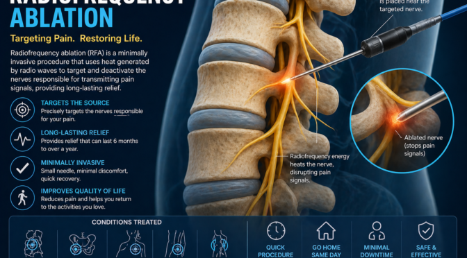
Radiofrequency Ablation is a minimally invasive nerve ablation procedure designed to target and disrupt the nerves responsible for transmitting pain signals. Unlike treatments that simply mask symptoms, this advanced technique focuses on eliminating pain at its source. As a widely used method of RFA for pain, it has become a preferred option for patients suffering from chronic joint conditions, especially in the spine, knees and hips. 

In this comprehensive guide, we will explore everything you need to know about Radiofrequency Ablation, including how it works, the different types available, its benefits, potential risks and what you can expect before, during and after the procedure. By the end of this article, you will have a clear understanding of how this innovative nerve ablation procedure can provide long-term relief and help you regain control of your daily life. 

What Is Radiofrequency Ablation? 

Understanding Radiofrequency Ablation in Detail 

Radiofrequency Ablation is a specialized medical technique used to treat chronic pain by targeting specific nerves and disrupting their ability to transmit pain signals. This procedure uses heat generated by radiofrequency energy to create a controlled lesion on the nerve, effectively reducing its function. 

Unlike surgical procedures, this nerve ablation procedure does not involve large incisions or removal of tissue. Instead, it is performed using a thin needle inserted through the skin, making it a minimally invasive option with a relatively quick recovery time. Patients who undergo RFA for pain often report significant improvement in their symptoms, especially when the source of pain has been accurately identified. 

This treatment is commonly recommended for individuals who have already tried conservative therapies such as medications or physical therapy without achieving lasting relief. It is also frequently used after successful diagnostic nerve blocks, which help confirm the exact nerve responsible for the pain. 

How Radiofrequency Ablation Works? 

The effectiveness of Radiofrequency Ablation lies in its ability to interrupt the transmission of pain signals. Pain travels through nerves to the brain, where it is interpreted as discomfort. By applying controlled heat to these nerves, the procedure disrupts their ability to send signals. 

During the nerve ablation procedure, a specialized needle is guided to the target nerve using imaging technology such as fluoroscopy. Once in position, radiofrequency energy is delivered through the needle, heating the nerve tissue and creating a lesion. This process reduces the nerve’s ability to function, thereby decreasing pain. 

Key mechanisms include: 

  • Blocking pain signals before they reach the brain  
  • Reducing nerve sensitivity in the affected area  
  • Providing sustained relief for months or longer  

This makes RFA for pain an effective solution for patients seeking long-term relief without relying on medications. 

Conditions Treated with Radiofrequency Ablation 

Radiofrequency Ablation is widely used to treat various chronic pain conditions, particularly those involving joints and the spine. 

Common conditions include: 

  • Facet joint arthritis in the spine  
  • Chronic neck and lower back pain  
  • Knee osteoarthritis  
  • Hip joint pain  
  • Sacroiliac joint dysfunction  

This versatile nerve ablation procedure is especially beneficial for patients whose pain originates from specific nerve pathways. 

Types of Radiofrequency Ablation Procedures 

Conventional Radiofrequency Ablation 

Conventional Radiofrequency Ablation is the most performed technique. It uses continuous radiofrequency energy to generate heat and create a precise lesion on the targeted nerve. 

This method of RFA for pain is highly effective for treating localized pain conditions, particularly those involving the spine. The precision of this technique ensures that only the targeted nerve is affected, minimizing the risk of damage to surrounding tissues. 

Patients who undergo this type of nerve ablation procedure often experience significant pain relief that lasts for several months. 

Pulsed Radiofrequency Ablation 

Pulsed Radiofrequency Ablation is a variation that uses intermittent bursts of energy instead of continuous heat. This approach is less aggressive and is often used for sensitive nerves or areas where preserving nerve function is important. 

Benefits include: 

  • Reduced risk of nerve damage  
  • Suitable for delicate or complex cases  
  • Provides pain modulation rather than complete nerve disruption  

This form of RFA for pain is ideal for patients who require a more conservative approach to treatment. 

Cooled Radiofrequency Ablation 

Cooled Radiofrequency Ablation is an advanced technique that uses water-cooled electrodes to create larger and more uniform lesions. This allows for better coverage of the targeted nerve area, increasing the likelihood of successful pain relief. 

Advantages include: 

  • Greater precision and effectiveness  
  • Ability to treat larger joints such as the knee  
  • Longer-lasting results  

This modern nerve ablation procedure has become increasingly popular due to its enhanced outcomes and reliability. 

Benefits of Radiofrequency Ablation 

Long-Lasting Pain Relief 

One of the key benefits of Radiofrequency Ablation is its ability to deliver long-lasting pain relief. Unlike temporary treatments such as injections, this nerve ablation procedure can reduce pain for several months or even up to a year. 

Patients undergoing RFA for pain often report a substantial decrease in discomfort, allowing them to return to normal activities with greater ease. 

Minimally Invasive and Quick Recovery 

Radiofrequency Ablation is a minimally invasive procedure, meaning it does not require major surgery or extended hospital stays. 

Key benefits include: 

  • Performed on an outpatient basis  
  • Minimal downtime  
  • Faster recovery compared to surgery  

This makes the nerve ablation procedure an attractive option for patients seeking effective treatment without the risks of invasive surgery. 

Reduced Dependence on Medications 

Chronic pain is often managed with medications that can cause side effects or lead to dependency. Radiofrequency Ablation addresses the root cause of pain, reducing the need for ongoing medication use. 

Benefits include: 

  • Lower risk of addiction  
  • Fewer side effects  
  • Improved overall health  

This makes RFA for pain a safer and more sustainable solution for long-term pain management. 

Procedure: What to Expect 

Before the Procedure 

Before undergoing Radiofrequency Ablation, patients undergo a thorough evaluation to ensure they are suitable candidates. This includes reviewing medical history, conducting imaging tests and performing diagnostic nerve blocks. 

These steps help confirm the source of pain and increase the likelihood of success for the nerve ablation procedure

During the Procedure 

The procedure itself is relatively quick and typically takes between 30 and 60 minutes. 

Steps include: 

  1. Cleaning and numbing the treatment area  
  1. Using imaging guidance to position the needle  
  1. Delivering radiofrequency energy to the target nerve  

Throughout the RFA for pain procedure, patients remain awake but comfortable. 

After the Procedure 

After Radiofrequency Ablation, patients are monitored briefly before being discharged. 

Post-procedure care includes: 

  • Resting for 24 hours  
  • Avoiding strenuous activities  
  • Gradually resuming normal routines  

Most patients notice pain relief within a few days to weeks. 

Risks and Side Effects 

Common Side Effects 

While Radiofrequency Ablation is generally safe, some mild side effects may occur. 

These include: 

  • Temporary soreness at the treatment site  
  • Mild swelling or bruising  
  • Slight numbness  

Potential Risks 

Although rare, complications can occur, including infection or nerve irritation. 

Choosing an experienced provider reduces risks associated with this nerve ablation procedure

How Are Risks Minimized? 

Doctors use imaging guidance and sterile techniques to ensure the safety of RFA for pain procedures. 

Is Radiofrequency Ablation Right for You? 

Ideal Candidates 

Radiofrequency Ablation is ideal for patients with chronic joint pain who have not responded to other treatments. 

Consultation and Decision-Making 

A thorough consultation helps determine whether this nerve ablation procedure is suitable for your condition. 

Conclusion 

Radiofrequency Ablation is a highly effective and minimally invasive solution for chronic joint pain. By targeting the nerves responsible for pain signals, this advanced nerve ablation procedure provides long-lasting relief and improves overall quality of life. As a proven method of RFA for pain, it offers a safe and reliable alternative to surgery and long-term medication use. 

If you are ready to take the next step toward pain relief, book a consultation today and discover how Radiofrequency Ablation can help you regain mobility and live a more comfortable life. 
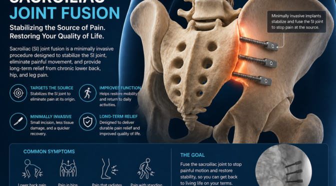
Sacroiliac Joint Fusion: When Chronic SI Joint Pain Requires Surgery 

Lower back pain is often assumed to originate from the spine, but in many cases, the true source lies elsewhere. One commonly overlooked cause is the sacroiliac (SI) joint-a small yet critical structure that connects the lower spine to the pelvis. When this joint becomes inflamed, unstable or damaged, it can lead to persistent discomfort that affects mobility, posture and overall quality of life. For individuals experiencing long-term pain that does not respond to conservative therapies, sacroiliac joint fusion may be the most effective path forward. 

Sacroiliac joint fusion is a specialized surgical procedure designed to stabilize the SI joint by permanently joining the bones together. This eliminates abnormal movement, which is often the primary source of pain. As an advanced form of SI joint pain treatment, it is typically recommended only after non-surgical approaches-such as medications, injections and physical therapy-have failed to provide adequate relief. 

Have you ever wondered why your lower back pain persists despite trying multiple treatments? Or why sitting, standing or even walking for short periods causes discomfort? These are common questions among patients with SI joint dysfunction. Understanding the underlying issue is the first step toward effective treatment. 

In this comprehensive guide, we will explore every aspect of sacroiliac joint fusion, including the anatomy of the SI joint, causes and symptoms of dysfunction, non-surgical options, surgical techniques, recovery expectations and long-term outcomes. By the end, you will have a clear and detailed understanding of whether this SI joint pain treatment could be the right solution for your condition. 

Understanding Sacroiliac Joint Pain 

What Is the Sacroiliac Joint and Why Is It Important? 

The sacroiliac joints are located at the base of the spine, where the sacrum (the triangular bone at the bottom of the spine) meets the iliac bones of the pelvis. There are two SI joints-one on each side-and they play a crucial role in maintaining stability and balance. 

Although these joints have limited movement compared to others in the body, their function is essential. They act as shock absorbers, distributing weight and forces between the upper body and lower extremities. Every time you walk, sit or lift something, the SI joints help stabilize your body. 

When these joints function properly, they operate almost unnoticed. However, when they become inflamed or unstable, even minor movements can trigger significant pain. In such cases, patients may eventually require sacroiliac joint fusion if other treatments do not restore stability. 

Common Causes of SI Joint Dysfunction 

SI joint dysfunction can result from a wide range of factors, often making diagnosis challenging. The condition may develop gradually or occur suddenly after an injury. 

Some of the most common causes include: 

  • Degenerative arthritis: Wear and tear over time can damage the joint surfaces, leading to inflammation and pain.  
  • Trauma or injury: Falls, car accidents or sports injuries can disrupt the joint’s alignment.  
  • Pregnancy-related changes: Hormonal shifts loosen ligaments, increasing joint instability.  
  • Previous spinal surgery: Procedures like lumbar fusion can place additional stress on the SI joints.  
  • Repetitive stress: Activities that involve heavy lifting or twisting motions can strain the joint.  

When these issues persist, they may require advanced SI joint pain treatment, including surgical intervention such as sacroiliac joint fusion

Symptoms and How They Progress Over Time 

SI joint pain often starts subtly but can worsen if left untreated. Early symptoms may be mild and intermittent, making them easy to ignore. 

Common symptoms include: 

  • Pain in the lower back or buttocks  
  • Discomfort radiating to the hips, groin or thighs  
  • Increased pain when sitting or standing for long periods  
  • Difficulty transitioning from sitting to standing  
  • Reduced range of motion  

Over time, the pain may become chronic and more intense, interfering with daily activities and sleep. When symptoms reach this stage and conservative treatments fail, sacroiliac joint fusion may be considered as a long-term SI joint pain treatment

Non-Surgical SI Joint Pain Treatment Options 

Physical Therapy and Lifestyle Modifications 

Before considering surgery, doctors typically recommend non-invasive approaches to manage SI joint pain. Physical therapy is often the first line of defence and focuses on strengthening the muscles surrounding the joint. 

Therapists design customized exercise programs to improve stability, flexibility and posture. Strengthening the core and pelvic muscles helps reduce strain on the SI joint, which can significantly alleviate pain. 

Lifestyle modifications also play an important role. Patients are often advised to: 

  • Avoid prolonged sitting or standing  
  • Use proper lifting techniques  
  • Maintain a healthy weight  
  • Incorporate low-impact exercises such as swimming or walking  

These strategies form the foundation of effective SI joint pain treatment and can delay or eliminate the need for sacroiliac joint fusion in many cases. 

Medications and Pain Management 

Medications are commonly used to manage inflammation and discomfort associated with SI joint dysfunction. Nonsteroidal anti-inflammatory drugs (NSAIDs) are frequently prescribed to reduce swelling and relieve pain. 

In some cases, muscle relaxants or prescription pain medications may be recommended. While these treatments can provide temporary relief, they do not address the underlying cause of the problem. 

As a result, patients with persistent symptoms may eventually explore more advanced SI joint pain treatment options, including sacroiliac joint fusion

Injections and Minimally Invasive Procedures 

Injection therapies are another important component of non-surgical treatment. Corticosteroid injections directly target inflammation within the SI joint, providing temporary relief. 

Diagnostic injections are also used to confirm whether the SI joint is the source of pain. If the injection significantly reduces pain, it indicates that the joint is likely responsible. 

While injections can be effective, their benefits are often short-lived. For patients who require repeated injections without lasting improvement, sacroiliac joint fusion may be recommended as a definitive SI joint pain treatment

What Is Sacroiliac Joint Fusion? 

Detailed Overview of the Procedure 

Sacroiliac joint fusion is a surgical technique designed to stabilize the SI joint by permanently joining the bones. This eliminates abnormal motion, which is often the root cause of pain. 

The procedure involves placing implants or bone grafts across the joint, allowing the bones to grow together over time. As the joint fuses, it becomes stable and no longer produces pain during movement. 

As a long-term SI joint pain treatment, this procedure is particularly beneficial for patients with severe or chronic symptoms. 

Step-by-Step Surgical Process 

Modern sacroiliac joint fusion is typically performed using minimally invasive techniques, which reduce recovery time and complications. 

The procedure generally includes: 

  1. Administering anaesthesia to ensure patient comfort  
  1. Making a small incision near the SI joint  
  1. Using imaging guidance to position instruments accurately  
  1. Inserting implants or bone graft material  
  1. Closing the incision  

This advanced SI joint pain treatment usually takes less than an hour and is often performed on an outpatient basis. 

Types of Fusion Techniques Explained 

There are different approaches to sacroiliac joint fusion, depending on the patient’s condition. 

These include: 

  • Minimally invasive fusion: Smaller incisions, faster recovery  
  • Open surgery: Used in complex cases, involves larger incisions  

Minimally invasive techniques are preferred because they offer better outcomes and fewer complications, making them a leading SI joint pain treatment option. 

Benefits of Sacroiliac Joint Fusion 

Long-Term Pain Relief and Stability 

One of the primary advantages of sacroiliac joint fusion is its ability to provide long-lasting pain relief. By stabilizing the joint, the procedure eliminates the source of discomfort. 

Patients often experience significant improvement in pain levels within weeks to months after surgery. 

Improved Mobility and Daily Function 

Once the joint is stabilized, patients can perform daily activities with greater ease and confidence. 

Benefits include: 

  • Easier walking and movement  
  • Reduced stiffness  
  • Improved posture  

This makes it a highly effective SI joint pain treatment for restoring quality of life. 

Reduced Need for Ongoing Treatments 

After successful sacroiliac joint fusion, many patients no longer require frequent medications or injections. This reduces the long-term burden of managing chronic pain. 

Risks and Considerations 

Potential Complications 

Although sacroiliac joint fusion is generally safe, potential risks include infection, bleeding and nerve irritation. 

Recovery Challenges and Expectations 

Recovery demands patience and careful adherence to medical guidance. Patients must follow activity restrictions and attend follow-up appointments. 

Minimizing Risks 

Choosing a skilled surgeon and following post-operative guidelines can significantly reduce complications associated with this SI joint pain treatment

Recovery and Rehabilitation 

Immediate Recovery Phase 

After sacroiliac joint fusion, patients are monitored and typically discharged within a day. Mild discomfort is common but manageable. 

Long-Term Healing Process 

Healing takes several weeks to months as the bones fuse together. Pain and mobility typically improve gradually over time. 

Role of Physical Therapy 

Physical therapy is essential for strengthening muscles and ensuring the success of this SI joint pain treatment

Conclusion 

Sacroiliac joint fusion is a highly effective solution for individuals suffering from chronic SI joint pain that does not respond to conservative treatments. By stabilizing the joint and eliminating painful motion, this advanced SI joint pain treatment offers long-term relief and improved quality of life. 

If you are struggling with persistent pain, now is the time to act. Book a consultation today to find out whether sacroiliac joint fusion is the right treatment for you and start your journey toward lasting relief. 

Peripheral Nerve Stimulation: A Modern Approach to Chronic Pain 

Chronic pain is a condition that affects millions of people worldwide, often limiting mobility, reducing productivity and significantly impacting emotional well-being. For many individuals, traditional treatments such as medications, physical therapy or even surgery may not provide sufficient or lasting relief. This ongoing struggle leads patients to seek innovative and more effective solutions. One such breakthrough treatment is Peripheral Nerve Stimulation, a cutting-edge technique designed to target pain at its source. 

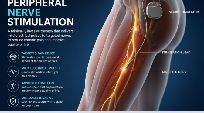
Peripheral Nerve Stimulation is a modern form of nerve stimulation therapy that focuses on interrupting pain signals before they reach the brain. Instead of simply masking discomfort, this therapy works by modifying how the nervous system processes pain, offering a more sustainable and targeted approach. It is particularly beneficial for individuals suffering from chronic nerve-related pain conditions that have not responded well to conventional treatments. 

In this in-depth guide, we will explore every aspect of Peripheral Nerve Stimulation, including how it works, the different types available, its benefits, risks and what patients can expect during and after the procedure. By the end of this article, you will have a complete understanding of how this advanced nerve stimulation therapy can transform pain management and improve overall quality of life. 

What Is Peripheral Nerve Stimulation? 

Understanding Peripheral Nerve Stimulation in Detail 

Peripheral Nerve Stimulation is a minimally invasive medical procedure that uses electrical impulses to target specific nerves responsible for pain. These peripheral nerves exist outside the brain and spinal cord and act as pathways for transmitting sensory information, including pain signals. When these nerves become irritated or damaged, they can send persistent pain signals to the brain, resulting in chronic discomfort. 

This innovative nerve stimulation therapy involves placing small electrodes near the affected nerve. These electrodes are linked to a device that emits regulated electrical impulses. The purpose of these pulses is to disrupt or alter the pain signals before they reach the brain. As a result, the brain either does not perceive the pain or interprets it differently, often as a mild tingling sensation. 

Unlike traditional treatments that rely heavily on medication, Peripheral Nerve Stimulation directly addresses the root cause of pain. This targeted approach makes it highly effective for managing chronic conditions, especially when other treatments have failed. 

How Peripheral Nerve Stimulation Works? 

The science behind Peripheral Nerve Stimulation is based on neuromodulation, a process that alters nerve activity through electrical stimulation. Pain signals pass through the nerves to the brain, where they are perceived as discomfort. By introducing electrical impulses, this nerve stimulation therapy modifies these signals, effectively reducing or eliminating the sensation of pain. 

The process works in several ways: 

  • It blocks pain signals from reaching the brain  
  • It replaces pain sensations with mild, non-painful tingling  
  • It reduces nerve hypersensitivity over time  

This combination of effects makes Peripheral Nerve Stimulation a powerful and long-lasting solution for chronic pain management. 

Conditions Treated with Peripheral Nerve Stimulation 

Peripheral Nerve Stimulation is used to treat a wide range of chronic pain conditions, particularly those involving nerve damage or irritation. It is often recommended when other treatments have not provided adequate relief. 

Common conditions include: 

  • Neuropathic pain caused by nerve damage  
  • Chronic back and neck pain  
  • Post-surgical pain syndromes  
  • Migraine and chronic headaches  
  • Complex Regional Pain Syndrome (CRPS)  

This versatile nerve stimulation therapy can be customized to target specific nerves, making it suitable for various pain conditions. 

Types of Peripheral Nerve Stimulation Systems 

Implantable Peripheral Nerve Stimulation Devices 

Implantable devices are one of the most widely used forms of Peripheral Nerve Stimulation. These systems involve surgically placing electrodes near the targeted nerve and connecting them to a small pulse generator implanted under the skin. 

This type of nerve stimulation therapy is designed for long-term pain management. The device continuously delivers electrical impulses, which can be adjusted based on the patient’s needs. Patients are often given a remote control or mobile app to modify the intensity and frequency of stimulation. 

Key advantages include: 

  • Continuous and consistent pain relief  
  • Customizable settings for individual needs  
  • Long-term solution for chronic pain  

Temporary Peripheral Nerve Stimulation Systems 

Temporary systems are often used as a trial phase before committing to permanent implantation. In this approach, electrodes are placed near the nerve for a short period, usually a few days to weeks. 

This trial allows patients and doctors to evaluate the effectiveness of Peripheral Nerve Stimulation before making a long-term decision. It is a crucial step in ensuring that the therapy will provide meaningful relief. 

Benefits include: 

  • Non-permanent and reversible  
  • Helps assess treatment success  
  • Minimally invasive  

Wireless and Advanced Technologies 

Recent advancements have significantly improved the design and functionality of Peripheral Nerve Stimulation devices. Modern systems are smaller, more efficient and often wireless, enhancing patient comfort and convenience. 

Innovations include: 

  • Wireless devices controlled via smartphones  
  • Miniaturized implants with minimal visibility  
  • Enhanced targeting for precise pain relief  

These developments have made nerve stimulation therapy more accessible and effective for a broader range of patients. 

Benefits of Peripheral Nerve Stimulation 

Targeted Pain Relief Without Medication 

One of the most significant benefits of Peripheral Nerve Stimulation is its ability to provide targeted pain relief without relying on medications. Traditional pain management often involves drugs that affect the entire body, leading to unwanted side effects. 

This nerve stimulation therapy focuses directly on the source of pain, reducing the need for systemic treatments and improving overall safety. 

Minimally Invasive and Reversible 

Unlike major surgical procedures, Peripheral Nerve Stimulation is minimally invasive. The implantation process involves small incisions and typically requires only local anaesthesia. 

Additionally, the treatment is reversible. If a patient decides to discontinue therapy, the device can be removed without permanent changes to the body. This flexibility makes nerve stimulation therapy a low-risk option. 

Improved Quality of Life 

Patients who undergo Peripheral Nerve Stimulation often report significant improvements in their daily lives. By reducing pain, the therapy enables individuals to engage in activities they previously avoided. 

Improvements include: 

  • Better sleep patterns  
  • Increased physical activity  
  • Enhanced emotional well-being  
  • Greater independence  

This transformative impact makes nerve stimulation therapy a valuable tool in modern pain management. 

Procedure: What to Expect 

Pre-Procedure Evaluation 

Before undergoing Peripheral Nerve Stimulation, patients undergo a comprehensive evaluation. This includes reviewing medical history, conducting physical examinations and sometimes performing imaging tests. 

The goal is to identify the exact source of pain and determine whether this nerve stimulation therapy is suitable for the patient. 

Trial Phase 

The trial phase is a critical step in the Peripheral Nerve Stimulation process. Temporary electrodes are placed near the targeted nerve and the patient monitors their pain relief over several days. 

This phase helps ensure that the therapy is effective before proceeding with permanent implantation. 

Permanent Implantation 

If the trial yields positive results, a permanent device is implanted. The procedure is generally quick and is typically performed on an outpatient basis. 

After implantation, the Peripheral Nerve Stimulation device can be adjusted to provide optimal pain relief. 

Risks and Considerations 

Common Side Effects 

While Peripheral Nerve Stimulation is generally safe, some patients may experience mild side effects. 

These include: 

  • Temporary discomfort at the implant site  
  • Mild swelling or bruising  
  • Skin irritation  

Potential Risks 

Although rare, there are potential risks associated with Peripheral Nerve Stimulation, such as infection or device malfunction. 

Working with an experienced specialist ensures that this nerve stimulation therapy is performed safely. 

Who Should Avoid This Therapy? 

Certain individuals may not be suitable candidates for Peripheral Nerve Stimulation, including those with active infections or specific medical conditions. 

Is Peripheral Nerve Stimulation Right for You? 

Ideal Candidates 

Peripheral Nerve Stimulation is ideal for patients with chronic pain that has not responded to conventional treatments. 

Consultation and Decision-Making 

A detailed consultation with a pain specialist is essential to determine whether this nerve stimulation therapy is the right choice. 

Conclusion 

Peripheral Nerve Stimulation is a modern, effective and minimally invasive solution for managing chronic pain. By targeting specific nerves and altering pain signals, this advanced nerve stimulation therapy offers long-lasting relief and improved quality of life. 

If you are tired of living with persistent pain and are looking for a reliable solution, it may be time to explore this innovative treatment. Book a consultation today to discover how Peripheral Nerve Stimulation can help you regain control and live pain-free. 

Laminectomy Surgery Explained: Indications, Benefits and Recovery 

Living with persistent back pain, numbness in the legs or difficulty walking can make even simple daily activities feel exhausting and overwhelming. Many individuals try medications, physical therapy and lifestyle changes, yet the pain continues to interfere with their quality of life. When conservative treatments fail, laminectomy surgery is often recommended as a reliable solution to relieve nerve pressure and restore mobility. 

Laminectomy surgery is a well-established form of spinal decompression surgery that involves removing a portion of the vertebra known as the lamina. This creates more space within the spinal canal, reducing pressure on the spinal cord and nerves. Conditions such as spinal stenosis, herniated discs and bone spurs often lead to nerve compression, making this procedure highly effective for long-term relief. 

If you’re considering surgical options, understanding the procedure is essential. In this comprehensive guide, you will learn everything about laminectomy surgery, including when it is needed, how it is performed, its benefits, potential risks and a detailed laminectomy recovery timeline. By the end, you will have a clear understanding of whether this treatment could be the right step toward a pain-free life. 

What Is Laminectomy Surgery? 

Understanding Laminectomy Surgery in Detail 

Laminectomy surgery is a surgical procedure designed to relieve pressure on the spinal cord or nerves by removing part or all the lamina, which is the back portion of a vertebra. This pressure often develops due to narrowing of the spinal canal, a condition known as spinal stenosis. Over time, this narrowing can compress nerves and lead to chronic pain, weakness and reduced mobility. 

As a widely performed spinal decompression surgery, this procedure directly targets the root cause of nerve compression rather than simply managing symptoms. By increasing the available space within the spine, it allows nerves to function properly again, reducing inflammation and discomfort. 

How Spinal Compression Develops? 

Spinal compression is typically a gradual process caused by aging or degenerative conditions. As the spine undergoes wear and tear, structures like discs and ligaments may thicken or shift, leading to reduced space within the spinal canal. 

Common causes include: 

  • Spinal stenosis, where the spinal canal narrows  
  • Herniated discs, pressing against nearby nerves  
  • Bone spurs, often caused by arthritis  
  • Thickened ligaments, reducing spinal flexibility  

These conditions often require spinal decompression surgery, especially when symptoms become severe and persistent. 

Types of Laminectomy Surgery 

The procedure can be performed on different parts of the spine depending on where the compression occurs. 

Types include: 

  • Cervical laminectomy – performed in the neck region  
  • Thoracic laminectomy – targets the mid-back  
  • Lumbar laminectomy – most common, performed in the lower back  

Each type of laminectomy surgery is tailored to the patient’s condition, ensuring precise and effective treatment. 

Indications for Laminectomy Surgery 

Medical Conditions That Require Laminectomy Surgery 

Doctors recommend laminectomy surgery when patients experience severe symptoms that do not respond to non-surgical treatments. The procedure is particularly effective in relieving nerve compression caused by structural issues in the spine. 

Conditions that may require surgery include: 

  • Spinal stenosis  
  • Degenerative disc disease  
  • Herniated discs  
  • Spinal tumours or cysts  

In such cases, spinal decompression surgery becomes necessary to prevent worsening symptoms and improve function. 

Symptoms That Signal the Need for Surgery 

Recognizing the symptoms of nerve compression is essential for timely treatment. Many patients initially ignore mild discomfort, but as the condition progresses, symptoms become more severe and persistent. 

Key symptoms include: 

  • Chronic back or neck pain  
  • Numbness or tingling in arms or legs  
  • Muscle weakness  
  • Difficulty standing or walking for long periods  

These symptoms often improve significantly after laminectomy recovery, making early diagnosis and treatment important. 

Diagnostic Evaluation Before Surgery 

Before recommending laminectomy surgery, doctors perform detailed evaluations to confirm the diagnosis and determine the exact location of nerve compression. 

Common diagnostic methods include: 

  • MRI scans to visualize soft tissues and nerves  
  • CT scans for detailed bone imaging  
  • X-rays to assess spinal alignment  
  • Physical examinations to evaluate symptoms  

These tests ensure that spinal decompression surgery is the most appropriate treatment option. 

Benefits of Laminectomy Surgery 

Effective Pain Relief 

One of the most significant advantages of laminectomy surgery is its ability to relieve chronic pain caused by nerve compression. Patients often experience noticeable improvement shortly after the procedure. 

Key benefits include: 

  • Reduced nerve pain  
  • Decreased inflammation  
  • Relief from radiating pain in limbs  

As a proven spinal decompression surgery, it addresses the root cause rather than masking symptoms. 

Improved Mobility and Function 

Many patients struggle with limited movement due to pain and stiffness. Following laminectomy recovery, they often regain the ability to perform daily activities with ease. 

Improvements may include: 

  • Better flexibility  
  • Increased walking ability  
  • Enhanced overall mobility  

Enhanced Quality of Life 

Chronic pain can affect mental health, sleep and overall well-being. Successful laminectomy surgery can significantly improve quality of life by restoring independence and comfort. 

Patients often report: 

  • Better sleep patterns  
  • Reduced stress and anxiety  
  • Increased participation in daily activities  

Risks and Complications of Laminectomy Surgery 

General Surgical Risks 

Like any surgical procedure, laminectomy surgery carries certain risks. However, these risks are generally low when performed by experienced professionals. 

Common risks include: 

  • Infection  
  • Bleeding  
  • Blood clots  
  • Reactions to anaesthesia  

Specific Risks of Spinal Decompression Surgery 

Some complications are specific to spinal decompression surgery and require careful consideration. 

These may include: 

  • Nerve damage  
  • Spinal instability  
  • Persistent pain  
  • Leakage of spinal fluid  

How Risks Are Minimized? 

Advancements in medical technology have improved the safety of laminectomy surgery. Surgeons now use minimally invasive techniques that reduce complications and speed up laminectomy recovery

Preventive measures include: 

  • Thorough preoperative assessment  
  • Advanced imaging techniques  
  • Postoperative monitoring  

Laminectomy Surgery Procedure Explained 

Preparation Before Surgery 

Preparation is a crucial step in ensuring the success of laminectomy surgery. Patients undergo medical evaluations and receive instructions to prepare for the procedure. 

Preparation steps include: 

  • Reviewing medical history  
  • Stopping certain medications  
  • Fasting before surgery  

Step-by-Step Procedure 

During laminectomy surgery, the surgeon carefully removes the lamina to relieve pressure on the nerves. 

Procedure steps: 

  1. Administration of anaesthesia  
  1. Making a small incision near the affected area  
  1. Removing part of the lamina  
  1. Relieving pressure on the spinal cord  

This spinal decompression surgery typically takes a few hours, depending on the complexity. 

Postoperative Care 

After surgery, proper care is essential for successful laminectomy recovery

Postoperative care includes: 

  • Pain management  
  • Wound care  
  • Gradual movement and rehabilitation  

Laminectomy Recovery Timeline 

Immediate Recovery Phase 

The first few days after laminectomy surgery focus on pain management and monitoring for complications. Patients may stay in the hospital for observation. 

Key aspects include: 

  • Controlled movement  
  • Pain relief medications  
  • Initial physical therapy  

Short-Term Recovery 

Within a few weeks, patients notice improvements in mobility and pain levels. Laminectomy recovery during this phase involves gradual return to daily activities. 

Progress includes: 

  • Increased activity levels  
  • Reduced discomfort  
  • Improved strength  

Long-Term Recovery 

Full laminectomy recovery may take several months, depending on the individual’s condition and overall health. 

Long-term outcomes include: 

  • Restored mobility  
  • Reduced symptoms  
  • Improved quality of life  

Conclusion 

Laminectomy surgery is a highly effective solution for relieving nerve compression and restoring mobility. As a trusted form of spinal decompression surgery, it addresses the root cause of pain and helps patients regain control over their lives. With proper care and a structured laminectomy recovery plan, most individuals experience significant improvements in both function and comfort. 

If chronic back pain is limiting your daily activities, don’t wait to seek help. Book a consultation today with a qualified spine specialist to explore whether this procedure is right for you and take the first step toward a healthier, pain-free future.

Spinal Cord Stimulation for Chronic Pain: Patient Benefits and Results 

Chronic pain is more than just a physical condition – it is a life-altering experience that affects mobility, sleep, emotional health and overall well-being. Millions of people struggle daily with persistent pain that does not respond to medications, physical therapy or even surgery. If you have tried multiple treatments without success, you may feel frustrated, exhausted and unsure of what to do next. This is where spinal cord stimulation emerges as a promising solution. 

Spinal cord stimulation is an advanced and minimally invasive therapy designed to manage chronic pain by interrupting pain signals before they reach the brain. Instead of masking the pain with medications, this treatment targets the nervous system directly, offering a more sustainable and effective approach. A spinal cord stimulator for pain is a small, implanted device that delivers controlled electrical impulses to the spinal cord, altering how pain is perceived. 

In this detailed guide, we will explore every aspect of spinal cord stimulation, including how it works, who qualifies for it, the procedure involved, benefits, risks and long-term results. By the end of this article, you will have a complete understanding of how this treatment works and whether it may be the right option for your chronic pain management journey. 

What Is Spinal Cord Stimulation? 

Understanding Spinal Cord Stimulation in Depth 

Spinal cord stimulation is a medical procedure used to treat chronic pain by delivering low-level electrical signals to the spinal cord. These signals interfere with pain messages traveling to the brain, effectively reducing the sensation of pain. This technique is particularly useful for nerve-related pain, which is often difficult to treat using conventional methods. 

Unlike medications that temporarily reduce symptoms, spinal cord stimulation addresses the root cause of pain perception. The therapy modifies how the brain interprets pain signals, allowing patients to experience relief without heavy reliance on drugs. A spinal cord stimulator for pain can be adjusted to meet individual needs, making it a highly personalized treatment option. 

How a Spinal Cord Stimulator for Pain Works? 

The system consists of several components that work together to deliver pain relief. Electrodes are placed near the spinal cord and a small device called a pulse generator is implanted under the skin. This generator sends electrical impulses through the electrodes to the spinal cord. 

These impulses disrupt pain signals, replacing them with a mild tingling sensation or in newer devices, a completely pain-free experience without any sensation. The flexibility of a spinal cord stimulator for pain allows patients to control the intensity and pattern of stimulation using a remote device. 

Conditions Treated with Spinal Cord Stimulation 

Doctors recommend spinal cord stimulation for a variety of chronic pain conditions, especially those involving nerve damage or dysfunction. 

Common conditions include: 

  • Failed Back Surgery Syndrome (FBSS) 
  • Complex Regional Pain Syndrome (CRPS)  
  • Peripheral neuropathy  
  • Chronic leg or arm pain  

For these conditions, a spinal cord stimulator for pain often provides significant relief when other treatments have failed. 

Who Is a Candidate for Spinal Cord Stimulation? 

Ideal Candidates for Spinal Cord Stimulation 

Not everyone with chronic pain is a suitable candidate for spinal cord stimulation. Doctors carefully evaluate each patient to ensure the treatment will be effective. Ideal candidates are typically those who have experienced chronic pain for more than six months and have not responded to other treatments. 

Patients who benefit most from this therapy often have nerve-related pain rather than muscle or joint pain. Additionally, individuals who are seeking to reduce their dependence on pain medications may find this treatment particularly appealing. 

Symptoms That Indicate Suitability 

Certain symptoms suggest that a spinal cord stimulator for pain may be an effective option. These symptoms are often related to nerve dysfunction and include persistent, radiating pain that does not improve with standard treatments. 

Common symptoms include: 

  • Burning or stabbing pain  
  • Tingling or “pins and needles” sensations  
  • Pain that worsens with movement  
  • Limited relief from medications  

These symptoms indicate that spinal cord stimulation may be able to target the underlying issue effectively. 

Evaluation and Trial Phase 

Before a permanent device is implanted, patients undergo a trial period to determine whether spinal cord stimulation will provide sufficient relief. During this phase, temporary electrodes are placed near the spinal cord and an external device is used to deliver electrical impulses. 

Patients typically use the trial system for several days while monitoring their pain levels and daily activities. If significant improvement is observed, a permanent spinal cord stimulator for pain is implanted. This trial phase ensures that patients only proceed with the treatment if it is likely to be beneficial. 

Benefits of Spinal Cord Stimulation 

Effective and Targeted Pain Relief 

One of the most significant advantages of spinal cord stimulation is its ability to provide targeted pain relief. Unlike medications that affect the entire body, this treatment focuses specifically on the affected nerves. 

Patients often report a substantial reduction in pain intensity, allowing them to perform daily activities with greater ease. A spinal cord stimulator for pain can also be adjusted to provide optimal relief at different times of the day. 

Reduced Dependence on Medications 

Chronic pain is often managed with strong medications, including opioids, which can lead to side effects and dependency. Spinal cord stimulation offers an alternative by reducing the need for these medications. 

Patients who undergo this treatment frequently experience fewer side effects and improved overall health. The ability to manage pain without relying heavily on drugs is a major benefit of a spinal cord stimulator for pain

Improved Quality of Life 

Chronic pain can impact all areas of life, including physical function and emotional health. Successful spinal cord stimulation can lead to significant improvements in quality of life. 

Patients often experience: 

  • Better sleep patterns  
  • Increased mobility  
  • Improved mental health  

These improvements make a spinal cord stimulator for pain a valuable option for long-term pain management. 

Spinal Cord Stimulation Procedure Explained 

Preparation Before the Procedure 

Preparation for spinal cord stimulation involves a thorough evaluation to ensure the patient is a suitable candidate. Doctors review medical history, conduct imaging tests and provide instructions for the procedure. 

Patients may be asked to stop certain medications and follow specific guidelines to reduce the risk of complications. Adequate preparation plays a crucial role in ensuring the success of the treatment. 

Trial Procedure 

The trial phase is a critical step in the process. Temporary electrodes are placed near the spinal cord and an external device is used to deliver electrical impulses. 

During this period, patients evaluate the effectiveness of the treatment in reducing pain and improving daily activities. This phase helps determine whether a permanent spinal cord stimulator for pain should be implanted. 

Permanent Implantation 

If the trial is successful, the permanent device is implanted during a minimally invasive procedure. Electrodes are carefully positioned along the spinal cord and the pulse generator is implanted just under the skin 

The device is then calibrated to ensure the correct level of stimulation. Patients can adjust the settings as needed, making spinal cord stimulation a flexible and customizable treatment option. 

Risks and Complications 

General Risks 

While spinal cord stimulation is considered safe, it does carry some risks. Overall, the risks are low and can be lessened through proper care. 

Common risks include: 

  • Infection  
  • Bleeding  
  • Pain at the implant site  

Device-Related Complications 

Certain complications are directly related to the device itself. A spinal cord stimulator for pain may occasionally require adjustments or repairs. 

Possible issues include: 

  • Movement of the electrodes  
  • Device malfunction  
  • Incomplete pain relief  

Managing and Reducing Risks 

Advancements in medical technology have significantly improved the safety of spinal cord stimulation. Regular follow-ups and proper device maintenance help reduce the risk of complications. 

Patients are encouraged to follow their doctor’s instructions and report any unusual symptoms promptly. 

Results and Long-Term Outcomes 

Short-Term Results 

Many patients experience immediate relief after starting spinal cord stimulation. Pain levels often decrease significantly within the first few days. 

This early improvement helps patients regain confidence and begin returning to normal activities. 

Long-Term Benefits 

Over time, a spinal cord stimulator for pain continues to provide consistent relief. Patients often report sustained improvements in mobility and overall well-being. 

Long-term benefits include: 

  • Reduced pain intensity  
  • Improved physical function  
  • Enhanced quality of life  

Success Rates 

Research shows that spinal cord stimulation can reduce pain by 50% or more in many patients. These results make it one of the most effective treatments for chronic nerve-related pain. 

Recovery and Lifestyle After Treatment 

Recovery Timeline 

Recovery from spinal cord stimulation is relatively quick compared to major surgeries. In most cases, patients can go back to their regular activities within a few weeks. 

The recovery process includes: 

  • Initial rest and healing  
  • Gradual increase in activity  
  • Follow-up appointments  

Lifestyle Adjustments 

After receiving a spinal cord stimulator for pain, patients may need to make minor lifestyle adjustments to ensure optimal results. 

Recommendations include: 

  • Avoiding heavy lifting initially  
  • Maintaining good posture  
  • Following medical advice  

Maintaining Long-Term Results 

Long-term success depends on proper care and regular monitoring. Patients should stay in contact with their healthcare providers and attend follow-up visits. 

By maintaining a healthy lifestyle and properly using the device, patients can maximize the benefits of spinal cord stimulation

Conclusion 

Spinal cord stimulation offers a powerful and effective solution for managing chronic pain, especially for patients who have not found relief through traditional treatments. By using advanced technology, a spinal cord stimulator for pain helps block pain signals and improve overall quality of life. 

If chronic pain is affecting your daily life, now is the time to explore your options. Book a consultation today with a pain specialist and take the first step toward a more comfortable, active and pain-free future. 

Nerve Blocks for Pain Relief: Types, Benefits and How Long They Last 

Chronic pain is one of the most challenging health conditions to live with, often affecting not just the body but also emotional well-being, productivity and overall quality of life. Whether it stems from injury, nerve damage, arthritis or post-surgical complications, persistent pain can make even simple daily activities feel overwhelming. Many patients rely on medications, physical therapy or lifestyle changes, but these treatments do not always provide lasting relief. This is where nerve blocks for pain come into focus as a highly targeted and effective solution. 

Nerve blocks for pain are medical procedures that interrupt pain signals traveling through the nervous system. By injecting medication directly near specific nerves, doctors can significantly reduce or even eliminate pain in a localized area. A pain management nerve block is not only used to relieve discomfort but also to diagnose the source of pain, making it a versatile and valuable tool in modern medicine. 

In this comprehensive guide, we will explore every aspect of nerve blocks for pain, including how they work, the different types available, their benefits, risks and how long the effects typically last. By the end of this article, you will have a clear and detailed understanding of whether this treatment option could be the right choice for your pain management journey. 

What Are Nerve Blocks for Pain? 

Understanding Nerve Blocks for Pain in Detail 

Nerve blocks for pain are specialized medical procedures designed to stop or reduce pain signals before they reach the brain. These procedures involve injecting a combination of medications-usually local anaesthetics and anti-inflammatory drugs-near a specific nerve or group of nerves responsible for transmitting pain signals. By doing so, the communication pathway between the affected area and the brain is temporarily interrupted. 

pain management nerve block can serve both therapeutic and diagnostic purposes. Therapeutically, it provides relief from acute or chronic pain, allowing patients to regain mobility and improve their quality of life. Diagnostically, it helps physicians identify the exact source of pain. For example, if a nerve block relieves pain in a specific area, it confirms that the targeted nerve is responsible for the discomfort. 

How Pain Signals Are Blocked? 

Pain is transmitted through a complex network of nerves that send signals from the affected area to the brain. When an injury or inflammation occurs, these signals become more frequent and intense, resulting in persistent pain. Nerve blocks for pain work by disrupting this communication process. 

The medications used in a pain management nerve block play different roles: 

  • Local anaesthetics numb the nerve, stopping pain signals instantly  
  • Steroids reduce inflammation around the nerve, providing longer-lasting relief  
  • Combination injections offer both immediate and extended benefits  

This dual-action approach makes nerve blocks for pain an effective solution for managing various types of pain conditions. 

Common Conditions Treated 

Doctors recommend nerve blocks for pain for a wide range of conditions, particularly those involving nerve irritation or inflammation. 

Common conditions include: 

  • Chronic back and neck pain  
  • Sciatica and nerve-related leg pain  
  • Migraines and cluster headaches  
  • Arthritis and joint pain  
  • Post-surgical pain  

pain management nerve block is often part of a comprehensive treatment plan that may also include physical therapy and lifestyle modifications. 

Types of Nerve Blocks for Pain 

Diagnostic vs Therapeutic Nerve Blocks for Pain 

There are two primary categories of nerve blocks for pain: diagnostic and therapeutic. Understanding the difference between these types is essential for determining the appropriate treatment approach. 

  • Diagnostic nerve blocks are used to identify the source of pain. By temporarily numbing a specific nerve, doctors can confirm whether it is responsible for the patient’s symptoms.  
  • Therapeutic nerve blocks are designed to provide pain relief, either temporarily or long-term.  

In many cases, a pain management nerve block begins as a diagnostic tool and then transitions into a therapeutic treatment once the source of pain is identified. 

Common Types of Nerve Blocks 

There are several types of nerve blocks for pain, each targeting specific areas of the body and addressing different conditions. 

Some of the most common types include: 

  • Epidural nerve blocks: Commonly used for back pain and childbirth, these injections target the epidural space around the spinal cord.  
  • Facet joint injections: These are used to treat pain originating from the joints of the spine.  
  • Peripheral nerve blocks: Target nerves outside the spinal cord, often used for limb pain or surgical procedures.  
  • Sympathetic nerve blocks: Used to treat conditions like complex regional pain syndrome (CRPS).  

Each pain management nerve block is tailored to the patient’s condition, ensuring precise and effective treatment. 

Advanced Techniques in Nerve Blocks 

Modern advancements have significantly improved the accuracy and effectiveness of nerve blocks for pain. Doctors now use imaging technologies to guide injections, ensuring that the medication is delivered exactly where it is needed. 

Advanced techniques include: 

  • Ultrasound-guided injections for real-time visualization  
  • Fluoroscopy (X-ray guidance) for precise placement  
  • Radiofrequency ablation, which uses heat to disable nerves for longer-lasting relief  

These innovations enhance the success rate of a pain management nerve block while minimizing risks and complications. 

Benefits of Nerve Blocks for Pain 

Targeted and Immediate Pain Relief 

One of the most significant advantages of nerve blocks for pain is their ability to provide targeted relief. Unlike oral medications, which affect the entire body, these injections focus directly on the source of pain. 

Patients often experience immediate relief due to the anesthetic component of the injection. A pain management nerve block can quickly reduce pain intensity, allowing patients to resume normal activities. 

Reduced Dependence on Medications 

Chronic pain is often managed with medications, including opioids, which can lead to side effects and dependency. Nerve blocks for pain offer an alternative that reduces the need for long-term medication use. 

Benefits include: 

  • Lower risk of addiction  
  • Fewer systemic side effects  
  • Improved overall health  

pain management nerve block can help patients transition away from heavy reliance on medications. 

Improved Quality of Life 

By reducing pain, nerve blocks for pain enable patients to regain control over their lives. Activities that were once difficult or impossible become manageable again. 

Patients often report: 

  • Better sleep quality  
  • Increased mobility  
  • Enhanced emotional well-being  

This improvement in daily life makes a pain management nerve block a valuable treatment option. 

How Long Do Nerve Blocks for Pain Last? 

Short-Term Relief 

The duration of relief provided by nerve blocks for pain can vary widely depending on the type of injection and the patient’s condition. Some nerve blocks provide relief for only a few hours, particularly those that use only local anaesthetics. 

Long-Term Pain Relief 

When combined with steroids or advanced techniques, nerve blocks for pain can provide longer-lasting relief, ranging from several weeks to months. Repeated treatments may further extend the duration of relief. 

pain management nerve block is often used as part of a long-term strategy to manage chronic pain effectively. 

Factors Influencing Duration 

Several factors determine how long nerve blocks for pain will last: 

  • Type of medication used  
  • Severity and cause of pain  
  • Patient’s overall health and response to treatment  

Understanding these factors helps set realistic expectations for a pain management nerve block

Procedure: What to Expect? 

Before the Procedure 

Before receiving nerve blocks for pain, patients undergo a thorough evaluation to determine the best treatment approach. This includes reviewing medical history, identifying the source of pain and discussing expectations. 

During the Procedure 

This procedure is commonly performed quickly and with minimal intrusion. Using imaging guidance, the doctor injects medication near the targeted nerve. 

Steps include: 

  1. Cleaning and preparing the injection site  
  1. Using imaging for precise placement  
  1. Injecting the medication  

pain management nerve block usually takes less than 30 minutes. 

After the Procedure 

After the injection, patients are monitored briefly before being discharged. In most cases, patients return home the very same day. 

Post-procedure care includes: 

  • Resting for a few hours  
  • Avoiding strenuous activities  
  • Monitoring for side effects  

Risks and Side Effects 

Common Side Effects 

While nerve blocks for pain are generally safe, some minor side effects may occur. 

These include: 

  • Temporary numbness  
  • Mild swelling  
  • Soreness at the injection site  

Potential Complications 

Serious complications are rare but possible. These may include infection, bleeding or nerve damage. 

pain management nerve block should always be performed by a qualified professional to minimize risks. 

How Are Risks Minimized? 

Doctors use advanced techniques and strict safety protocols to ensure the success of nerve blocks for pain

Conclusion 

Nerve blocks for pain are an effective and targeted solution for managing both acute and chronic pain conditions. By interrupting pain signals at their source, they provide relief that is both immediate and long-lasting. A pain management nerve block not only reduces discomfort but also improves mobility, sleep and overall quality of life. 

If you are struggling with persistent pain that does not respond to traditional treatments, it may be time to explore advanced options. Book a consultation today with a pain specialist to determine whether this treatment is right for you and take the first step toward lasting relief. suitable treatment option and take the first step toward recovery and long-term spinal health. 

Kyphoplasty vs Vertebroplasty: Which Spine Procedure Is Right for You? 

Severe back pain can feel debilitating, especially when it stems from a spinal fracture that limits your ability to move, sit or even sleep comfortably. If you’ve recently been diagnosed with a spinal fracture, you may have come across the terms kyphoplasty vs vertebroplasty while researching treatment options. These procedures are often recommended when conservative treatments fail, but understanding which one is right for you can feel overwhelming. 

The comparison of kyphoplasty vs vertebroplasty is essential when choosing the most effective vertebral compression fracture treatment. Both procedures are minimally invasive and aim to stabilize fractured vertebrae, reduce pain and restore mobility. However, they differ in technique, recovery outcomes and overall benefits, which makes selecting the right option a critical decision for long-term spine health. 

In this comprehensive guide, we will explore the key differences between kyphoplasty vs vertebroplasty, how each procedure works, their benefits, risks and recovery timelines. By the end of this article, you will have a clear understanding of both procedures and be better equipped to discuss your treatment plan with your healthcare provider confidently. 

Understanding Kyphoplasty vs Vertebroplasty 

What Is Kyphoplasty vs Vertebroplasty? 

When discussing kyphoplasty vs vertebroplasty, both procedures are designed to treat spinal fractures known as vertebral compression fractures. These fractures occur when one or more vertebrae collapse, leading to severe pain, reduced mobility and potential spinal deformity. 

Both techniques fall under the category of vertebral compression fracture treatment, as they aim to stabilize the damaged vertebra and alleviate pain. However, the key difference lies in how the stabilization is achieved and whether the structure of the vertebra is restored. 

Causes of Vertebral Compression Fractures 

Understanding the underlying causes of spinal fractures helps clarify why procedures like kyphoplasty vs vertebroplasty are necessary. These fractures often develop due to weakened bones or sudden trauma. 

Common causes include: 

  • Osteoporosis, which reduces bone density and strength  
  • Falls or accidents that place pressure on the spine  
  • Cancer that weakens spinal bones  
  • Long-term use of medications like steroids  

In many cases, patients require advanced vertebral compression fracture treatment because non-surgical options are not sufficient to relieve pain or restore function. 

Goals of Treatment 

Both procedures share similar goals despite their differences. When comparing kyphoplasty vs vertebroplasty, the focus is on improving the patient’s quality of life. 

Primary goals include: 

  • Stabilizing the fractured vertebra  
  • Reducing pain quickly and effectively  
  • Improving mobility and function  
  • Preventing further spinal collapse  

These goals make both procedures effective options for vertebral compression fracture treatment

Kyphoplasty Procedure Explained 

How Kyphoplasty Works 

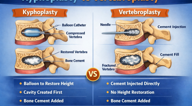
In the debate of kyphoplasty vs vertebroplasty, kyphoplasty stands out due to its ability to restore the shape of the vertebra. The procedure begins with the insertion of a small balloon into the fractured vertebra. This balloon is gently inflated to create space and elevate the collapsed bone. 

Once the space is created, medical-grade bone cement is injected into the cavity to stabilize the vertebra. This dual-action approach makes kyphoplasty a highly effective vertebral compression fracture treatment, especially in cases where structural correction is needed. 

Benefits of Kyphoplasty 

Kyphoplasty offers several advantages that make it a preferred option in certain cases. 

Key benefits include: 

  • Restoration of vertebral height and alignment  
  • Reduction in spinal deformity such as kyphosis  
  • Significant and rapid pain relief  
  • Lower risk of cement leakage due to controlled placement  

These benefits play a major role in the kyphoplasty vs vertebroplasty decision-making process. 

When Kyphoplasty Is Recommended 

Doctors typically recommend kyphoplasty for patients with more severe fractures or noticeable spinal deformity. 

Common indications include: 

  • Collapsed vertebrae with height loss  
  • Severe, persistent pain  
  • Postural changes due to fracture  

As a specialized vertebral compression fracture treatment, kyphoplasty is ideal for patients needing both stabilization and structural correction. 

Vertebroplasty Procedure Explained 

How Vertebroplasty Works 

When analysing kyphoplasty vs vertebroplasty, vertebroplasty is considered a simpler and faster procedure. It involves injecting bone cement directly into the fractured vertebra without creating space beforehand. 

The cement rapidly hardens, stabilizing the fracture and alleviating pain. This straightforward approach makes vertebroplasty an effective vertebral compression fracture treatment for many patients. 

Benefits of Vertebroplasty 

Vertebroplasty offers several advantages, particularly for patients seeking quick relief. 

Key benefits include: 

  • Shorter procedure time  
  • Immediate stabilization of the fracture  
  • Rapid pain relief  
  • Minimally invasive with small incisions  

These benefits make vertebroplasty a strong contender in the kyphoplasty vs vertebroplasty comparison. 

When Vertebroplasty Is Recommended 

Vertebroplasty is often recommended for patients with stable fractures that do not require structural correction. 

Suitable cases include: 

  • Mild to moderate compression fractures  
  • Patients with minimal spinal deformity  
  • Individuals needing quick recovery  

It remains a reliable vertebral compression fracture treatment for pain management and stabilization. 

Kyphoplasty vs Vertebroplasty: Key Differences 

Technique Comparison 

Understanding the technical differences between kyphoplasty vs vertebroplasty is essential for choosing the right procedure. 

Key differences include: 

  • Balloon insertion in kyphoplasty vs direct injection in vertebroplasty  
  • Height restoration vs stabilization only  
  • Slightly longer procedure vs shorter duration  

Both methods are effective forms of vertebral compression fracture treatment, but their approach varies significantly. 

Effectiveness and Outcomes 

Both procedures have high success rates, but the outcomes differ depending on the severity of the fracture. 

Comparison highlights: 

  • Kyphoplasty improves spinal alignment and posture  
  • Vertebroplasty provides faster pain relief  
  • Both reduce pain significantly and improve mobility  

These factors are central to the kyphoplasty vs vertebroplasty decision. 

Risks and Complications 

Although both procedures are generally safe, they still involve some level of risk. 

Common risks include: 

  • Cement leakage  
  • Infection  
  • Bleeding  
  • Nerve irritation  

Selecting the appropriate vertebral compression fracture treatment helps minimize these risks and ensures better outcomes. 

Recovery Timeline: Kyphoplasty vs Vertebroplasty 

Immediate Recovery 

Patients undergoing kyphoplasty vs vertebroplasty typically experience noticeable pain relief shortly after the procedure. 

Immediate recovery includes: 

  • Monitoring in a recovery room  
  • Limited activity for 24 – 48 hours  
  • Pain management with medications  

Short-Term Recovery 

Within a few weeks, most patients can return to light activities and daily routines. 

Typical progress includes: 

  • Improved mobility  
  • Reduced dependence on pain medication  
  • Gradual return to normal activities  

Both procedures offer efficient vertebral compression fracture treatment with relatively quick recovery. 

Long-Term Recovery 

Long-term results are typically favourable for both procedures. 

Key results include: 

  • Enhanced spinal stability  
  • Improved posture (especially with kyphoplasty)  
  • Better quality of life  

Choosing the Right Procedure for You 

Factors to Consider 

Choosing between kyphoplasty vs vertebroplasty requires careful evaluation of multiple factors. 

Important considerations include: 

  • Severity and type of fracture  
  • Bone density and overall health  
  • Pain level and symptoms  
  • Age and lifestyle  

Consulting a Specialist 

A spine specialist will use diagnostic tools such as MRI or X-rays to determine the best vertebral compression fracture treatment for your condition. 

Personalized Treatment Plan 

Every patient is different and the choice between kyphoplasty vs vertebroplasty should be tailored to individual needs. A personalized approach ensures optimal results and long-term recovery. 

Conclusion 

When deciding between kyphoplasty vs vertebroplasty, it’s important to consider your specific condition, symptoms and treatment goals. Both procedures are highly effective forms of vertebral compression fracture treatment, offering significant pain relief, improved mobility and enhanced quality of life. 

If you are struggling with persistent back pain caused by a spinal fracture, don’t delay seeking expert advice. Book a consultation today with a spine specialist to determine the most suitable treatment option and take the first step toward recovery and long-term spinal health. 

Intrathecal Pain Pumps: Advanced Treatment for Severe Chronic Pain 

Living with relentless, severe pain can make even the simplest tasks feel impossible. For individuals who have tried medications, physical therapy and other treatments without success, advanced solutions like the intrathecal pain pump may offer a new sense of hope. But how does this innovative treatment work and why is it considered one of the most effective options for managing chronic pain? 

An intrathecal pain pump is a small, surgically implanted device that delivers medication directly into the fluid surrounding the spinal cord. This targeted approach allows for significantly lower doses of medication compared to oral treatments, while providing more effective relief. As a result, pain pump therapy has become a preferred option for patients dealing with severe, long-term conditions that are resistant to traditional treatments. 

In this detailed guide, we’ll cover everything you need to know about the intrathecal pain pump-how it works, who it’s suitable for, its benefits and risks and what to expect before, during and after the procedure. Whether you are considering a chronic pain pump or simply looking for advanced pain management options, this article will help you make an informed and confident decision about your care. 

What Is an Intrathecal Pain Pump? 

Understanding Intrathecal Pain Pump Technology 

An intrathecal pain pump is a medical device designed to deliver medication directly into the intrathecal space, which surrounds the spinal cord. This method bypasses the digestive system and bloodstream, allowing medication to reach pain receptors more efficiently. 

Unlike traditional pain management methods, pain pump therapy uses a controlled system that releases precise doses of medication over time. This ensures consistent relief while minimizing side effects commonly associated with oral medications. 

How Pain Pump Therapy Works 

The effectiveness of pain pump therapy lies in its targeted delivery system. The pump is implanted under the skin, usually in the abdomen and connected to a catheter that delivers medication directly to the spinal cord. 

Key components include: 

  • A small programmable pump  
  • A catheter for medication delivery  
  • A reservoir that stores the medication  

This system allows the chronic pain pump to provide continuous pain relief tailored to the patient’s needs. 

Conditions Treated with a Chronic Pain Pump 

The intrathecal pain pump is commonly used to treat severe and persistent pain conditions that do not respond to other treatments. 

Common conditions include: 

  • Cancer-related pain  
  • Failed back surgery syndrome  
  • Complex regional pain syndrome  
  • Severe spinal cord injuries  

These conditions often require long-term management, making pain pump therapy an effective solution. 

Benefits of Intrathecal Pain Pump Therapy 

Targeted Pain Relief 

One of the most significant advantages of the intrathecal pain pump is its ability to deliver medication directly to the source of pain. This targeted approach results in faster and more effective relief. 

Benefits include: 

  • Reduced pain intensity  
  • Improved comfort levels  
  • Consistent medication delivery  

This makes pain pump therapy a reliable option for managing severe pain. 

Lower Medication Dosage 

Because the medication is delivered directly to the spinal cord, patients require much smaller doses compared to oral medications. This reduces the risk of side effects. 

Advantages of lower dosage: 

  • Less drowsiness  
  • Reduced nausea  
  • Lower risk of dependency  

The chronic pain pump helps improve overall patient safety and comfort. 

Improved Quality of Life 

Chronic pain can significantly impact emotional and physical well-being. By providing effective relief, the intrathecal pain pump allows patients to regain control over their lives. 

Improvements may include: 

  • Better sleep quality  
  • Increased mobility  
  • Enhanced daily functioning  

Customizable Treatment 

The programmable nature of pain pump therapy allows doctors to adjust medication levels based on the patient’s needs. This ensures optimal pain control at all times. 

Who Is a Candidate for a Chronic Pain Pump? 

Ideal Candidates for Intrathecal Pain Pump 

Not everyone with chronic pain is a candidate for an intrathecal pain pump. Doctors carefully evaluate patients to determine if this treatment is appropriate. 

Ideal candidates include: 

  • Patients with severe, chronic pain  
  • Individuals who have not responded to other treatments  
  • Patients who experience significant side effects from oral medications  

Symptoms That Indicate the Need for Pain Pump Therapy 

Certain symptoms may indicate that pain pump therapy could be beneficial. 

These include: 

  • Persistent, severe pain  
  • Limited response to medications  
  • Difficulty performing daily activities  

In such cases, a chronic pain pump may provide long-term relief. 

Evaluation and Trial Process 

Before receiving a permanent implant, patients typically undergo a trial phase. This helps determine whether the intrathecal pain pump will be effective. 

Trial steps include: 

  1. Temporary medication delivery  
  1. Monitoring pain relief  
  1. Assessing side effects  

This process ensures that pain pump therapy is the right choice. 

Intrathecal Pain Pump Procedure: Step-by-Step 

Preparation for the Procedure 

Before receiving an intrathecal pain pump, patients undergo thorough evaluations to ensure they are suitable candidates. 

Preparation includes: 

  • Medical history review  
  • Imaging tests  
  • Medication adjustments  

These steps help ensure the success of the procedure. 

Implantation Process 

The implantation of the intrathecal pain pump is a surgical procedure performed under anaesthesia. 

Steps include: 

  1. Making a small incision in the abdomen  
  1. Implanting the pump under the skin  
  1. Inserting a catheter into the spinal canal  
  1. Connecting the system  

This process allows the chronic pain pump to deliver medication effectively. 

Post-Procedure Care 

After surgery, patients are monitored to ensure proper healing and device function. 

Post-care includes: 

  • Pain management  
  • Monitoring for complications  
  • Adjusting medication levels  

This ensures the success of pain pump therapy

Risks and Complications of Pain Pump Therapy 

General Surgical Risks 

Like any surgical procedure, implanting an intrathecal pain pump carries certain risks. 

Common risks include: 

  • Infection  
  • Bleeding  
  • Reaction to anaesthesia  

These risks are typically minimal when the procedure is carried out by experienced professionals. 

Device-Related Complications 

Specific risks associated with a chronic pain pump may include: 

  • Catheter displacement  
  • Pump malfunction  
  • Medication delivery issues  

Regular monitoring helps prevent these complications. 

Managing Risks Effectively 

Doctors take several precautions to minimize risks associated with pain pump therapy

Preventive measures include: 

  • Routine follow-ups  
  • Device monitoring  
  • Proper patient education  

These steps ensure safe and effective treatment. 

Recovery and Lifestyle After Intrathecal Pain Pump 

Immediate Recovery Phase 

After receiving an intrathecal pain pump, patients may experience mild discomfort as the body adjusts to the device. 

Initial recovery includes: 

  • Rest and limited movement  
  • Wound care  
  • Gradual return to activities  

Long-Term Recovery and Adjustments 

Over time, patients adapt to living with a chronic pain pump and experience improved pain control. 

Long-term benefits include: 

  • Reduced reliance on oral medications  
  • Increased activity levels  
  • Better overall health  

Lifestyle Considerations 

Living with an intrathecal pain pump requires certain lifestyle adjustments. 

Important considerations: 

  • Regular refill appointments  
  • Avoiding extreme physical strain  
  • Monitoring for symptoms  

These practices ensure effective pain pump therapy

Conclusion 

The intrathecal pain pump represents a breakthrough in advanced pain management, offering targeted relief for individuals suffering from severe chronic conditions. By delivering medication directly to the spinal cord, this innovative approach reduces pain, minimizes side effects and improves overall quality of life. 

If traditional treatments have not provided the relief you need, it may be time to consider pain pump therapy as a long-term solution. Book a consultation today with a pain management specialist to find out if this advanced treatment is right for you and take the first step toward a more comfortable, pain-free life. me to take the next step. Book a consultation with a pain management specialist today and discover how thermal ablation therapy can help you live pain-free. 

Foraminotomy Procedure: Benefits, Risks, and Recovery Timeline 

Living with persistent back or neck pain can feel overwhelming, especially when it begins to interfere with your daily routine, sleep and overall well-being. If you’ve been dealing with symptoms like radiating pain, tingling sensations or muscle weakness, your doctor may have suggested a foraminotomy as a possible treatment. But what does this procedure involve and how can it help you regain control over your life? 

foraminotomy is a specialized surgical approach designed to relieve pressure on spinal nerves by enlarging the openings in the spine through which these nerves pass. Over time, factors such as aging, spinal degeneration or injuries can narrow these openings, leading to nerve compression. This is where foraminotomy surgery and other forms of nerve decompression surgery become essential in restoring function and reducing discomfort. 

In this in-depth guide, we will walk you through everything you need to know about the foraminotomy procedure. From understanding how the surgery works to exploring its benefits, potential risks and detailed recovery timeline, this article is designed to give you clarity and confidence. Whether you are considering foraminotomy surgery or simply researching treatment options, this comprehensive resource will help you make an informed decision about your health. 

What Is a Foraminotomy Procedure? 

Understanding the Purpose of Foraminotomy 

The foraminotomy procedure is a surgical technique aimed at relieving nerve compression in the spine. It focuses on widening the neural foramen, which are small openings between the vertebrae that allow spinal nerves to exit the spinal column. When these spaces become narrowed, nerves can become compressed, causing pain and other neurological symptoms. 

This type of nerve decompression surgery is particularly effective for patients who have not found relief through conservative treatments such as medication, physical therapy or injections. By removing the structures causing the compression, foraminotomy surgery restores space for the nerves and helps alleviate symptoms. 

How Spinal Nerve Compression Develops? 

Nerve compression can occur due to several underlying conditions that affect the structure of the spine. As the body ages, natural wear and tear can lead to degeneration of spinal discs and joints, which reduces the available space for nerves. 

Common causes include: 

  • Herniated or bulging discs  
  • Bone spurs caused by arthritis  
  • Thickened ligaments  
  • Degenerative disc disease  

These conditions gradually narrow the neural foramen, making foraminotomy an effective solution for restoring normal nerve function. 

Types of Foraminotomy Surgery 

Depending on the location of the affected nerve, foraminotomy surgery can be categorized into different types. Each type targets a specific region of the spine and addresses unique symptoms. 

  • Cervical foraminotomy – targets the neck region  
  • Thoracic foraminotomy – focuses on the mid-back  
  • Lumbar foraminotomy – treats the lower back  

Each variation of nerve decompression surgery is carefully planned based on diagnostic imaging and patient symptoms. 

Benefits of Foraminotomy Surgery 

Effective Pain Relief 

One of the most significant advantages of foraminotomy is its ability to relieve chronic nerve pain. By decompressing the affected nerve, patients often experience immediate or gradual relief from symptoms. 

Key benefits include: 

  • Reduced radiating pain in arms or legs  
  • Decreased numbness and tingling  
  • Improved nerve function  

This makes foraminotomy surgery a highly effective option for long-term pain management. 

Improved Mobility and Function 

Patients who undergo nerve decompression surgery often notice a significant improvement in mobility. Activities that were once difficult – such as walking, bending or lifting – become easier after recovery. 

Improvements may include: 

  • Better range of motion  
  • Increased strength  
  • Enhanced physical performance  

Minimally Invasive Options 

Modern foraminotomy surgery techniques often use minimally invasive methods, which involve smaller incisions and less disruption to surrounding tissues. 

Advantages of minimally invasive approaches: 

  • Shorter hospital stays  
  • Reduced scarring  
  • Faster healing time  

This evolution in nerve decompression surgery has made the procedure safer and more accessible. 

Enhanced Quality of Life 

Chronic pain can affect mental health, productivity and relationships. By addressing the root cause of nerve compression, foraminotomy can significantly improve overall quality of life. 

Patients often report: 

  • Better sleep  
  • Reduced stress and anxiety  
  • Increased independence  

Who Needs Foraminotomy Surgery? 

Medical Conditions That Require Foraminotomy 

Doctors typically recommend foraminotomy for patients experiencing nerve compression caused by specific spinal conditions. When non-surgical treatments fail to provide relief, foraminotomy surgery becomes a viable option. 

Common conditions include: 

  • Spinal stenosis  
  • Herniated discs  
  • Osteoarthritis of the spine  
  • Degenerative spine disorders  

These conditions often lead to nerve irritation, making nerve decompression surgery necessary. 

Symptoms That Indicate the Need for Surgery 

Recognizing the symptoms of nerve compression is crucial for timely treatment. Patients who may benefit from foraminotomy often experience persistent and worsening symptoms. 

Warning signs include: 

  • Radiating pain down the arms or legs  
  • Numbness or tingling sensations  
  • Muscle weakness  
  • Difficulty standing or walking  

If these symptoms persist despite conservative care, foraminotomy surgery may be recommended. 

Diagnostic Process 

Before proceeding with nerve decompression surgery, doctors conduct thorough evaluations to confirm the diagnosis and determine the best treatment plan. 

Diagnostic tools include: 

  1. MRI scans for detailed imaging  
  1. CT scans for bone structure analysis  
  1. X-rays to assess spinal alignment  
  1. Physical examinations to evaluate symptoms  

These assessments ensure that foraminotomy is the most appropriate solution. 

Risks and Complications of Foraminotomy 

General Surgical Risks 

Like any medical procedure, foraminotomy surgery carries certain risks. While complications are relatively rare, it is important for patients to be aware of potential issues. 

Common risks include: 

  • Infection  
  • Bleeding  
  • Blood clots  
  • Reactions to anaesthesia  

These risks are minimized through proper surgical techniques and post-operative care. 

Specific Risks of Nerve Decompression Surgery 

Certain risks are unique to nerve decompression surgery, particularly when operating near delicate spinal structures. 

Possible complications include: 

  • Nerve injury  
  • Persistent or recurring pain  
  • Spinal instability  
  • Leakage of spinal fluid  

Although uncommon, these complications highlight the importance of choosing an experienced surgeon for foraminotomy

Risk Reduction Strategies 

Advancements in medical technology have significantly improved the safety of foraminotomy surgery. Surgeons now use advanced imaging and precision tools to enhance outcomes. 

Preventive measures include: 

  • Preoperative planning  
  • Minimally invasive techniques  
  • Careful monitoring during surgery  

These strategies ensure safer and more effective nerve decompression surgery outcomes. 

Foraminotomy Procedure: Step-by-Step Process 

Preoperative Preparation 

Preparing for a foraminotomy involves several important steps to ensure a smooth procedure and recovery. Patients are typically advised to undergo medical evaluations and follow specific guidelines before surgery. 

Preparation steps include: 

  • Reviewing medical history  
  • Completing diagnostic tests  
  • Adjusting medications  
  • Fasting before surgery  

These steps help optimize the success of foraminotomy surgery

Surgical Procedure 

During the procedure, the surgeon performs precise steps to relieve nerve compression. The goal of nerve decompression surgery is to create more space for the affected nerve. 

Procedure steps: 

  1. Administration of anaesthesia  
  1. Small incision near the affected area  
  1. Removal of bone or tissue causing compression  
  1. Enlargement of the neural foramen  

The entire foraminotomy process typically takes one to two hours. 

Postoperative Care 

After foraminotomy surgery, patients are closely monitored to ensure proper healing. Recovery begins immediately after the procedure. 

Post-surgery care includes: 

  • Pain management  
  • Monitoring for complications  
  • Gradual movement and activity  

This phase is critical for successful outcomes in nerve decompression surgery

Recovery Timeline After Foraminotomy 

Immediate Recovery Phase 

The first few days after foraminotomy are focused on rest and healing. Patients may experience mild discomfort, which is managed with medication. 

Key aspects: 

  • Limited physical activity  
  • Wound care  
  • Pain control  

Short-Term Recovery 

Within a few weeks, patients who undergo foraminotomy surgery begin to regain mobility and resume light activities. 

Typical progress includes: 

  • Starting physical therapy  
  • Increasing daily movement  
  • Gradual pain reduction  

Long-Term Recovery 

Full recovery from nerve decompression surgery may take several weeks to months, depending on individual health and the complexity of the procedure. 

Long-term outcomes include: 

  • Improved strength and flexibility  
  • Return to normal activities  
  • Minimal or no pain  

Tips for Faster Recovery 

To maximize the benefits of foraminotomy, patients should follow medical advice and maintain a healthy lifestyle. 

Helpful tips: 

  • Attend follow-up appointments  
  • Avoid heavy lifting  
  • Stay active within limits  
  • Maintain proper posture  

Conclusion 

The foraminotomy procedure is a highly effective solution for relieving nerve compression and restoring mobility. By addressing the root cause of pain, foraminotomy surgery offers long-term relief and improved quality of life for many patients. While there are risks involved, advancements in nerve decompression surgery have made the procedure safer and more reliable than ever. 

If chronic pain is affecting your daily life, it may be time to explore your options. Book a consultation today with a qualified spine specialist to determine whether this procedure is right for you and take the first step toward a pain-free future. 

Thermal Ablation Therapy: How It Works for Long-Term Pain Relief

Living with chronic pain can interfere with daily routines and impact emotional well-being. For patients struggling with persistent discomfort, thermal ablation therapy has emerged as a highly effective, minimally invasive solution for long-term relief. But how does this treatment work and why is it becoming a preferred choice for both patients and healthcare providers? 

Many individuals dealing with conditions like arthritis, nerve damage or spinal issues often rely on medications or temporary treatments that only mask symptoms. Over time, these approaches may lose effectiveness or come with unwanted side effects. This is where thermal ablation therapy stands out by targeting the root cause of pain rather than simply managing it. 

Using controlled heat to disrupt pain signals, this advanced form of pain ablation treatment offers a longer-lasting alternative compared to traditional therapies. It is particularly effective in procedures like nerve ablation for pain, where specific nerves responsible for transmitting pain signals are precisely treated. 

In this comprehensive guide, you will learn how thermal ablation therapy works, who can benefit from it, the types of procedures available and what to expect before, during and after treatment. By the end, you will have a clear understanding of whether this innovative solution could be the key to achieving lasting pain relief. 

What Is Thermal Ablation Therapy and How Does It Work? 

Understanding Thermal Ablation Therapy in Modern Pain Management 

Thermal ablation therapy is a minimally invasive medical procedure that uses heat to destroy or deactivate nerve fibres responsible for transmitting pain signals. This technique is widely used in pain ablation treatment, particularly for chronic conditions that do not respond well to medication or physical therapy. 

The procedure typically involves inserting a thin probe into the targeted area. Once positioned, the probe delivers controlled heat energy, effectively interrupting the nerve’s ability to send pain signals to the brain. This makes nerve ablation for pain a highly targeted and efficient solution. 

The Science Behind Pain Signal Interruption 

The human body relies on a complex network of nerves to communicate sensations, including pain. In chronic conditions, these signals can become overactive or misdirected. Thermal ablation therapy works by disrupting these signals at their source. 

Key mechanisms include: 

  • Heat generation: Radiofrequency or laser energy produces controlled heat  
  • Nerve disruption: Heat damages the nerve fibres responsible for pain transmission  
  • Signal blockage: The brain no longer receives pain signals from the treated area  

This approach ensures that pain ablation treatment addresses the root cause rather than just the symptoms. 

Types of Thermal Ablation Techniques 

There are several forms of thermal ablation therapy, each suited for different conditions: 

  1. Radiofrequency Ablation (RFA): 
    Uses electrical currents to generate heat and target nerves.  
  1. Microwave Ablation: 
    Leverages electromagnetic waves to access deeper tissue structures. 
  1. Laser Ablation: 
    Applies concentrated light energy for exact targeting. 

Each method plays a crucial role in nerve ablation for pain, depending on the patient’s condition and treatment goals. 

Conditions Treated with Thermal Ablation Therapy 

Chronic Back and Neck Pain 

One of the most common uses of thermal ablation therapy is for chronic back and neck pain. Conditions such as facet joint arthritis or degenerative disc disease often respond well to this treatment. 

Joint Pain and Arthritis 

Patients suffering from osteoarthritis in the knees, hips or shoulders can benefit from pain ablation treatment. By targeting the nerves around the affected joint, nerve ablation for pain provides significant relief. 

Neuropathic Pain Disorders 

When nerves are damaged, the resulting neuropathic pain can be complex and tough to treat. Thermal ablation therapy offers a targeted approach that reduces abnormal nerve activity. 

Cancer-Related Pain 

In some cases, pain ablation treatment is used to manage pain associated with tumours. By destroying nerve pathways, it helps improve the patient’s quality of life. 

Benefits of Thermal Ablation Therapy for Long-Term Pain Relief 

Minimally Invasive and Safe 

One of the biggest advantages of thermal ablation therapy is that it is minimally invasive. Unlike major surgeries, it requires only small incisions and has a lower risk of complications. 

Long-Lasting Pain Relief 

Compared to temporary treatments, nerve ablation for pain can provide relief lasting several months to years. This makes it a preferred option for chronic pain sufferers. 

Reduced Dependence on Medication 

Patients undergoing pain ablation treatment often experience a significant reduction in their need for pain medications, minimizing side effects and dependency risks. 

Quick Recovery Time 

In most cases, patients can quickly return to their usual routines within a few days. This makes thermal ablation therapy a convenient option for those with busy lifestyles. 

Procedure Overview: What to Expect 

Before the Procedure 

Patients are typically evaluated through physical exams and imaging tests to determine if thermal ablation therapy is appropriate. To verify the pain source, doctors may carry out diagnostic nerve blocks. 

During the Procedure 

Local anaesthesia is commonly administered during the procedure. A specialized needle is guided to the target area using imaging techniques. 

Steps include: 

  1. Positioning the probe  
  1. Delivering controlled heat  
  1. Monitoring nerve response  

This ensures precise nerve ablation for pain with minimal discomfort. 

After the Procedure 

Recovery is generally quick. Patients may experience mild soreness, but this subsides within a few days. The full effects of pain ablation treatment may take a few weeks to become noticeable. 

Risks and Considerations of Thermal Ablation Therapy 

Potential Side Effects 

While thermal ablation therapy is considered safe, some patients may experience: 

  • Temporary discomfort  
  • Mild swelling or bruising  
  • Rare complications such as infection  

Who Should Avoid the Procedure 

Not all patients are suitable candidates for pain ablation treatment. Those with certain medical conditions or infections may need alternative therapies. 

Importance of Experienced Professionals 

Choosing a skilled specialist is crucial for successful nerve ablation for pain. Accurate technique minimizes complications and boosts effectiveness. 

Comparing Thermal Ablation Therapy with Other Pain Treatments 

Medication vs. Thermal Ablation Therapy 

Medications provide temporary relief, while thermal ablation therapy addresses the root cause of pain. 

Physical Therapy vs. Pain Ablation Treatment 

Physical therapy improves mobility but may not eliminate pain. Pain ablation treatment offers a more direct solution. 

Surgery vs. Nerve Ablation for Pain 

Surgery is invasive and requires longer recovery. Nerve ablation for pain is less invasive and has fewer risks. 

Future of Thermal Ablation Therapy in Pain Management 

Advancements in Technology 

New technologies are making thermal ablation therapy more precise and effective than ever before. 

Expanding Applications 

The use of pain ablation treatment is expanding to treat a wider range of conditions. 

Improved Patient Outcomes 

With ongoing research, nerve ablation for pain continues to deliver better and longer-lasting results. 

Conclusion 

For individuals struggling with chronic pain, thermal ablation therapy offers a powerful and long-lasting solution. By targeting the source of pain through advanced pain ablation treatment techniques, patients can experience significant relief and improved quality of life. 

If you’re ready to explore a safer, minimally invasive option for lasting relief, now is the time to take the next step. Book a consultation with a pain management specialist today and discover how thermal ablation therapy can help you live pain-free. 

Facet Joint Injections: How They Reduce Chronic Back and Neck Pain

Chronic back and neck pain affect millions of people worldwide, often interfering with daily life, mobility and overall well-being. While many individuals find relief through medications, physical therapy or lifestyle changes, others continue to struggle with persistent discomfort that does not respond to conventional treatments. In such cases, facet joint injections have emerged as a highly effective, minimally invasive solution for targeting the root cause of pain. But what exactly are these injections and how do they work to provide relief? 

The spine is a complex structure, and even small components like facet joints can have a significant impact on movement and comfort. When these joints become inflamed due to aging, injury or degenerative conditions, they can cause localized or radiating pain. This is where spinal injections play a crucial role, offering targeted treatment that directly addresses inflammation and nerve irritation. Understanding the full scope of facet injection benefits can help patients make informed decisions about their care. 

For many patients, the idea of undergoing any type of injection near the spine can feel intimidating. Questions about safety, effectiveness and recovery are common. However, advancements in imaging technology and pain management techniques have made facet joint injections safer and more precise than ever before. 

In this detailed guide, we will explore everything you need to know about facet joint injections, including how they work, the conditions they treat, the procedure itself, recovery expectations, risks, benefits and long-term outcomes. 

Understanding Facet Joint Injections and Spinal Anatomy 

What Are Facet Joints? 

Facet joints are small joints located at the back of the spine, connecting each vertebra to the one above and below it. These joints play a critical role in providing stability while allowing flexibility and movement. Without properly functioning facet joints, everyday movements such as bending, twisting or turning would become difficult. 

Over time, these joints can experience wear and tear, especially due to aging or repetitive stress. Conditions such as osteoarthritis can cause the cartilage within the facet joints to break down, leading to inflammation and pain. This is one of the primary reasons patients seek facet joint injections

How Facet Joint Injections Work 

Facet joint injections involve delivering medication directly into the affected joint. Typically, the injection contains a combination of a local anesthetic and a corticosteroid. Immediate relief comes from the anesthetic, while the steroid targets inflammation over time. 

This targeted approach is what makes spinal injections particularly effective compared to oral medications, which affect the entire body. By focusing on the exact source of pain, these injections provide faster and more efficient relief. 

Why Targeted Treatment Matters 

One of the key facet injection benefits is precision. Instead of masking symptoms, the treatment addresses the root cause of pain. This not only improves effectiveness but also reduces the need for higher doses of systemic medications. 

Conditions Treated with Facet Joint Injections 

Chronic Lower Back Pain 

Chronic lower back pain is one of the most common reasons patients undergo facet joint injections. Conditions such as degenerative disc disease and arthritis often affect the lumbar facet joints, leading to persistent discomfort. 

Neck Pain and Cervical Issues 

Facet joint problems are not limited to the lower back. Cervical facet joints in the neck can also become inflamed, causing stiffness, headaches and reduced range of motion. In such cases, spinal injections provide targeted relief. 

Arthritis and Degenerative Conditions 

Arthritis is a leading cause of facet joint pain. As the joints deteriorate, inflammation increases, leading to chronic discomfort. Facet injection benefits include reducing this inflammation and improving joint function. 

Post-Injury or Post-Surgical Pain 

Patients who have experienced spinal injuries or undergone surgery may develop facet joint pain as a secondary condition. Facet joint injections can help manage this pain effectively. 

The Procedure: What Happens During Facet Joint Injections 

Preparation Before the Procedure 

Before undergoing facet joint injections, patients typically undergo imaging tests such as X-rays or MRI scans. These procedures help detect the precise location of the issue. 

Patients may also receive instructions regarding medications, fasting and transportation, as the procedure may involve mild sedation. 

Step-by-Step Procedure 

The process of administering spinal injections is straightforward and usually takes less than 30 minutes. Here’s what typically happens: 

  1. The patient is positioned comfortably on a procedure table  
  1. The skin is cleaned and numbed with a local anesthetic  
  1. A needle is guided into the facet joint using imaging technology  
  1. The medication is injected into the joint  

This precise approach ensures maximum effectiveness and safety. 

After the Procedure 

Following facet joint injections, patients are monitored for a short period before being discharged. Most individuals can return home the same day and resume light activities. 

Recovery and Expected Results 

Immediate Relief and Initial Response 

Many patients experience immediate relief due to the anesthetic used in facet joint injections. However, this relief may be temporary as the anesthetic wears off. 

Long-Term Benefits 

The corticosteroid component begins to reduce inflammation within a few days, providing longer-lasting relief. The duration of relief varies but can last from weeks to several months. 

Maximizing Facet Injection Benefits 

To enhance the effectiveness of spinal injections, patients are often advised to: 

  • Participate in physical therapy  
  • Maintain proper posture  
  • Engage in regular exercise  

These steps help sustain the facet injection benefits over time. 

Risks and Safety of Facet Joint Injections 

Common Side Effects 

While facet joint injections are generally safe, some patients may experience mild side effects such as soreness or swelling at the injection site. 

Rare Complications 

Serious complications rarely occur but can involve infection, bleeding or nerve damage. Engaging experienced professionals can significantly reduce these risks. 

Ensuring Safety 

The use of imaging guidance significantly improves the safety and accuracy of spinal injections, making them a reliable option for pain management. 

Benefits of Facet Joint Injections 

Targeted Pain Relief 

One of the most significant facet injection benefits is the ability to deliver medication directly to the source of pain. This targeted approach leads to faster and more effective relief. 

Minimally Invasive Treatment 

Unlike surgical options, facet joint injections are minimally invasive and require little to no downtime. This makes them an attractive option for many patients. 

Improved Mobility and Function 

By reducing pain and inflammation, spinal injections help patients regain mobility and perform daily activities more comfortably. 

Diagnostic Advantages 

In addition to providing relief, facet joint injections can help diagnose the source of pain, guiding further treatment decisions. 

Who Should Consider Facet Joint Injections 

Ideal Candidates 

Patients who may benefit from facet joint injections include those with: 

  • Chronic back or neck pain  
  • Limited response to conservative treatments  
  • Evidence of facet joint involvement  

Consultation and Evaluation 

A thorough evaluation is essential to determine if spinal injections are appropriate. This can involve conducting physical exams alongside imaging tests. 

Personalized Treatment Approach 

Each patient’s condition is unique, and treatment plans should be tailored to maximize facet injection benefits and ensure optimal outcomes. 

Conclusion 

Facet joint injections are a powerful and minimally invasive solution for managing chronic back and neck pain caused by facet joint inflammation. By delivering targeted treatment directly to the source of pain, these injections provide both immediate and long-term relief. 

Understanding the full range of facet injection benefits and the role of spinal injections can help patients make confident decisions about their care. With proper guidance and follow-up, many individuals experience significant improvements in mobility, comfort and overall quality of life. 

It’s time to take charge of your pain and reclaim your quality of life. 
Schedule a consultation today to discover how facet joint injections can support your recovery and mobility. pain-free life. to learn whether Botox for chronic pain could be the right treatment option for you.

Discectomy Surgery: When It’s Needed and What to Expect 

Back pain is one of the leading causes of disability worldwide, but when that pain becomes severe, persistent and radiates into the arms or legs, it may indicate a deeper spinal issue such as a herniated disc. In such cases, discectomy surgery is often considered a highly effective solution to relieve nerve pressure and restore quality of life. But how do you know when surgery is truly necessary and what does the journey look like from diagnosis to recovery? 

Many patients initially try conservative treatments such as physical therapy, medications or injections. While these methods can provide relief for some, others continue to experience debilitating symptoms that interfere with daily activities. This is where procedures like lumbar discectomy and herniated disc surgery come into play, offering targeted relief by addressing the root cause of the problem. 

The thought of undergoing spinal surgery can be intimidating. Questions about safety, recovery time and long-term outcomes are common. However, advancements in medical technology have made discectomy surgery safer, less invasive and more effective than ever before. 

In this comprehensive guide, we will explore in detail what discectomy surgery involves, when it is recommended, the different techniques used, recovery expectations, risks, benefits and how patients can achieve the best possible outcomes. 

What Is Discectomy Surgery and How It Works 

Understanding the Spine and Disc Problems 

The human spine is made up of vertebrae separated by intervertebral discs, which act as cushions and shock absorbers. When a disc becomes damaged or herniated, its inner material pushes outward and compresses nearby nerves. This condition often leads to pain, numbness and weakness. 

Discectomy surgery is designed to remove the portion of the disc that is causing this compression. By relieving pressure on the nerve, the procedure helps reduce pain and restore normal function. 

Types of Discectomy Surgery 

There are several forms of discectomy surgery, depending on the location and severity of the condition: 

  • Lumbar discectomy: Targets the lower back and is the most common type  
  • Cervical discectomy: Performed in the neck region  
  • Microdiscectomy: A minimally invasive approach with smaller incisions  

Each type of herniated disc surgery is tailored to the patient’s specific needs and condition. 

How the Procedure Relieves Symptoms 

The effectiveness of discectomy surgery lies in its ability to directly address nerve compression. Once the pressure is removed, inflammation decreases and the nerve begins to heal. This leads to significant pain relief, especially in patients undergoing lumbar discectomy

When Is Discectomy Surgery Needed 

Recognizing the Warning Signs 

Not all back pain requires surgery, but certain symptoms strongly indicate the need for discectomy surgery

  • Persistent pain lasting more than 6–8 weeks  
  • Radiating pain (sciatica)  
  • Numbness or tingling  
  • Muscle weakness  

These symptoms often point to conditions requiring herniated disc surgery

When Conservative Treatments Fail 

Doctors typically recommend non-surgical treatments first. However, if these approaches fail to provide relief, lumbar discectomy may be the next step. 

Common non-surgical treatments include: 

  1. Physical therapy  
  1. Anti-inflammatory medications  
  1. Steroid injections  

If symptoms persist, discectomy surgery becomes a viable option. 

Emergency Situations 

In rare cases, immediate herniated disc surgery is required, especially when there is: 

  • Loss of bladder or bowel control  
  • Severe nerve damage  

These conditions require urgent lumbar discectomy to prevent permanent complications. 

Types of Discectomy Surgery Techniques 

Open Discectomy 

Traditional discectomy surgery involves a larger incision and direct access to the spine. While effective, it may involve longer recovery times. 

Microdiscectomy 

Microdiscectomy is a minimally invasive form of lumbar discectomy. It uses specialized instruments and a microscope to remove the damaged disc portion. 

Benefits include: 

  • Smaller incisions  
  • Reduced pain  
  • Faster recovery  

This technique is widely used in modern herniated disc surgery

Endoscopic Discectomy 

Endoscopic discectomy surgery is performed using a tiny camera and instruments inserted through small incisions. It is one of the least invasive options available. 

Choosing the Right Approach 

The choice of technique depends on: 

  • Severity of the condition  
  • Patient health  
  • Surgeon expertise  

Your doctor will recommend the most suitable lumbar discectomy method. 

What to Expect During Discectomy Surgery 

Preoperative Preparation 

Before undergoing discectomy surgery, patients will: 

  • Undergo imaging tests like MRI  
  • Receive medical clearance  
  • Follow fasting and medication guidelines  

These steps ensure a safe and successful procedure. 

Step-by-Step Procedure 

During discectomy surgery, the surgeon: 

  1. Makes a small incision  
  1. Accesses the affected disc  
  1. Removes the damaged portion  
  1. Relieves nerve pressure  

The procedure typically takes 1–2 hours. 

Hospital Stay and Immediate Recovery 

After lumbar discectomy, many patients go home the same day or within 24 hours. Minimally invasive herniated disc surgery often allows quicker discharge. 

Recovery After Discectomy Surgery 

Early Recovery Phase 

In the first few days after discectomy surgery, patients focus on pain management and gentle movement. Walking is encouraged to improve circulation. 

Rehabilitation and Physical Therapy 

Physical therapy is essential after lumbar discectomy. It helps: 

  • Strengthen core muscles  
  • Improve flexibility  
  • Prevent recurrence  

Returning to Daily Activities 

Most patients can return to light activities within a few weeks. Full recovery after herniated disc surgery may take several months. 

Tips for Better Recovery 

  • Avoid heavy lifting  
  • Maintain proper posture  
  • Follow medical advice  

These practices improve outcomes after discectomy surgery

Risks and Complications of Discectomy Surgery 

Common Risks 

Although discectomy surgery is generally safe, potential risks include: 

  • Infection  
  • Bleeding  
  • Nerve injury  

Risk of Recurrence 

There is a small chance that the disc may herniate again, requiring additional lumbar discectomy

Reducing Risks 

Patients can reduce risks by: 

  • Choosing experienced surgeons  
  • Following postoperative care  
  • Attending follow-up appointments  

Proper care ensures better outcomes in herniated disc surgery

Benefits and Success Rates of Discectomy Surgery 

Immediate Pain Relief 

One of the biggest advantages of discectomy surgery is rapid pain relief. Noticeable improvement is common among patients soon after the procedure. 

High Success Rates 

Studies indicate that lumbar discectomy has a success rate of 80–90% in relieving symptoms. 

Improved Quality of Life 

Patients who undergo herniated disc surgery often experience: 

  • Better mobility  
  • Reduced pain  
  • Enhanced daily functioning  

Conclusion 

Discectomy surgery is a proven and effective solution for individuals suffering from severe pain caused by herniated discs. By understanding when it is needed, how it is performed and what to expect during recovery, patients can approach the procedure with confidence. 

Whether you are considering lumbar discectomy or another form of herniated disc surgery, consulting with a qualified specialist is the first step toward recovery. Are you ready to improve and take control of your spine health? Book a consultation with a spine expert today and begin your journey toward a pain-free life. to learn whether Botox for chronic pain could be the right treatment option for you.