Botox for Chronic Pain: Medical Uses Beyond Cosmetic Treatment

For decades, Botox has been widely recognized for its cosmetic benefits, particularly its ability to reduce wrinkles and fine lines. However, many people are unaware that Botox for chronic pain has become an increasingly important medical treatment option for a variety of debilitating conditions. From migraine headaches to muscle disorders, this injectable therapy is now being used in innovative ways to improve patients’ quality of life.

Chronic pain affects millions of individuals worldwide and can significantly interfere with daily activities, sleep, and mental well-being. Traditional treatments such as oral medications, physical therapy, and surgery may not always provide lasting relief. As a result, healthcare providers continue searching for safer and more effective solutions for long-term pain relief. This is where Botox pain management is gaining recognition as a promising alternative.

Botox works by temporarily blocking nerve signals that trigger muscle contractions and pain responses. This mechanism allows physicians to target specific muscles or nerve pathways responsible for chronic discomfort. In recent years, the use of Botox medical treatment for pain conditions has expanded significantly, supported by clinical research and FDA approvals for certain indications.

In this comprehensive guide, we will explore how Botox is used beyond cosmetic treatments. You will learn how Botox for chronic pain works, which medical conditions it treats, what to expect during treatment, and how it compares to other pain management options. By the end of this article, you will have a clear understanding of whether Botox therapy could be an effective solution for managing persistent pain.

Understanding Botox for Chronic Pain

The growing interest in Botox for chronic pain reflects a broader shift in how physicians approach pain management. Rather than simply masking symptoms with medication, modern treatments focus on targeting the underlying nerve pathways responsible for pain signals.

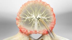
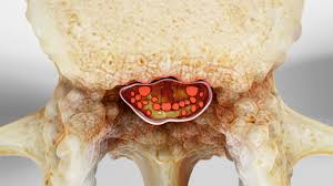
Botox, derived from botulinum toxin type A, works by temporarily interrupting communication between nerves and muscles. When injected into specific areas, it prevents muscles from contracting excessively and reduces the transmission of pain signals.

How Botox Works in Pain Treatment

In medical settings, Botox injections are carefully administered by trained healthcare professionals. The treatment targets nerves that release chemical messengers responsible for muscle contractions and pain responses.

The mechanism behind Botox pain management involves several key processes:

  1. Blocking nerve signals
    Botox prevents the release of neurotransmitters responsible for activating pain pathways.
  2. Reducing muscle tension
    Many chronic pain conditions involve persistent muscle contractions that contribute to discomfort.
  3. Interrupting inflammatory responses
    Botox may reduce inflammation around nerves and muscles, helping relieve long-term pain.
  4. Preventing recurring pain cycles
    By disrupting pain signalling patterns, Botox can help break the cycle of chronic pain.

The use of Botox medical treatment for pain is particularly beneficial for conditions involving nerve hypersensitivity or muscle overactivity.

Conditions Treated with Botox for Chronic Pain

Healthcare providers increasingly rely on Botox for chronic pain as part of comprehensive treatment strategies. Although it is most commonly associated with migraines, Botox injections are now used to manage several other pain conditions.

Chronic Migraine Treatment

Chronic migraine is one of the most well-known conditions treated using Botox therapy. Patients suffering from migraines often experience severe headaches multiple times per month, which can disrupt work, relationships, and daily routines.

Clinical research shows that Botox pain management injections can significantly reduce migraine frequency and severity. Patients typically receive injections around the forehead, temples, neck, and shoulders to relax muscles and block pain signals.

Key benefits for migraine patients

  • Reduced frequency of migraine attacks
  • Decreased headache intensity
  • Improved daily functioning

Because of its proven effectiveness, Botox is now considered an established Botox medical treatment option for individuals with chronic migraines.

Neck and Shoulder Pain

Chronic neck and shoulder pain often results from muscle tension, poor posture, or repetitive strain. When muscles remain contracted for long periods, they can create persistent discomfort and stiffness.

Doctors may recommend Botox for chronic pain to relax overactive muscles in the neck and shoulders. By reducing muscle contractions, Botox injections help restore mobility and decrease pain levels.

Patients undergoing Botox pain management for neck pain often report:

  • Reduced muscle tightness
  • Improved range of motion
  • Less frequent pain flare-ups

This approach is particularly helpful for individuals whose symptoms have not responded well to traditional therapies.

Neuropathic Pain Disorders

Neuropathic pain occurs when nerves become damaged or overly sensitive, causing burning, tingling, or stabbing sensations. Conditions such as post-herpetic neuralgia or nerve injuries may produce persistent neuropathic pain.

Research suggests that Botox medical treatment may help reduce nerve hypersensitivity in certain neuropathic conditions. The injections work by blocking chemical signals involved in pain transmission.

For patients experiencing nerve-related pain, Botox for chronic pain can offer an alternative when medications fail to provide adequate relief.

How Botox Pain Management Is Performed

Understanding the treatment process can help patients feel more comfortable before undergoing Botox therapy. While procedures may vary slightly depending on the condition being treated, most follow a similar structure.

Step-by-Step Treatment Process

The Botox pain management procedure typically includes several stages designed to ensure safety and effectiveness.

  1. Medical evaluation

A healthcare provider reviews the patient’s medical history and performs a physical examination to determine whether Botox injections are appropriate.

  1. Targeted injection planning

Specific muscles or nerve areas responsible for pain are identified to ensure precise treatment.

  1. Injection procedure

Botox is injected using a fine needle into targeted locations. The treatment plan, including the number of injections, is based on the condition.

Post-treatment observation

Patients are monitored briefly after the procedure to ensure there are no immediate side effects.

The full procedure usually takes fewer than 30 minutes to complete. Because Botox medical treatment is minimally invasive, most patients can resume normal activities shortly afterward.

Benefits of Botox Medical Treatment for Pain

The increasing popularity of Botox for chronic pain is largely due to its ability to deliver targeted relief without the need for invasive surgery or long-term medication use.

Many patients who struggle with persistent pain conditions experience noticeable improvements after Botox therapy.

Major Advantages of Botox Therapy

Minimally invasive treatment

Unlike surgical interventions, Botox injections require no incisions or extended recovery time.

Long-lasting relief

Results from Botox pain management typically last three to six months, allowing patients to maintain relief between treatment sessions.

Reduced medication dependency

Patients receiving Botox medical treatment may rely less on daily pain medications.

Improved mobility

Relaxing overactive muscles can restore flexibility and reduce stiffness.

Better quality of life

Many individuals report improved sleep, productivity, and overall well-being after undergoing Botox therapy.

Because of these benefits, physicians increasingly consider Botox for chronic pain as part of integrated pain management programs.

Risks and Safety Considerations

While Botox injections are generally considered safe, patients should still understand the potential risks associated with the procedure.

Side effects are typically mild and temporary. However, discussing concerns with a healthcare provider is essential before beginning treatment.

Possible Side Effects

Some patients undergoing Botox pain management may experience temporary reactions such as:

  • Mild swelling at injection sites
  • Headache or fatigue
  • Temporary muscle weakness

Serious complications are rare when Botox medical treatment is performed by experienced medical professionals.

Who Should Avoid Botox

Certain individuals may not be suitable candidates for Botox for chronic pain, including:

  • Pregnant or breastfeeding patients
  • Individuals with neuromuscular disorders
  • Patients with allergies to botulinum toxin

A thorough consultation helps determine whether Botox therapy is safe and appropriate for each patient.

Conclusion: Chronic pain can significantly disrupt daily life, affecting everything from physical activity to emotional well-being. Fortunately, innovative therapies are helping patients find new pathways to relief. Botox for chronic pain has emerged as a valuable medical option for individuals suffering from migraines, muscle disorders, and certain nerve-related conditions.

Through targeted injections, Botox pain management works by interrupting nerve signals responsible for persistent discomfort. As a result, many patients experience reduced pain, improved mobility, and a better quality of life. With growing clinical evidence supporting its effectiveness, Botox medical treatment is becoming an increasingly important part of modern pain care.

Book Your Consultation Today: If chronic pain is limiting your lifestyle, professional guidance can help you explore effective solutions. Schedule a consultation with a qualified specialist today to learn whether Botox for chronic pain could be the right treatment option for you.

Basivertebral Nerve Ablation for Chronic Back Pain: A Complete Guide

Chronic back pain is one of the most common medical conditions affecting adults worldwide. Millions of people struggle daily with persistent discomfort that limits mobility, reduces productivity, and significantly impacts quality of life. Traditional approaches like medication, physical therapy, and even spinal injections often provide only temporary relief. For patients who continue to suffer despite conservative treatments, newer interventional techniques such as Basivertebral nerve ablation are gaining attention as a promising long-term solution.

Basivertebral nerve ablation is a minimally invasive procedure designed specifically to treat vertebrogenic pain — a form of chronic low back pain originating from damaged vertebral endplates. By targeting the basivertebral nerve inside the vertebral body, physicians can interrupt the pain signals being transmitted to the brain. This targeted approach helps many patients experience long-lasting pain relief without major surgery.

One commonly used technique for this treatment is the Intracept procedure, which has been clinically studied and widely adopted for patients suffering from persistent vertebrogenic pain. As awareness grows, more individuals are exploring this treatment option as part of their chronic low back pain treatment journey.

In this comprehensive guide, we will explore everything you need to know about basivertebral nerve ablation. You will learn how the procedure works, who qualifies for it, what to expect during recovery, and how it compares to other chronic low back pain treatment options. By the end of this article, you will have a clear understanding of whether this innovative therapy could be the solution to managing your chronic back pain.

Understanding Chronic Back Pain and Vertebrogenic Pain

Chronic back pain is typically defined as pain that persists for more than three months. While many people experience occasional back discomfort, persistent pain can significantly affect daily activities, work performance, and overall well-being. Understanding the root cause of chronic back pain is essential in selecting the most effective treatment.

One specific type of pain that has gained increasing attention in recent years is vertebrogenic pain. This condition originates from the vertebral endplates—the thin layers of cartilage that separate the spinal discs from the vertebrae. When these endplates become damaged or inflamed, they can trigger persistent pain signals through the basivertebral nerve.

Unlike muscle-related back pain or nerve compression from herniated discs, vertebrogenic pain stems from structural changes inside the vertebra itself. These changes can be identified through imaging studies such as MRI scans, where physicians may detect Modic changes in the vertebral endplates.

Many patients suffering from vertebrogenic pain struggle for years with limited relief from conventional therapies. Common treatments include:

  • Physical therapy programs designed to strengthen spinal muscles
  • Pain medications such as NSAIDs or muscle relaxants
  • Steroid injections to reduce inflammation
  • Lifestyle modifications including weight management and posture correction

While these strategies can help manage symptoms, they may not always address the underlying pain source. This is where basivertebral nerve ablation becomes an important advancement in chronic low back pain treatment.

By targeting the basivertebral nerve responsible for transmitting pain signals, physicians can directly treat the root cause of vertebrogenic pain. For patients who meet the eligibility criteria, this approach offers a promising alternative to more invasive spinal surgeries.

What Is Basivertebral Nerve Ablation?

Basivertebral nerve ablation is a minimally invasive spinal procedure designed to treat chronic vertebrogenic back pain by disabling the basivertebral nerve. This nerve runs inside the vertebral body and carries pain signals from damaged endplates to the brain.

The procedure uses radiofrequency energy to heat and deactivate the nerve, preventing it from transmitting pain signals. Because the nerve is responsible for chronic pain originating from the vertebral body, its ablation can significantly reduce discomfort for eligible patients.

This treatment has become increasingly popular due to its targeted approach and favourable outcomes compared to some traditional interventions. The procedure is typically performed by interventional pain specialists or spine surgeons in an outpatient setting.

How the Intracept Procedure Works

The Intracept procedure is one of the most widely used methods for performing basivertebral nerve ablation. It is FDA-cleared and supported by clinical research demonstrating long-term pain reduction in appropriately selected patients.

The procedure generally involves the following steps:

  1. Patient preparation
    The patient is positioned comfortably and given sedation or local anesthesia.
  2. Guided access to the vertebra
    Using fluoroscopic imaging, the physician inserts a small cannula through the back into the targeted vertebral body.
  3. Placement of the radiofrequency probe
    A specialized probe is advanced through the cannula toward the basivertebral nerve.
  4. Radiofrequency ablation
    Controlled heat is applied to ablate the nerve, preventing it from sending pain signals.
  5. Completion and recovery
    The instruments are removed, and the small incision is covered with a bandage.

The Intracept procedure typically takes about 60 to 90 minutes, and most patients can return home the same day.

Who Qualifies for Basivertebral Nerve Ablation Treatment?

Not every patient with back pain qualifies for basivertebral nerve ablation. The procedure is specifically designed for individuals suffering from vertebrogenic pain associated with structural changes in the vertebral endplates.

Candidates for this chronic low back pain treatment typically meet several criteria.

Common eligibility factors include

  • Chronic low back pain lasting more than six months
  • Pain that has not improved with conservative therapies
  • MRI evidence of Modic type 1 or type 2 changes
  • Pain localized primarily in the lower back rather than legs

Patients who may not qualify include

  • Individuals with severe spinal instability
  • Patients with spinal infections or tumours
  • Those with nerve compression causing leg pain (sciatica)

A thorough evaluation by a spine specialist is necessary before determining eligibility for basivertebral nerve ablation. Diagnostic imaging and clinical history help physicians confirm whether the basivertebral nerve is the primary pain source.

When appropriate patients undergo the Intracept procedure, many experience substantial improvement in pain levels and daily functioning.

Benefits of Basivertebral Nerve Ablation

The growing popularity of basivertebral nerve ablation is largely due to the benefits it offers compared to traditional treatments for chronic back pain.

Its targeted approach is one of its most notable advantages. Instead of broadly addressing symptoms, the procedure directly treats the nerve responsible for transmitting vertebrogenic pain signals.

Key benefits include

Minimally invasive treatment

The procedure requires only a small incision and typically does not involve extensive tissue disruption. This reduces surgical risks and shortens recovery time.

Long-lasting pain relief

Clinical studies have shown that many patients experience sustained improvement in pain and functional outcomes after undergoing basivertebral nerve ablation.

Reduced reliance on medications

By addressing the underlying cause of pain, patients may reduce their dependence on long-term pain medications.

Outpatient procedure

Most individuals undergoing the Intracept procedure return home the same day, making it convenient and less disruptive to daily life.

Improved quality of life

Patients often report increased mobility, better sleep, and improved ability to perform everyday activities after successful treatment.

For individuals who have exhausted other chronic low back pain treatment options, basivertebral nerve ablation can represent a major turning point in their pain management journey.

What to Expect During Recovery

Recovery from basivertebral nerve ablation is generally faster compared to many other spinal procedures. Because the treatment is minimally invasive, most patients experience only mild discomfort immediately after the procedure.

During the first few days following the Intracept procedure, patients may notice soreness near the treatment site. This is typically temporary and can be managed with mild pain relievers or rest.

The majority of patients can get back to light activities in just a few days. However, physicians may recommend avoiding heavy lifting or strenuous physical activity for a short period.

Typical recovery timeline

First week

Patients may experience mild soreness or fatigue. Short, gentle walks can help promote better blood flow.

Two to six weeks

Gradual improvement in pain levels may occur as the body adjusts and inflammation subsides.

Two to three months

Many patients begin experiencing noticeable relief from chronic back pain as the effects of the nerve ablation take hold.

While results vary, many individuals report significant improvement in their chronic low back pain treatment outcomes after undergoing basivertebral nerve ablation.

Risks and Considerations

Although basivertebral nerve ablation is generally considered safe, it is important for patients to understand the potential risks associated with any medical procedure.

Complications are relatively rare but may include infection, bleeding, or temporary nerve irritation. Proper patient selection and experienced physicians significantly reduce these risks.

Patients should also understand that while the Intracept procedure can provide substantial relief, it may not completely eliminate pain in every case. Some individuals may still require complementary treatments as part of a comprehensive chronic low back pain treatment plan.

Discussing expectations with a healthcare provider is essential before deciding whether basivertebral nerve ablation is the right option.

Conclusion: Chronic back pain can be life-altering, especially when conventional treatments fail to provide lasting relief. Basivertebral nerve ablation offers a promising solution for patients suffering from vertebrogenic pain by targeting the nerve responsible for transmitting pain signals.

Through advanced techniques such as the Intracept procedure, physicians can treat the root cause of chronic pain using a minimally invasive approach. Many patients experience long-term improvements in mobility, comfort, and overall quality of life after this innovative chronic low back pain treatment.

If you have been struggling with persistent back pain despite conservative therapies, consulting a spine specialist about basivertebral nerve ablation could be an important step toward lasting relief.

Book Your Consultation Today: If chronic back pain is affecting your daily life, it may be time to explore advanced treatment options. Schedule a consultation today to learn whether basivertebral nerve ablation or the Intracept procedure is the right chronic low back pain treatment for you

Thoracic Outlet Syndrome: Symptoms, Diagnosis, and Treatment Options

Thoracic outlet syndrome is a complex condition that can cause persistent discomfort in the neck, shoulders, and arms, often interfering with everyday activities such as typing, lifting, or even holding objects. Many people who experience unexplained arm pain, numbness, or weakness may not immediately realize that the issue could be related to nerve or blood vessel compression in a narrow passageway near the collarbone known as the thoracic outlet. Because this area contains vital nerves and blood vessels that connect the spine to the arms, any pressure or restriction in this space can result in a wide range of physical symptoms.

For many individuals, the earliest thoracic outlet syndrome symptoms begin subtly. Mild tingling in the fingers, aching in the shoulder, or a feeling of weakness in the arm may appear occasionally and then gradually become more frequent. Over time, these symptoms can progress and affect posture, mobility, and overall quality of life. The condition may develop due to repetitive motions, poor posture, traumatic injuries, or anatomical variations such as an extra rib. Athletes, office workers, and individuals who perform repetitive arm movements are particularly susceptible to developing this condition.

Understanding how thoracic outlet syndrome develops is the first step toward effective management and recovery. When recognized early, many cases can be successfully treated with non-surgical therapies such as physical rehabilitation, posture correction, and targeted pain management strategies. However, accurate diagnosis is essential because the symptoms can often resemble other nerve or vascular conditions.

This comprehensive guide explores thoracic outlet syndrome, its causes, symptoms, diagnostic methods, and available treatment options. By the end of this article, readers will gain a clear understanding of how the condition develops, how it is diagnosed, and which treatment strategies can help relieve pain and restore normal function.

Understanding Thoracic Outlet Syndrome and Its Causes

Thoracic outlet syndrome occurs when nerves or blood vessels in the thoracic outlet become compressed. The thoracic outlet is the narrow space between the collarbone and the first rib, through which major nerves and blood vessels travel from the spine to the arms.

When pressure develops in this area, it can interfere with nerve signals or blood circulation, leading to discomfort, weakness, or vascular complications. The condition may develop gradually or suddenly depending on the underlying cause.

Types of Thoracic Outlet Syndrome

Medical professionals generally classify thoracic outlet syndrome into three primary types based on the structures affected.

Neurogenic thoracic outlet syndrome

This is the most common form and occurs when the brachial plexus nerves become compressed. These nerves control movement and sensation in the shoulders, arms, and hands.

Venous thoracic outlet syndrome

This type develops when veins in the thoracic outlet become compressed, potentially causing swelling or blood clots in the arm.

Arterial thoracic outlet syndrome

This rare but serious type involves compression of the arteries, which can affect blood flow to the arms and hands.

Causes of Thoracic Outlet Compression

Several factors can contribute to the development of thoracic outlet syndrome symptoms.

Common causes include:

  • Poor posture that places pressure on the shoulder region
  • Repetitive arm movements or overhead activities
  • Traumatic injuries such as whiplash or fractures
  • Congenital abnormalities like an extra rib
  • Muscle enlargement or inflammation around the neck and shoulders

Each of these factors can narrow the thoracic outlet and create pressure on nearby nerves or blood vessels.

Risk Factors

Certain individuals may face a higher risk of developing thoracic outlet syndrome.

These include:

  1. Athletes involved in repetitive overhead sports
  2. Individuals who perform repetitive arm motions at work
  3. People with poor posture or prolonged desk work habits
  4. Individuals recovering from neck or shoulder injuries

Understanding these risk factors can help patients and healthcare providers take preventive measures and reduce the likelihood of developing thoracic outlet syndrome symptoms.

Recognizing Thoracic Outlet Syndrome Symptoms

Early recognition of thoracic outlet syndrome symptoms is essential for preventing complications and initiating appropriate treatment. Because the thoracic outlet contains both nerves and blood vessels, symptoms may vary depending on which structure is compressed.

In many cases, symptoms begin gradually and may initially appear mild or intermittent.

Common Thoracic Outlet Syndrome Symptoms

Patients with thoracic outlet syndrome may experience a combination of nerve-related and circulation-related symptoms.

Common symptoms include:

  • Pain in the neck, shoulder, or arm
  • Numbness or tingling in the fingers
  • Weak grip strength
  • Shoulder fatigue or heaviness
  • Limited range of motion

These thoracic outlet syndrome symptoms often worsen with repetitive arm movements or prolonged overhead activities.

Neurological Symptoms

When nerves are affected, additional symptoms may develop.

These may include:

  • Burning sensations in the arm
  • Muscle weakness in the hand
  • Difficulty gripping objects
  • Reduced coordination in the fingers

These neurological symptoms often occur in cases of neurogenic thoracic outlet syndrome.

Circulatory Symptoms

Compression of blood vessels can produce a different set of symptoms.

Examples include:

  • Swelling in the arm or hand
  • Discoloration of the skin
  • Cold sensitivity in the fingers
  • Reduced pulse in the wrist

Recognizing these thoracic outlet syndrome symptoms early allows healthcare providers to differentiate the condition from other nerve disorders.

When Symptoms Worsen

If thoracic outlet syndrome progresses without treatment, symptoms may become persistent and significantly interfere with daily activities.

Patients who notice worsening numbness, swelling, or weakness should seek medical evaluation to prevent complications.

Diagnosing Thoracic Outlet Syndrome

Accurate diagnosis is essential for developing an effective treatment plan for thoracic outlet syndrome. Because the symptoms can resemble other conditions such as carpal tunnel syndrome or cervical spine disorders, healthcare providers must perform thorough evaluations.

The diagnostic process typically involves several steps.

Medical History and Physical Examination

Doctors begin by reviewing the patient’s medical history and discussing thoracic outlet syndrome symptoms.

During the physical examination, physicians may assess:

  • Posture and shoulder alignment
  • Arm strength and flexibility
  • Sensory responses in the hands and arms
  • Circulation in the affected limb

Special physical tests may also be performed to reproduce symptoms and confirm nerve compression.

Imaging Tests

Imaging studies help identify structural abnormalities contributing to thoracic outlet syndrome.

Common imaging tests include:

X-rays

X-rays can detect bone abnormalities, such as an extra rib, that may compress nerves or blood vessels.

MRI scans

MRI imaging provides detailed views of soft tissues, including muscles and nerves.

CT scans

CT scans help visualize bone structures and detect narrowing of the thoracic outlet.

Vascular and Nerve Tests

Additional tests may be used to confirm the cause of thoracic outlet syndrome symptoms.

These may include:

  • Nerve conduction studies
  • Electromyography (EMG)
  • Ultrasound imaging of blood vessels

These diagnostic tools help physicians determine whether the condition affects nerves, veins, or arteries.

Non-Surgical Treatment Options for Thoracic Outlet Syndrome

Many patients with thoracic outlet syndrome can successfully manage their condition using conservative treatment methods. These therapies focus on relieving pressure in the thoracic outlet and restoring proper nerve and blood flow.

Non-surgical treatment often produces significant improvement when symptoms are diagnosed early.

Physical Therapy

Physical therapy is one of the most effective approaches for managing thoracic outlet syndrome symptoms.

Therapy programs typically focus on:

  • Strengthening shoulder muscles
  • Improving posture
  • Increasing flexibility in the neck and chest
  • Expanding the thoracic outlet space

These exercises help reduce nerve compression and improve arm mobility.

Pain Management Techniques

Pain management may also play a role in thoracic outlet syndrome treatment.

Common methods include:

  • Anti-inflammatory medications
  • Muscle relaxants
  • Targeted injections to reduce inflammation

These treatments can help relieve discomfort while patients undergo rehabilitation.

Lifestyle Adjustments

Lifestyle changes can significantly reduce the severity of thoracic outlet syndrome symptoms.

Recommended adjustments include:

  • Improving workstation ergonomics
  • Avoiding repetitive overhead activities
  • Taking regular breaks during prolonged work tasks
  • Maintaining proper posture throughout the day

These changes help reduce stress on the shoulder region and prevent further compression.

Surgical Treatment for Severe Thoracic Outlet Syndrome

While conservative therapies work for many patients, some individuals with severe thoracic outlet syndrome may require surgical intervention.

Surgery is typically considered when symptoms persist despite extensive non-surgical treatment.

Types of Surgical Procedures

Common surgical options include:

First rib resection

This procedure removes a portion of the first rib to create more space in the thoracic outlet.

Scalenectomy

In this surgery, certain neck muscles are removed to relieve nerve compression.

Vascular repair

If blood vessels are damaged, surgeons may repair or reconstruct affected arteries or veins.

Recovery After Surgery

Recovery from thoracic outlet syndrome surgery varies depending on the procedure and individual health factors.

Patients often participate in rehabilitation programs that focus on restoring strength and mobility while preventing recurrence of thoracic outlet syndrome symptoms.

Long-Term Management and Prevention

Managing thoracic outlet syndrome effectively often requires ongoing lifestyle adjustments and preventive strategies.

Long-term management helps reduce the risk of recurring thoracic outlet syndrome symptoms.

Posture and Ergonomic Improvements

Maintaining proper posture helps keep the thoracic outlet open and prevents nerve compression.

Helpful habits include:

  • Keeping shoulders relaxed
  • Positioning computer screens at eye level
  • Supporting arms during desk work

Regular Exercise

Strengthening exercises improve muscle balance around the shoulders and spine.

These exercises support spinal alignment and reduce the likelihood of thoracic outlet syndrome recurrence.

Monitoring Symptoms

Patients should remain aware of any returning thoracic outlet syndrome symptoms and consult healthcare providers if discomfort reappears.

Addressing the condition early helps reduce complications and supports lasting recovery.

Conclusion: Living with persistent shoulder or arm discomfort can be frustrating, especially when the cause is not immediately clear. Thoracic outlet syndrome occurs when nerves or blood vessels become compressed in the narrow space between the collarbone and first rib, leading to a variety of neurological and circulatory symptoms. Recognizing thoracic outlet syndrome symptoms early allows individuals to pursue timely treatment and prevent long-term complications.

With proper diagnosis, many patients successfully manage the condition through physical therapy, posture correction, and lifestyle changes. These treatments help relieve nerve compression, improve mobility, and restore normal function.

If you are experiencing ongoing shoulder, arm, or neck discomfort, book a consultation with a healthcare specialist today to explore personalized treatment options for thoracic outlet syndrome.

Spinal Stenosis Treatment Options: When Surgery Isn’t the First Choice

Spinal stenosis treatment is often the first question people ask when persistent back or leg pain begins to interfere with everyday life. Many individuals worry that surgery may be the only solution, but that assumption is not always accurate. In reality, a wide range of conservative and minimally invasive approaches can successfully manage symptoms and improve quality of life without requiring an operation.

Spinal stenosis occurs when spaces within the spine narrow, placing pressure on the spinal cord or surrounding nerves. This condition most commonly develops in the lower back and is often referred to as lumbar spinal stenosis. The narrowing may occur gradually due to aging, degenerative changes, arthritis, or thickened ligaments. As the condition progresses, people may begin to notice discomfort that radiates from the lower back to the legs, along with numbness, tingling, or weakness.

Understanding the early warning signs is essential. Recognizing spinal stenosis symptoms at an early stage allows patients and healthcare providers to choose effective treatments before the condition becomes severe. Fortunately, modern medicine now offers a variety of non-surgical solutions that can help reduce inflammation, improve mobility, and relieve nerve compression.

This article explores the most effective spinal stenosis treatment options available today when surgery is not the first choice. You will learn about the causes of the condition, the symptoms to watch for, conservative therapies that may help manage pain, and lifestyle strategies that support long-term spinal health. By the end, you will have a clear understanding of how non-surgical care can provide meaningful relief and help individuals return to a more active and comfortable life.

Understanding Spinal Stenosis and Its Causes

Spinal stenosis develops when the spinal canal gradually becomes narrower, creating pressure on the nerves that travel through the spine. This compression can lead to discomfort, mobility limitations, and nerve-related symptoms that affect daily functioning.

When healthcare professionals evaluate spinal stenosis treatment, they first examine the underlying cause of the narrowing. Identifying the source of the problem allows providers to design a treatment strategy that targets both pain relief and long-term stability.

The spine is composed of vertebrae, discs, joints, ligaments, and nerves that work together to support the body. Over time, natural wear and tear can alter these structures. In many cases, the condition progresses slowly and may not cause symptoms for years.

Common Causes of Spinal Stenosis

Several factors contribute to spinal canal narrowing and nerve compression.

Age-related degeneration

As individuals age, the spinal discs gradually lose hydration and elasticity. This process reduces disc height and may allow surrounding structures to shift slightly, narrowing the spinal canal. Degenerative changes are the most common cause of lumbar spinal stenosis.

Osteoarthritis and bone spurs

Arthritis in the spine can trigger the growth of bone spurs. These bony projections may extend into the spinal canal and compress nearby nerves.

Thickened ligaments

Over time, spinal-supporting ligaments can become thicker. This thickening can reduce the available space in the spinal canal and contribute to nerve pressure.

Herniated discs

Discs act as cushions between vertebrae. When a disc bulges or ruptures, it may press against spinal nerves and contribute to spinal stenosis symptoms.

Congenital conditions

Certain individuals have a spinal canal that is naturally narrow from birth. In these cases, even minor structural changes can cause noticeable symptoms earlier in life.

Why Early Evaluation Matters

Identifying these causes early can influence the effectiveness of spinal stenosis treatment. Diagnostic tools such as MRI scans, CT imaging, and physical examinations help clinicians determine the degree of nerve compression and select appropriate therapies.

A personalized treatment plan can often prevent symptoms from worsening and reduce the likelihood of surgical intervention. For many patients, a combination of lifestyle adjustments and therapeutic care provides meaningful relief and helps restore mobility.

Recognizing Spinal Stenosis Symptoms Early

Detecting spinal stenosis symptoms early plays a critical role in determining the most appropriate treatment strategy. Because the condition develops gradually, many individuals initially dismiss mild discomfort as routine back pain. However, early recognition allows for timely spinal stenosis treatment, which can significantly reduce long-term complications.

Spinal stenosis can affect different areas of the spine, but the lower back is the most frequently impacted region. This condition, known as lumbar spinal stenosis, often leads to symptoms that extend beyond the spine and into the legs.

Common Spinal Stenosis Symptoms

People may experience a wide range of symptoms depending on the severity of nerve compression.

Lower back pain

Persistent discomfort in the lower back is often one of the earliest warning signs. The pain may intensify during prolonged standing or walking.

Leg pain or cramping

Nerve compression associated with lumbar spinal stenosis can cause pain that radiates down the legs. Many patients describe a heavy or burning sensation when walking.

Numbness or tingling

Some individuals notice numbness or tingling in the legs, feet, or buttocks. These sensations occur when nerves responsible for sensation become compressed.

Weakness in the legs

Muscle weakness may develop as nerve signals to the muscles become disrupted. This can make walking or climbing stairs more difficult.

Relief when bending forward

Interestingly, many individuals experience relief when leaning forward or sitting. This posture slightly opens the spinal canal and reduces pressure on the nerves.

Progressive Spinal Stenosis Symptoms

With the progression of the condition, symptoms can become more severe.

Numbered progression often includes:

  1. Reduced walking distance due to leg pain
  2. Increased numbness or weakness
  3. Balance issues
  4. Difficulty standing for extended periods

Recognizing these patterns helps healthcare providers determine when spinal stenosis treatment should be initiated.

When to Seek Medical Evaluation

If symptoms persist for several weeks or begin to interfere with daily activities, medical evaluation is recommended. Early diagnosis enables providers to implement conservative treatments that can slow progression and minimize discomfort.

Addressing spinal stenosis symptoms early improves the likelihood that non-surgical therapies will be effective, helping individuals maintain independence and mobility.

Non-Surgical Spinal Stenosis Treatment Approaches

Many patients are surprised to learn that spinal stenosis treatment often begins with conservative approaches rather than surgery. These therapies focus on reducing inflammation, improving spinal mobility, and strengthening the muscles that support the spine.

Non-surgical treatments are particularly effective during the early stages of lumbar spinal stenosis. With consistent care and proper guidance, patients frequently experience significant symptom relief.

Physical Therapy as a Primary Treatment

Physical therapy is one of the most widely recommended strategies for managing spinal stenosis symptoms. A trained therapist can design exercises that improve posture, strengthen core muscles, and reduce pressure on spinal nerves.

Benefits of physical therapy include:

  • Improved flexibility and mobility
  • Strengthened muscles supporting the spine
  • Reduced nerve compression
  • Better posture and spinal alignment

Therapists often incorporate stretching routines, strengthening exercises, and guided movement patterns to support long-term recovery.

Medications for Pain and Inflammation

Medication may also play a role in managing symptoms associated with lumbar spinal stenosis. These treatments typically focus on reducing inflammation and relieving discomfort.

Common medications include:

  • Nonsteroidal anti-inflammatory drugs (NSAIDs)
  • Muscle relaxants
  • Nerve pain medications

These medications are usually recommended as part of a comprehensive spinal stenosis treatment plan rather than as a standalone solution.

Lifestyle Adjustments

Lifestyle modifications can significantly influence symptom severity and recovery outcomes.

Important adjustments may include:

  • Maintaining a healthy body weight
  • Practicing proper posture
  • Engaging in low-impact exercise such as walking or swimming
  • Avoiding prolonged standing when possible

When combined with medical therapy, these changes can help slow the progression of spinal stenosis symptoms and improve overall spinal health.

Advanced Non-Surgical Treatments for Lumbar Spinal Stenosis

In some cases, conservative therapy alone may not fully relieve symptoms. Fortunately, several advanced non-surgical techniques are available to enhance spinal stenosis treatment outcomes.

These options aim to reduce inflammation around compressed nerves and improve mobility without requiring invasive surgery.

Epidural Steroid Injections

Epidural injections deliver anti-inflammatory medication directly around the affected nerves. This approach can significantly reduce swelling and relieve spinal stenosis symptoms for many patients.

Key benefits include:

  • Targeted pain relief
  • Reduced inflammation
  • Improved ability to participate in physical therapy

For individuals with lumbar spinal stenosis, injections can provide relief lasting several weeks or months.

Image-Guided Therapies

Modern imaging technologies allow physicians to deliver treatments with high precision. Fluoroscopy or ultrasound guidance ensures medications reach the exact location of nerve compression.

This targeted approach enhances the effectiveness of spinal stenosis treatment and minimizes potential side effects.

Minimally Invasive Procedures

In some cases, small procedures can help relieve nerve pressure without traditional surgery. Examples include decompression techniques that remove small portions of tissue contributing to the narrowing.

These minimally invasive procedures can improve symptoms associated with lumbar spinal stenosis while requiring shorter recovery periods than traditional surgery.

Lifestyle Strategies to Support Long-Term Recovery

Lifestyle management plays a crucial role in maintaining spinal health after beginning spinal stenosis treatment. While medical therapies can address immediate symptoms, daily habits often determine long-term outcomes.

Patients diagnosed with lumbar spinal stenosis benefit greatly from proactive strategies that reduce spinal strain and support mobility.

Maintaining a Healthy Weight

Excess body weight increases pressure on the spine and may worsen spinal stenosis symptoms. Even modest weight reduction can decrease stress on spinal structures.

Healthy weight management strategies include:

  • Balanced nutrition
  • Regular physical activity
  • Portion control

Reducing weight often improves the effectiveness of spinal stenosis treatment.

Strengthening Core Muscles

The muscles surrounding the abdomen and lower back provide essential support for the spine. Strengthening these muscles can stabilize the spinal column and reduce nerve compression.

Common exercises include:

  • Pelvic tilts
  • Bridges
  • Gentle abdominal strengthening routines

These exercises can be integrated into daily routines to support recovery from lumbar spinal stenosis.

Improving Posture and Movement

Posture plays an important role in spinal alignment. Poor posture can increase pressure on already compressed nerves.

Patients are often encouraged to:

  • Maintain neutral spine alignment
  • Avoid prolonged sitting or standing
  • Use ergonomic seating

Improving posture helps reduce spinal stenosis symptoms and supports long-term spinal health.

Conclusion: Managing spinal conditions does not always require immediate surgery. With the right approach, spinal stenosis treatment can effectively reduce pain, improve mobility, and support long-term spinal health through conservative care. Recognizing spinal stenosis symptoms early and addressing underlying factors associated with lumbar spinal stenosis allows patients to explore a variety of non-surgical options before considering invasive procedures. If you or someone you know is experiencing persistent back or leg discomfort, consulting a healthcare professional can help determine the most appropriate treatment path. Book a consultation today to explore personalized spinal stenosis treatment options and take the first step toward lasting relief.

Spondylolisthesis: Understanding Spinal Slippage and Pain Management

Spondylolisthesis is a spinal condition that often develops gradually but can lead to persistent discomfort, mobility limitations, and nerve-related complications if left untreated. Many people who experience ongoing lower back pain may not realize that the underlying cause could be a structural issue in the spine, particularly a condition known as spinal slippage. When one vertebra slips forward over the vertebra beneath it, pressure may build on surrounding nerves and tissues, leading to a range of physical symptoms that interfere with daily activities.

This condition is more common than many individuals realize and can affect people of different ages for different reasons. Some cases develop due to natural aging and degenerative changes within the spine, while others may arise from stress fractures, congenital conditions, or repetitive strain from physical activities. Because the spine plays such a vital role in supporting the body and protecting the spinal cord, even small shifts in spinal alignment can lead to noticeable discomfort or neurological symptoms.

Recognizing spondylolisthesis symptoms early can make a significant difference in how effectively the condition is managed. When diagnosed at the right stage, many individuals can control symptoms through conservative treatment methods, lifestyle adjustments, and guided rehabilitation programs. Understanding the causes, risk factors, and available treatment options empowers patients to take an active role in protecting their spinal health.

This article provides a comprehensive overview of spondylolisthesis, explaining how spinal slippage occurs, the symptoms to watch for, and the most effective strategies for pain management and recovery. By the end of this guide, readers will have a clear understanding of how the condition develops and how modern treatments can help restore mobility and improve quality of life.

What Is Spondylolisthesis and How Spinal Slippage Occurs

Spondylolisthesis occurs when a vertebra moves forward or slips out of its normal position relative to the vertebra below it. This displacement can affect the alignment of the spine and may lead to compression of nearby nerves, muscles, and ligaments. Over time, this structural change can contribute to pain, stiffness, and reduced mobility.

The condition is frequently associated with spinal slippage, which describes the movement of one vertebra over another. This shift can occur gradually due to degenerative changes or suddenly as a result of injury or stress fractures. While mild cases may not produce noticeable symptoms, more advanced cases often lead to discomfort and neurological issues.

Types of Spondylolisthesis

Medical professionals classify spondylolisthesis into several categories based on its underlying cause.

Degenerative spondylolisthesis

This type commonly develops in adults as part of the natural aging process. Degenerative changes in the spinal discs and joints can weaken the structures that maintain alignment, increasing the risk of spinal slippage.

Isthmic spondylolisthesis

This form often occurs due to a stress fracture in a specific portion of the vertebra called the pars interarticularis. Athletes and individuals who participate in activities that involve repetitive spinal extension may be at greater risk.

Congenital spondylolisthesis

Some individuals are born with spinal structures that are more susceptible to displacement. In these cases, abnormal bone formation can increase the likelihood of spinal slippage later in life.

Traumatic spondylolisthesis

Injuries such as fractures or severe trauma to the spine can lead to displacement of the vertebrae.

Severity Levels of Vertebral Slippage

Doctors often measure the severity of spondylolisthesis using a grading system that reflects how far the vertebra has shifted.

  1. Grade I: Mild slippage (less than 25%)
  2. Grade II: Moderate slippage (25–50%)
  3. Grade III: Significant slippage (50–75%)
  4. Grade IV: Severe slippage (75–100%)

Understanding the grade of displacement helps physicians determine the most effective treatment plan and evaluate the risk of worsening spondylolisthesis symptoms.

Common Spondylolisthesis Symptoms and Warning Signs

Recognizing spondylolisthesis symptoms early is essential for preventing the condition from progressing. Although some individuals may remain symptom-free for extended periods, many eventually develop discomfort as spinal slippage begins to affect surrounding nerves and muscles.

The severity of symptoms often depends on the degree of vertebral displacement and whether nerve compression is present.

Early Symptoms of Spinal Slippage

In the early stages, individuals may notice subtle changes in their physical comfort and mobility.

Common early spondylolisthesis symptoms include:

  • Persistent lower back pain
  • Stiffness in the lower spine
  • Muscle tightness in the hamstrings
  • Pain that worsens with physical activity
  • Relief when sitting or bending forward

These symptoms may initially appear mild but can gradually worsen if spinal slippage continues to progress.

Progressive Symptoms

As spondylolisthesis advances, nerve compression may occur, leading to additional neurological symptoms.

Patients may experience:

  • Pain radiating into the legs
  • Numbness or tingling sensations
  • Muscle weakness
  • Difficulty walking long distances
  • Reduced flexibility in the lower back

When spondylolisthesis symptoms reach this stage, medical evaluation becomes particularly important.

When to Seek Medical Care

Certain warning signs should prompt immediate medical attention.

These include:

  • Severe or persistent back pain
  • Loss of bladder or bowel control
  • Progressive leg weakness
  • Significant mobility limitations

Early diagnosis allows healthcare providers to initiate treatment strategies that may prevent further spinal slippage and reduce long-term complications.

Diagnosing Spondylolisthesis and Evaluating Spinal Slippage

Accurate diagnosis plays a critical role in managing spondylolisthesis effectively. Healthcare professionals use a combination of physical examinations, imaging tests, and medical history reviews to determine the extent of spinal slippage and identify the underlying cause.

A comprehensive evaluation allows clinicians to develop a personalized treatment strategy based on the severity of the condition and the patient’s symptoms.

Physical Examination

The diagnostic process often begins with a physical assessment. During this evaluation, doctors check spinal alignment, flexibility, and muscle strength.

The examination may also include:

  • Testing reflex responses
  • Evaluating nerve sensitivity
  • Assessing walking patterns
  • Identifying areas of pain or tenderness

These observations help determine whether spondylolisthesis symptoms are affecting nerve function.

Imaging Tests

Medical imaging plays an essential role in confirming spondylolisthesis.

Common imaging techniques include:

X-rays

X-rays provide a clear view of spinal alignment and can reveal the degree of spinal slippage.

Magnetic Resonance Imaging (MRI)

MRI scans help doctors evaluate soft tissues, nerves, and spinal discs. This imaging technique is particularly useful for identifying nerve compression associated with spondylolisthesis symptoms.

Computed Tomography (CT) scans

CT scans offer detailed cross-sectional images of spinal structures and may be used when additional anatomical detail is required.

Importance of Accurate Diagnosis

Identifying the precise location and severity of spinal slippage allows physicians to select appropriate treatment options.

In many cases, early diagnosis enables patients to manage spondylolisthesis symptoms using conservative therapies before the condition progresses to a stage requiring surgical intervention.

Non-Surgical Treatment Options for Spondylolisthesis

Many individuals diagnosed with spondylolisthesis can achieve significant relief through non-surgical treatment methods. These approaches focus on reducing inflammation, strengthening supporting muscles, and improving spinal stability.

For patients with mild to moderate spinal slippage, conservative care often represents the first line of treatment.

Physical Therapy and Rehabilitation

Physical therapy is widely considered one of the most effective ways to manage spondylolisthesis symptoms.

Therapists prepare specific exercise plans aimed at strengthening:

  • Strengthening core muscles
  • Improving flexibility
  • Enhancing spinal stability
  • Correcting posture

These exercises help reduce pressure on the affected vertebrae and prevent further spinal slippage.

Pain Management Strategies

Pain management techniques may also be incorporated into treatment plans for spondylolisthesis.

Common options include:

  • Anti-inflammatory medications
  • Muscle relaxants
  • Epidural steroid injections

These treatments can reduce inflammation and provide relief from nerve irritation associated with spondylolisthesis symptoms.

Activity Modification

Lifestyle adjustments can significantly influence recovery outcomes.

Healthcare providers often recommend:

  • Avoiding heavy lifting
  • Limiting high-impact activities
  • Practicing proper body mechanics

These modifications help minimize stress on the spine and reduce the risk of worsening spinal slippage.

Lifestyle Strategies for Managing Spinal Slippage

Long-term management of spondylolisthesis often requires lifestyle adjustments that support spinal health and prevent symptom recurrence.

Patients who adopt proactive strategies are more likely to maintain mobility and reduce discomfort associated with spinal slippage.

Maintaining a Healthy Weight

Excess body weight places additional stress on the spine and may worsen spondylolisthesis symptoms.

Healthy weight management involves:

  • Balanced nutrition
  • Regular physical activity
  • Consistent exercise routines

Reducing excess weight can significantly improve spinal stability.

Strengthening Core Muscles

The muscles surrounding the abdomen and lower back provide essential support for the spine.

Strengthening these muscles helps stabilize vertebrae and reduces the likelihood of further spinal slippage.

Common exercises include:

  • Pelvic tilts
  • Planks
  • Gentle back extensions

These exercises may also alleviate spondylolisthesis symptoms.

Improving Posture and Ergonomics

Proper posture plays a critical role in maintaining spinal alignment.

Patients are often encouraged to:

  • Use ergonomic seating
  • Maintain neutral spine alignment
  • Take regular breaks from prolonged sitting

Improving posture can help prevent worsening spondylolisthesis and support overall spinal health.

Conclusion: Living with back pain can significantly affect daily life, but understanding the underlying cause is the first step toward effective treatment. Spondylolisthesis occurs when vertebrae shift out of alignment, leading to discomfort, reduced mobility, and potential nerve compression. Recognizing spondylolisthesis symptoms early and addressing the causes of spinal slippage can help prevent the condition from progressing.

With proper diagnosis, many individuals successfully manage symptoms through physical therapy, lifestyle adjustments, and non-surgical treatments. These strategies can stabilize the spine, relieve pain, and restore mobility. If you are experiencing persistent back pain or suspect spinal slippage, schedule a consultation with a spine specialist today to explore personalized treatment options and begin your path to recovery.

Sciatica Pain: Causes, Symptoms, and Long-Term Relief Solutions

Sciatica pain is a common condition that affects millions of individuals worldwide and can significantly disrupt daily life. It typically begins as discomfort in the lower back and radiates down through the hips, buttocks, and legs. This type of nerve-related discomfort can make routine tasks such as walking, sitting, or bending extremely challenging. Many people experience occasional back pain, but sciatica pain is different because it follows the path of the sciatic nerve, the largest nerve in the human body.

The sciatic nerve extends from the lower spine, passing through the hips and traveling down each leg. When this nerve becomes compressed, inflamed, or irritated, it can trigger a variety of physical sensations ranging from mild tingling to severe shooting pain. Although the condition is often associated with aging, it can also affect younger adults due to lifestyle habits, posture issues, or spinal injuries.

Recognizing sciatica symptoms early can make a significant difference in how the condition is managed. Some individuals may notice numbness, weakness, or burning sensations that gradually worsen over time. Without proper care, these symptoms can interfere with mobility and overall quality of life.

Fortunately, several modern sciatica treatment options are available to help reduce nerve pressure, manage inflammation, and restore movement. With the right combination of medical guidance, lifestyle adjustments, and targeted therapies, long-term relief is achievable.

In this article, we will explore the causes of the condition, identify the most common sciatica symptoms, and discuss practical strategies that support long-term recovery and spinal health.

Understanding Sciatica Pain and How It Develops

Compression or irritation of the sciatic nerve can lead to sciatica pain. Because this nerve extends from the lower spine through the legs, any disruption along its pathway can trigger discomfort that radiates downward from the back.

The condition usually develops gradually as spinal structures change or experience stress. In many cases, individuals initially feel stiffness or mild lower back discomfort before the pain begins to travel through the leg.

Medical research shows that nerve compression is the most common reason for sciatica pain. When pressure builds around the nerve root, it can disrupt normal nerve signalling and cause inflammation. As a result, patients may experience sharp sensations, muscle weakness, or persistent discomfort.

Structure of the Sciatic Nerve

The sciatic nerve originates from nerve roots in the lower spine and travels through the pelvis and buttocks before extending into each leg. Because of its length and size, it is particularly vulnerable to compression from nearby structures.

Several spinal components can influence the health of the sciatic nerve. These include the intervertebral discs, vertebrae, surrounding muscles, and supporting ligaments.

Factors That Contribute to Sciatica Pain

Various structural and lifestyle factors can contribute to the development of sciatica pain. These include spinal injuries, poor posture, and repetitive strain on the lower back.

Several common factors that may lead to this condition include:

  • Progressive spinal degeneration due to aging
  • Muscle inflammation near the nerve
  • Prolonged sitting combined with improper posture

Understanding these triggers can help individuals take proactive steps to prevent nerve irritation.

Major Causes of Sciatica Pain

Although sciatica pain is associated with nerve irritation, the underlying causes often vary from person to person. Most cases involve structural changes within the spine that place pressure on the sciatic nerve.

Age-related degeneration is one of the leading contributors. Over time, spinal discs lose flexibility and height, making them more vulnerable to injury or bulging.

Herniated Discs

A herniated disc is one of the most common causes of nerve compression. The discs that sit between spinal vertebrae act as cushions, absorbing shock and supporting flexibility.

When the inner material of a disc pushes through its outer layer, it may press directly against the sciatic nerve root. This pressure can trigger sciatica pain that radiates through the leg.

Symptoms associated with disc herniation often include tingling, numbness, and difficulty standing or walking for extended periods.

Compression of nerves due to a narrowed spinal canal

The condition called spinal stenosis involves the narrowing of the spinal canal. When the canal becomes restricted, nerves traveling through the spine may become compressed.

Common causes of spinal stenosis include arthritis, bone spurs, and thickened ligaments. As these changes progress, pressure on the sciatic nerve may increase.

Degeneration of the discs in the spine due to aging

Degenerative disc disease occurs when spinal discs gradually wear down with age. As the discs lose cushioning ability, vertebrae may shift closer together.

This structural change can increase the risk of nerve irritation and persistent sciatica pain, particularly during physical activity or prolonged sitting.

An Overview of Sciatica Symptoms

Recognizing sciatica symptoms early is important for preventing the condition from worsening. Symptoms typically appear along the pathway of the sciatic nerve and may vary in intensity depending on the level of nerve compression.

Many individuals initially notice mild discomfort in the lower back or hip area. Over time, this discomfort may travel down the leg and affect mobility.

Key Symptoms Associated with Sciatica

The most frequently reported sciatica symptoms include radiating pain from the lower back into the leg, numbness or tingling sensations, and muscle weakness in the affected limb. Some individuals also experience burning or electric-like sensations that intensify with movement.

Symptoms often worsen when sitting for long periods or when attempting sudden movements such as bending or twisting.

Gradually Developing Sciatica Symptoms

In more advanced cases, sciatica symptoms can interfere with everyday activities. Over time, tasks like walking, climbing stairs, or standing for extended periods may become harder to perform.

Severe nerve compression may cause reduced muscle control in the leg or foot. When symptoms reach this stage, medical evaluation is strongly recommended.

Warning Signs That Require Medical Attention

Persistent or severe sciatica symptoms should not be ignored. Individuals should seek professional care if symptoms continue for several weeks or begin affecting mobility.

Early medical intervention can prevent nerve damage and improve recovery outcomes.

Trusted Approaches for Sciatica Relief

Managing nerve irritation often requires a combination of medical care, physical therapy, and lifestyle changes. Modern sciatica treatment strategies focus on relieving nerve pressure while strengthening the spine and surrounding muscles.

The approach to sciatica treatment usually depends on the severity of the condition and the underlying cause.

Initial Treatment Methods for Sciatica

Most patients begin with conservative sciatica treatment methods that aim to reduce inflammation and improve mobility.

These methods may include physical therapy exercises, posture correction programs, and anti-inflammatory medications prescribed by a healthcare professional.

Physical therapy is particularly effective because it strengthens muscles that support the spine and improves flexibility.

Non-Surgical Sciatica Treatment

In many cases, non-surgical sciatica treatment approaches provide significant relief without requiring invasive procedures.

Therapies may involve guided stretching routines, mobility exercises, and specialized rehabilitation programs designed to reduce nerve pressure.

Regular therapy sessions can gradually restore movement and reduce discomfort.

Advanced Sciatica Treatment

For severe cases where nerve compression persists, doctors may recommend more advanced sciatica treatment options.

These may include corticosteroid injections that reduce inflammation or minimally invasive procedures that relieve nerve pressure.

Although surgery is rarely necessary, it may be considered when other treatments fail to provide relief.

Lifestyle Changes That Help Reduce Sciatica Pain

Managing sciatica pain effectively often requires long-term lifestyle adjustments that support spinal health. Simple daily habits can play a significant role in reducing nerve irritation and preventing recurring discomfort.

Maintaining proper posture is one of the most important steps. Poor sitting or standing posture can place excessive pressure on the lower spine and contribute to nerve compression.

Physical Activity and Movement

Regular movement helps maintain flexibility and strengthens muscles that support the spine.

Activities such as walking, swimming, and stretching exercises can reduce stiffness and improve circulation around the sciatic nerve.

Consistent physical activity can gradually reduce sciatica pain while improving overall mobility.

Ergonomic-Friendly Workspaces

For individuals who spend long hours sitting, creating an ergonomic workspace can significantly reduce spinal strain.

Adjusting chair height, maintaining proper screen positioning, and taking regular breaks to stretch can help prevent recurring sciatica pain.

Practical Strategies for Maintaining Healthy Weight

Excess body weight places additional pressure on the spine and surrounding joints. Maintaining a healthy weight can reduce strain on spinal structures and help prevent nerve irritation.

A balanced diet combined with regular exercise supports long-term spinal health.

Preventing Sciatica Symptoms in the Future

Preventing recurring sciatica symptoms requires consistent attention to posture, exercise habits, and spinal health. Many individuals experience recurring episodes because they return to the same lifestyle habits that originally triggered the condition.

Strengthening the muscles that support the lower back and maintaining flexibility are essential for long-term prevention.

Optimizing Core Muscle Strength

Strong core muscles help keep the spine stable and properly aligned. Strengthening these muscles helps maintain proper alignment and reduces pressure on the sciatic nerve.

Exercises such as planks, pelvic tilts, and controlled abdominal workouts can help reduce the likelihood of future sciatica symptoms.

Conclusion: Living with sciatica pain can be challenging, but understanding the condition and its causes can make a meaningful difference in recovery. Early recognition of sciatica symptoms allows individuals to seek medical guidance before the condition becomes severe. With proper care, many people successfully manage the condition and regain normal mobility.

Modern sciatica treatment approaches focus on reducing nerve pressure, improving spinal strength, and promoting long-term healing. By combining medical care, physical therapy, and healthy lifestyle habits, individuals can reduce discomfort and prevent future flare-ups.

If you are experiencing persistent nerve discomfort or mobility issues, consider speaking with a healthcare professional for personalized guidance. Book an appointment today to explore effective treatment options and take the first step toward lasting relief from sciatica pain.

Foraminal Stenosis Explained: Symptoms, Diagnosis, and Treatment

Foraminal stenosis is a spinal condition that can silently progress until nerve pain begins to interfere with daily life. If you have been experiencing radiating arm or leg pain, tingling, or weakness that worsens with movement, you may be dealing with foraminal stenosis. This condition occurs when the small openings in the spine—called neural foramina—become narrowed and compress nearby spinal nerves.

Unlike general spinal stenosis, which involves central canal narrowing, foraminal stenosis specifically affects the passageways where nerve roots exit the spine. Foraminal narrowing may develop gradually due to age-related degeneration, disc problems, arthritis, or injury. As the space tightens, nerve irritation can cause persistent discomfort that extends beyond the back or neck.

In this comprehensive guide, you will learn what foraminal stenosis is, what causes foraminal stenosis symptoms, how physicians diagnose the condition, and which treatment options provide effective relief. Whether you are newly diagnosed or seeking better answers for chronic nerve pain, this article will provide clarity, evidence-based insights, and practical next steps.

What Is Foraminal Stenosis?

Foraminal stenosis refers to the narrowing of the intervertebral foramina, which are small openings located between each pair of vertebrae. These openings allow spinal nerves to exit the spinal canal and travel to the arms, chest, abdomen, and legs.

When foraminal narrowing reduces available space, the affected nerve root can become compressed or inflamed. This compression leads to pain, numbness, weakness, or tingling along the nerve pathway.

Anatomy Behind Foraminal Stenosis

Each spinal level contains two foramina—one on the left and one on the right. Healthy foramina provide sufficient space for nerve roots to function without restriction. Over time, structural changes may reduce that space.

Common contributors to foraminal stenosis include:

  • Degenerative disc disease
  • Bone spur formation (osteophytes)
  • Herniated or bulging discs
  • Facet joint arthritis
  • Thickened ligaments

Foraminal narrowing may occur in the cervical spine (neck), thoracic spine (mid-back), or lumbar spine (lower back). Lumbar involvement is particularly common because this region bears significant weight and mechanical stress.

How Foraminal Stenosis Differs from Central Stenosis

Central spinal stenosis affects the main spinal canal, whereas foraminal stenosis impacts the nerve exit points. While both conditions can coexist, foraminal stenosis symptoms are typically more localized along a specific nerve distribution.

Understanding this distinction helps guide targeted treatment strategies.

Foraminal Stenosis Symptoms: Recognizing the Signs

Foraminal stenosis symptoms vary depending on the location and severity of nerve compression. Some individuals experience mild intermittent discomfort, while others develop persistent neurological deficits.

Cervical Foraminal Stenosis Symptoms

When foraminal narrowing occurs in the neck, symptoms may include:

  • Radiating arm pain
  • Shoulder discomfort
  • Numbness in fingers
  • Weak grip strength

These symptoms follow a dermatomal pattern, meaning they correspond to a specific nerve root.

Lumbar Foraminal Stenosis Symptoms

In the lower back, foraminal stenosis symptoms often present as:

  • Sciatic-type leg pain
  • Tingling in the feet
  • Muscle weakness
  • Pain that worsens when standing or walking

Lumbar foraminal narrowing may cause unilateral or bilateral leg symptoms depending on nerve involvement.

Progressive and Severe Symptoms

In advanced cases, patients may experience:

  • Persistent weakness
  • Loss of coordination
  • Bowel or bladder dysfunction (rare but serious)

Immediate medical evaluation is required if neurological deficits worsen.

Causes and Risk Factors of Foraminal Narrowing

Foraminal narrowing develops gradually in most cases. Age-related spinal degeneration is the leading cause.

Degenerative Disc Disease

As discs lose hydration and height over time, the space between vertebrae decreases. Reduced disc height directly contributes to foraminal stenosis by shrinking the nerve exit space.

Bone Spurs and Arthritis

Osteoarthritis leads to bone spur formation along the vertebrae and facet joints. These spurs may encroach upon the foraminal space.

According to the American Academy of Orthopaedic Surgeons (AAOS), spinal osteoarthritis becomes increasingly common after age 50, increasing the likelihood of foraminal narrowing.

Herniated Discs

A herniated or bulging disc can protrude into the neural foramen. This mechanical compression frequently triggers acute foraminal stenosis symptoms.

Trauma or Spinal Instability

Injuries or previous spinal surgeries may alter alignment and reduce foraminal dimensions.

Lifestyle Risk Factors

Certain factors increase susceptibility:

  • Sedentary lifestyle
  • Poor posture
  • Smoking
  • Obesity

Addressing these risk factors plays a role in long-term management of foraminal stenosis.

Diagnosing Foraminal Stenosis

Accurate diagnosis is essential for effective treatment planning. Physicians combine clinical evaluation with imaging studies.

Physical Examination

A physician assesses:

  • Reflexes
  • Muscle strength
  • Sensory changes
  • Pain distribution

Provocative tests may reproduce foraminal stenosis symptoms when certain movements narrow the neural foramen.

Imaging Studies

MRI is the gold standard for diagnosing foraminal stenosis. It provides detailed images of soft tissues, nerve roots, and disc structures.

Other imaging options include:

  • CT scans
  • X-rays to evaluate alignment
  • Nerve conduction studies

MRI findings confirm the degree of foraminal narrowing and help determine severity.

Diagnostic Injections

Selective nerve root blocks may be used to confirm the exact pain source. Relief following injection suggests that foraminal stenosis is responsible for symptoms.

Treatment Options for Foraminal Stenosis

Treatment depends on severity, symptom duration, and patient health status.

Conservative Management

Most mild to moderate cases responds to non-surgical care.

Common options include:

  • Physical therapy focused on spinal stabilization
  • Anti-inflammatory medications
  • Neuropathic pain medications
  • Activity modification

Physical therapy improves posture and reduces mechanical stress contributing to foraminal narrowing.

Epidural Steroid Injections

Steroid injections reduce inflammation around compressed nerves. These injections may significantly improve foraminal stenosis symptoms for weeks or months.

They are often recommended before considering surgical intervention.

Surgical Treatment

Surgery may be necessary when conservative treatments fail or neurological deficits progress.

Procedures include:

  • Foraminotomy (widening the foramen)
  • Discectomy
  • Spinal fusion if instability exists

Foraminotomy directly addresses foraminal narrowing by removing bone or tissue compressing the nerve.

Recovery and Rehabilitation

Post-treatment rehabilitation focuses on strengthening supportive spinal muscles. Long-term lifestyle adjustments reduce recurrence risk.

Living with Foraminal Stenosis: Long-Term Management

Managing foraminal stenosis involves ongoing care and prevention strategies.

Posture and Ergonomics

Maintaining spinal alignment reduces nerve compression. Proper workstation setup and supportive seating can ease foraminal stenosis symptoms.

Regular Exercise

Low-impact exercises such as swimming or walking improve circulation and spinal flexibility.

Weight Management

Reducing excess body weight decreases spinal load and minimizes further foraminal narrowing.

Smoking Cessation

Smoking accelerates disc degeneration. Quitting supports spinal health and improves recovery outcomes.

Conclusion

Foraminal stenosis can significantly affect mobility and quality of life, but early diagnosis and comprehensive treatment make a meaningful difference. By understanding foraminal stenosis symptoms, identifying the causes of foraminal narrowing, and exploring both conservative and surgical treatment options, patients can take proactive steps toward relief. If you are experiencing persistent nerve-related pain, do not ignore the warning signs. Book a consultation today with a spine specialist to evaluate your symptoms and develop a personalized treatment plan for foraminal stenosis that restores comfort and function.

Facet Joint Syndrome: What It Is and How Pain Specialists Treat It

Have you been dealing with persistent neck or back discomfort that worsens when bending, twisting, or standing for long periods? You may be experiencing facet joint syndrome, a common spinal condition that affects the small stabilizing joints between your vertebrae. These joints play a critical role in helping your spine move smoothly, and when they become inflamed or damaged, they can cause significant pain and mobility issues.

Many people assume spinal pain is caused only by disc problems, but facet joint disorders are a leading contributor to chronic spinal discomfort. According to the Cleveland Clinic, facet joint degeneration is one of the most common causes of persistent lower back and neck pain, particularly in aging adults. Without early treatment, symptoms may worsen over time and begin to interfere with daily activities such as sitting, walking, sleeping, or working.

The good news is that pain specialists offer effective, non-surgical approaches to manage this condition and restore spinal function. Early diagnosis, personalized therapy, and targeted interventions can significantly reduce inflammation and improve mobility. In this guide, you will learn the causes, symptoms, diagnostic methods, and advanced treatment options available to help you regain comfort and improve your quality of life.

Understanding Facet Joint Syndrome and Spinal Function

Facet joint syndrome develops when the small joints connecting your vertebrae become inflamed, irritated, or degenerated. These joints are essential for maintaining spinal stability while allowing smooth and controlled movements. When they become damaged, it disrupts normal spinal mechanics and causes localized or radiating pain.

Over time, joint degeneration can worsen due to aging, repetitive stress, or injury. This leads to inflammation, stiffness, and reduced mobility. Understanding how these joints function helps explain why proper care is essential for long-term spinal health and pain prevention.

What Is facet joint syndrome

Facet joint syndrome occurs when the cartilage within the spinal facet joints breaks down, leading to inflammation and friction between bones. This condition can affect different regions of the spine, including the neck and lower back, depending on the location of joint degeneration.

As joint damage progresses, movement becomes more difficult and painful. Early detection and treatment help reduce inflammation, improve mobility, and prevent long-term complications.

Key characteristics include:

  • Inflammation in spinal joints
  • Reduced flexibility
  • Pain during movement
  • Stiffness after inactivity

How facet joint syndrome Affects Spinal Stability

Facet joints play a crucial role in maintaining proper spinal alignment and movement. When these joints are damaged, the spine becomes less stable, and surrounding muscles must work harder to compensate. This can lead to muscle fatigue, inflammation, and worsening pain over time.

Joint degeneration also increases stress on nearby nerves and tissues. Without treatment, this may result in chronic discomfort and reduced physical function.

Effects on the spine include:

  • Increased spinal instability
  • Reduced range of motion
  • Muscle tension and fatigue
  • Higher risk of chronic pain

Recognizing facet joint pain: Symptoms and Warning Signs

Facet joint pain often develops gradually and may initially appear as mild discomfort or stiffness. Many people ignore early symptoms, assuming they are temporary muscle strain. However, untreated joint inflammation can worsen over time and lead to persistent pain.

Recognizing symptoms early allows pain specialists to implement effective treatment strategies. Early intervention reduces inflammation, restores mobility, and prevents long-term complications.

Early Signs of facet joint pain

Early symptoms typically involve localized discomfort in the neck or lower back. Physical movement may intensify the pain, whereas rest can reduce it. These early warning signs are important indicators of joint inflammation.

Ignoring early symptoms may allow the condition to progress and cause more severe discomfort.

Common early symptoms include:

  • Localized neck or back discomfort
  • Stiffness after sitting or sleeping
  • Pain during twisting or bending
  • Reduced flexibility

Progressive facet joint pain Symptoms

As inflammation increases, symptoms may become more frequent and intense. Pain can spread to nearby areas such as the shoulders, hips, or upper back. This progression can significantly affect daily activities.

Chronic inflammation may also lead to muscle tension and fatigue, worsening discomfort and mobility limitations.

Progressive symptoms include:

  • Radiating pain
  • Increased stiffness
  • Muscle tightness
  • Reduced mobility

When facet joint pain Requires Medical Attention

Persistent symptoms indicate worsening joint damage and require professional evaluation. Early medical intervention helps prevent further degeneration and improves recovery outcomes.

Delaying treatment may result in chronic pain and long-term mobility issues.

Warning signs include:

  • Pain lasting several weeks
  • Difficulty performing daily activities
  • Sleep disturbances
  • Increasing pain intensity

Causes and Risk Factors of facet joint syndrome

Facet joint syndrome develops due to multiple factors, including aging, lifestyle habits, and physical stress. These factors accelerate joint degeneration and increase inflammation. Understanding risk factors helps patients take preventive measures.

Addressing these causes early helps reduce symptoms and protect spinal health.

Aging and facet joint syndrome Development

Natural aging causes cartilage to wear down over time. Reduced cartilage increases friction between joints, causing inflammation and pain.

Age-related degeneration is one of the most common causes of spinal joint problems.

Age-related risk factors include:

  • Cartilage breakdown
  • Reduced joint lubrication
  • Increased inflammation
  • Joint stiffness

Lifestyle Factors That Increase Risk

Poor posture and inactivity increase stress on spinal joints. Sedentary lifestyles weaken muscles that support the spine, increasing joint strain.

Lifestyle improvements can significantly reduce the risk of joint degeneration.

Lifestyle risks include:

  • Poor posture
  • Lack of exercise
  • Obesity
  • Prolonged sitting

Injury and Mechanical Stress

Physical injuries and repetitive movements can damage facet joints. Occupational strain and sports injuries increase the risk of degeneration.

Preventive care reduces injury risk and protects spinal function.

Common injury-related causes include:

  • Sports injuries
  • Falls
  • Heavy lifting
  • Repetitive strain

Facet joint treatment: Conservative Treatment Options

Facet joint treatment focuses on reducing inflammation, relieving pain, and restoring spinal mobility. Pain specialists typically begin with non-surgical treatments that are safe and effective. These approaches improve joint function and reduce symptoms.

Early conservative care provides excellent outcomes for most patients.

Physical Therapy as facet joint treatment

Physical therapy strengthens muscles that support the spine. Strong muscles reduce stress on joints and improve spinal stability.

Therapy programs are customized based on individual needs and symptoms.

Benefits include:

  • Improved flexibility
  • Reduced inflammation
  • Better posture
  • Increased mobility

Medication-Based facet joint treatment

Medications help control inflammation and reduce discomfort. Pain specialists prescribe medications based on symptom severity.

Medication improves comfort and supports rehabilitation.

Common medications include:

  • Anti-inflammatory drugs
  • Muscle relaxants
  • Pain relievers
  • Topical treatments

Lifestyle-Based facet joint treatment

Lifestyle changes support long-term recovery and prevent further damage. Healthy habits reduce joint stress and improve spinal health.

Consistent lifestyle changes enhance treatment outcomes.

Helpful lifestyle changes include:

  • Regular exercise
  • Weight management
  • Proper posture
  • Ergonomic adjustments

Advanced facet joint treatment From Pain Specialists

Advanced facet joint treatment options provide relief when conservative care is insufficient. Pain specialists use minimally invasive procedures to reduce inflammation and block pain signals. These treatments offer long-term relief and improved mobility.

Modern interventional treatments are safe, effective, and widely used.

Injection-Based facet joint treatment

Injections deliver anti-inflammatory medication directly into affected joints. This reduces inflammation and relieves pain quickly.

These procedures are minimally invasive and performed by specialists.

Common injection treatments include:

  • Corticosteroid injections
  • Facet joint injections
  • Medial branch blocks
  • Diagnostic injections

Radiofrequency Ablation facet joint treatment

Radiofrequency ablation blocks pain signals by targeting affected nerves. This treatment ensures long-lasting comfort and greater mobility.

It is recommended for patients with chronic pain.

Benefits include:

  • Long-term pain relief
  • Improved mobility
  • Reduced inflammation
  • Minimally invasive treatment

Diagnosis and Specialist Evaluation of facet joint syndrome

Accurate diagnosis is essential for effective treatment. Pain specialists use clinical evaluations and imaging tests to identify joint inflammation and degeneration. Proper diagnosis ensures targeted treatment and faster recovery.

Early evaluation improves treatment success.

Clinical Evaluation of facet joint syndrome

Pain specialists assess symptoms, medical history, and spinal function. Physical examination helps identify joint inflammation and mobility limitations.

Accurate assessment guides treatment planning.

Evaluation methods include:

  • Physical examination
  • Range of motion testing
  • Pain assessment
  • Medical history review

Imaging Tests for facet joint syndrome

Imaging tests confirm diagnosis and identify joint damage. These tests help specialists develop personalized treatment plans.

Imaging improves diagnostic accuracy.

Common imaging tests include:

  • MRI scans
  • X-rays
  • CT scans
  • Diagnostic injections

Conclusion

Chronic spinal discomfort can significantly affect daily life, but early diagnosis and proper care provide effective relief. Understanding symptoms, causes, and treatment options empowers patients to take control of their spinal health. Pain specialists offer personalized care plans that improve mobility and reduce inflammation. Most importantly, facet joint syndrome can be effectively managed using non-surgical treatments and minimally invasive procedures. Addressing the issue early helps ensure better recovery and sustained relief. Schedule your consultation today to receive expert diagnosis and personalized treatment for lasting spinal pain relief.

Degenerative Disc Disease: Early Signs & Non-Surgical Care

Back pain is one of the most common health complaints worldwide, but what happens when the cause is more than just muscle strain? Degenerative disc disease is a progressive condition that affects the spinal discs, leading to discomfort, reduced mobility, and a lower quality of life. Many people assume it only affects older adults, but it can begin much earlier than expected, especially in individuals with sedentary lifestyles, repetitive strain, or previous injuries.

Have you ever experienced stiffness in your back after sitting for long hours, or pain that worsens when bending or lifting? These could be early warning signs of spinal disc degeneration. Ignoring these symptoms may allow the condition to progress, potentially leading to chronic pain and functional limitations. According to the National Institute of Arthritis and Musculoskeletal and Skin Diseases, spinal disc degeneration is a natural part of aging, but its severity and symptoms vary significantly among individuals.

Surgery is not always necessary to treat the condition. Early identification and conservative care can slow progression, reduce pain, and improve function. In this article, you will learn about early warning signs, risk factors, diagnostic methods, and proven non-surgical approaches that help manage the condition effectively. You will also discover when to seek professional care and how lifestyle changes can protect your spine long term.

Understanding Degenerative Disc Disease and Spinal Health

What Happens to the Spinal Discs Over Time

Your spine is made up of vertebrae separated by soft, gel-like discs that act as shock absorbers. These discs allow flexibility and protect the spine during movement. Over time, these discs naturally lose hydration, elasticity, and structural strength.

As the discs weaken, they become less effective at cushioning the spine. This leads to increased friction between vertebrae, inflammation, and nerve irritation. The condition develops gradually, often over years, before noticeable symptoms appear.

Common structural changes include:

  • Reduced disc height
  • Small tears in the outer disc layer
  • Loss of flexibility
  • Increased spinal stiffness

These changes can affect daily activities such as sitting, walking, lifting, or even sleeping comfortably.

Why Disc Degeneration Causes Pain

Pain develops when disc damage affects surrounding nerves or spinal structures. Inflammation and instability contribute significantly to discomfort.

Key pain mechanisms include:

  • Inflammatory response: Damaged discs release inflammatory chemicals.
  • Nerve compression: Reduced disc height may compress nearby nerves.
  • Muscle strain: Supporting muscles compensate for spinal instability.
  • Joint stress: Increased pressure on facet joints causes additional pain.

According to Mayo Clinic, disc degeneration may not always cause pain, but when nerves are involved, symptoms can become persistent and disruptive.

Early Degenerative Disc Disease Symptoms You Should Not Ignore

Recognizing degenerative disc disease symptoms Early

Identifying degenerative disc disease symptoms early allows timely intervention and prevents complications. Symptoms often start mild and worsen gradually, making them easy to overlook.

Common early warning signs include:

  • Persistent low back or neck pain
  • Pain that worsens after sitting
  • Stiffness in the morning
  • Pain during bending or lifting
  • Reduced spinal flexibility

These symptoms may come and go initially, but their frequency increases over time.

How degenerative disc disease symptoms Progress Over Time

As the condition advances, degenerative disc disease symptoms become more noticeable and frequent. Pain may become chronic and affect daily activities.

Progressive symptoms may include:

  • Radiating pain into arms or legs
  • Tingling or numbness
  • Muscle weakness
  • Increased pain after physical activity

These changes occur because disc degeneration can affect nerve function and spinal stability.

When degenerative disc disease symptoms Affect Daily Life

Ignoring degenerative disc disease symptoms may lead to long-term functional limitations. Tasks like sitting at a desk, driving, or exercising may become difficult.

Signs that professional care is needed include:

  • Pain lasting more than several weeks
  • Reduced mobility affecting work
  • Sleep disruption due to discomfort
  • Increasing intensity of symptoms

Causes and Risk Factors That Accelerate Disc Degeneration

Natural Aging and Disc Changes

Aging is the primary factor behind disc degeneration. As you age, discs lose water content, reducing their ability to absorb shock.

Age-related degeneration affects nearly everyone to some degree, but severity varies based on lifestyle and genetics.

Lifestyle Factors That Increase Risk

Certain habits accelerate disc damage and worsen spinal health.

Major lifestyle risks include:

  • Prolonged sitting
  • Poor posture
  • Lack of exercise
  • Obesity
  • Smoking

These factors reduce blood supply and weaken spinal support structures.

Occupational and Injury-Related Risk Factors

Physical stress plays a major role in disc degeneration.

High-risk activities include:

  • Heavy lifting
  • Repetitive bending
  • High-impact sports
  • Previous spinal injuries

According to Cleveland Clinic, repetitive spinal stress significantly increases the risk of chronic disc degeneration.

DDD Treatment: Non-Surgical Options That Relieve Pain

Conservative DDD treatment Approaches

Early DDD treatment focuses on reducing inflammation and improving spinal stability without surgery. These methods help control symptoms and restore function.

Common conservative treatments include:

  • Physical therapy
  • Pain management medications
  • Activity modification
  • Heat and cold therapy

These approaches improve mobility while reducing discomfort.

Physical Therapy as a Core DDD treatment Strategy

Physical therapy is one of the most effective forms of DDD treatment. It strengthens supporting muscles and improves spinal alignment.

Benefits include:

  • Improved flexibility
  • Reduced pain
  • Enhanced posture
  • Better mobility

Therapists design customized exercise programs to support recovery.

Lifestyle-Based DDD treatment Methods

Lifestyle changes play a crucial role in managing disc degeneration.

Recommended adjustments include:

  • Regular low-impact exercise
  • Weight management
  • Proper posture
  • Ergonomic workstations

These changes reduce stress on spinal discs.

Exercise, Posture, and Prevention Strategies

Strengthening Exercises That Protect the Spine

Exercise improves spinal stability and reduces stress on discs. Strengthening core muscles is especially important.

Recommended exercises include:

  • Walking
  • Swimming
  • Core strengthening
  • Gentle stretching

These activities improve circulation and spinal support.

Posture Correction and Ergonomics

Proper posture reduces unnecessary spinal strain.

Key posture tips include:

  • Keep your back straight while sitting
  • Use ergonomic chairs
  • Avoid slouching
  • Maintain proper desk height

Postural awareness prevents further degeneration.

Daily Habits That Support Long-Term Spine Health

Simple lifestyle habits help prevent progression.

Helpful habits include:

  • Staying physically active
  • Avoiding prolonged sitting
  • Maintaining healthy weight
  • Practicing safe lifting techniques

When to Seek Professional Care and degenerative disc disease Management

Signs That Professional Treatment Is Needed

Medical care becomes necessary when symptoms interfere with daily life.

Warning signs include:

  • Persistent pain
  • Nerve symptoms
  • Reduced mobility
  • Functional limitations

Professional evaluation ensures accurate diagnosis.

Personalized DDD treatment Plans Improve Outcomes

Customized DDD treatment plans address individual symptoms and lifestyle factors. Early intervention improves long-term outcomes and reduces complications.

Treatment plans may include:

  • Physical therapy
  • Lifestyle modification
  • Pain management
  • Rehabilitation programs

Conclusion

Spinal disc degeneration can significantly impact daily life, but early recognition and conservative care offer excellent outcomes. Understanding risk factors, recognizing warning signs, and adopting preventive strategies help protect your spine and maintain mobility. Most importantly, degenerative disc disease can often be managed successfully without surgery through therapy, lifestyle changes, and professional guidance. Booking an early evaluation allows accurate diagnosis and personalized treatment, helping you regain comfort, function, and long-term spinal health. If you are experiencing back or neck discomfort, schedule your consultation today to receive expert evaluation and a personalized recovery plan.

Bulging vs Herniated Disc: Symptoms, Causes, and Treatment Options

Back pain is one of the most common health complaints worldwide, and many people who suffer from persistent discomfort eventually hear medical terms like bulging disc vs herniated disc during diagnosis. These conditions affect the spinal discs that act as cushions between your vertebrae, but they differ significantly in severity, progression, and treatment requirements. Understanding these differences is not just helpful—it can be essential for protecting your long-term mobility and preventing permanent nerve damage.

The spine is a complex structure that supports your entire body, enables movement, and protects the spinal cord. When spinal discs weaken or become damaged, they may press on nearby nerves, causing pain, numbness, weakness, or tingling sensations. According to Mayo Clinic, disc injuries are among the leading causes of chronic back pain, particularly in adults over the age of 30. These problems often develop gradually due to aging, poor posture, repetitive strain, or injury, and they can worsen over time if left untreated.

Many people ignore early warning signs, assuming their pain is temporary or related to muscle strain. However, untreated spinal disc conditions can lead to serious complications, including nerve compression and reduced mobility. In this comprehensive guide, you will learn how spinal discs function, what happens when they become damaged, how symptoms develop, and which treatments can restore your quality of life. By understanding the causes, warning signs, and recovery options, you can take proactive steps to protect your spine and prevent long-term complications.

Understanding Spinal Disc Anatomy and Function

Your spine consists of 33 vertebrae stacked on top of each other, and between most of these bones are soft, flexible discs that serve as cushions. These discs absorb shock, reduce friction, and allow smooth movement. Without these discs, simple actions like walking, bending, or lifting would cause painful bone-on-bone contact. Each disc contains a tough outer layer called the annulus fibrosus and a gel-like inner core called the nucleus pulposus, which provides flexibility and shock absorption.

Over time, spinal discs naturally lose water content and elasticity. This process, known as disc degeneration, makes them more vulnerable to damage. According to National Institutes of Health, age-related disc degeneration affects a large percentage of adults and is a major contributor to spinal pain. As discs weaken, they may begin to bulge outward or rupture, increasing the risk of nerve compression and chronic pain.

When a disc becomes damaged, its ability to cushion and protect the spine decreases. This can result in inflammation, nerve irritation, and reduced mobility. Even small changes in disc structure can affect surrounding nerves, causing symptoms that range from mild discomfort to severe, debilitating pain. Understanding these structural changes is the first step toward recognizing and treating spinal disc conditions effectively.

Bulging Disc vs Herniated Disc: Understanding the Core Difference

When comparing a bulging disc vs herniated disc, the key difference lies in the condition of the disc’s outer layer and whether the inner gel leaks out. Both conditions involve structural changes in the spinal disc, but they represent different stages of damage and require different treatment approaches depending on severity.

A bulging disc occurs when the disc protrudes outward due to pressure or degeneration, but the outer layer remains intact. This means the inner gel stays contained, although the disc may press against nearby nerves. In many cases, this condition develops gradually and may not cause severe symptoms immediately. However, it can worsen over time if the underlying cause is not addressed.

A herniated disc, on the other hand, involves a tear in the outer layer, allowing the inner gel to leak out. This leakage can irritate nearby nerves, triggering inflammation and pain. Herniated discs are generally more severe and more likely to cause nerve compression. The progression from a bulging disc to a herniated disc can occur slowly over months or years, especially if preventive measures are not taken.

Understanding this distinction helps healthcare providers determine the most appropriate treatment plan and helps patients recognize the seriousness of their condition early.

Herniated Disc Symptoms: Recognizing the Warning Signs

Identifying herniated disc symptoms early is essential for preventing permanent nerve damage and ensuring timely treatment. These symptoms occur when the leaked disc material presses on nearby nerves, disrupting normal nerve function and triggering inflammation. The severity of symptoms depends on the location and extent of the disc injury.

One of the most common herniated disc symptoms is sharp or radiating pain that travels from the spine into the arms or legs. This occurs because compressed nerves send pain signals along their pathways. For example, a herniated disc in the lower back may cause sciatic pain that travels down the leg, while a herniated disc in the neck may cause pain in the shoulder or arm. This type of nerve-related pain is often described as burning, stabbing, or electric-like.

Another major group of herniated disc symptoms includes numbness, tingling, or weakness in affected areas. These sensations occur when nerves lose their ability to transmit signals properly. Muscle weakness may make it difficult to lift objects, maintain balance, or perform everyday activities. In severe cases, prolonged nerve compression can lead to permanent muscle damage if treatment is delayed.

Some herniated disc symptoms may worsen during certain movements, such as bending, twisting, coughing, or sitting for long periods. These activities increase pressure on the spine, intensifying nerve compression. Patients may also experience reduced flexibility and stiffness, which limits mobility and affects daily function.

Advanced herniated disc symptoms may include loss of bladder or bowel control, which indicates a serious medical emergency requiring immediate attention. Prompt diagnosis and treatment significantly improve recovery outcomes and reduce the risk of permanent complications.

Bulging Disc Causes and Risk Factors

Understanding bulging disc causes is essential for preventing spinal problems and maintaining long-term spine health. One of the most common bulging disc causes is age-related degeneration, which naturally occurs as discs lose hydration and flexibility. As discs weaken, they become less capable of absorbing shock and more likely to bulge outward under pressure.

Poor posture is another major bulging disc causes factor, particularly among people who spend long hours sitting. Slouching places uneven pressure on spinal discs, gradually weakening their structure. Over time, this repetitive stress can cause discs to deform and press against nearby nerves, leading to discomfort and mobility issues.

Physical strain and improper lifting techniques are also significant bulging disc causes. Lifting heavy objects without proper support from the legs places excessive pressure on spinal discs, increasing the risk of structural damage. Repetitive movements, especially in physically demanding jobs, can accelerate disc degeneration and increase vulnerability to injury.

Obesity is another important contributor to bulging disc causes, as excess body weight increases pressure on the spine. This additional stress accelerates disc wear and tear, making bulging more likely. Sedentary lifestyles further weaken supporting muscles, reducing spinal stability and increasing injury risk.

Trauma from accidents, falls, or sports injuries represents another category of bulging disc causes. Sudden impact can damage disc structure, leading to immediate or delayed symptoms. Preventive measures, including proper posture, regular exercise, and safe lifting techniques, can significantly reduce these risks.

Diagnosis and Medical Evaluation

Accurate diagnosis is essential for distinguishing between disc conditions and determining the most effective treatment plan. Doctors begin with a physical examination to assess muscle strength, reflexes, and nerve function. These tests help identify areas of nerve compression and determine symptom severity.

Imaging tests play a critical role in confirming diagnosis. MRI scans are considered the most effective diagnostic tool because they provide detailed images of soft tissues, including spinal discs and nerves. According to Cleveland Clinic, MRI imaging allows doctors to identify disc damage, nerve compression, and inflammation with high accuracy.

Diagnostic evaluation also helps confirm whether reported herniated disc symptoms are caused by disc damage or other spinal conditions. Early diagnosis improves treatment effectiveness and reduces the risk of complications, ensuring better long-term outcomes.

Treatment Options for Lasting Relief

Treatment strategies vary depending on severity, but many patients recover without surgery. When evaluating treatment options for a bulging disc vs herniated disc, healthcare providers consider symptom severity, nerve involvement, and overall health.

Non-surgical treatments include rest, physical therapy, and anti-inflammatory drugs. Physical therapy strengthens muscles, improves posture, and reduces pressure on the spine. These therapies help restore function and prevent recurrence.

Advanced treatments may include steroid injections, which reduce inflammation and relieve nerve compression. If symptoms persist or get worse even with conservative treatment, surgery may be recommended. Surgical procedures aim to remove damaged disc material and relieve nerve pressure, restoring normal function.

Early treatment significantly improves recovery outcomes and reduces the risk of permanent nerve damage.

Prevention and Long-Term Spine Protection

Preventing spinal disc damage requires lifestyle adjustments that support spinal health. Maintaining proper posture, exercising regularly, and avoiding excessive strain help protect discs from damage. Preventive care is especially important when managing risks related to a bulging disc vs herniated disc, as early intervention can stop progression.

Exercise strengthens muscles that support the spine, improving stability and reducing injury risk. Weight management also reduces pressure on spinal discs, lowering the likelihood of degeneration. Preventive care addresses underlying bulging disc causes and promotes long-term spinal health.

Developing healthy habits early can significantly reduce the likelihood of spinal disc problems and improve overall mobility.

Recovery Timeline and Prognosis

Recovery time varies depending on condition severity, treatment approach, and individual health factors. Mild cases may improve within weeks, while severe cases may require months of treatment and rehabilitation.

Consistency with treatment and lifestyle changes improves recovery outcomes. Early diagnosis, proper care, and preventive measures ensure better long-term spinal health and reduced risk of recurrence.

Conclusion

Understanding the difference between a bulging disc vs herniated disc is essential for recognizing symptoms, seeking timely treatment, and preventing long-term complications. While both conditions affect spinal discs, their severity, symptoms, and treatment approaches differ significantly. Early diagnosis, proper medical care, and preventive lifestyle changes can improve recovery outcomes and protect your spine from further damage.

If you are experiencing persistent back pain, numbness, or weakness, seeking medical evaluation is the most important step toward recovery.

Book Your Spine Consultation Today

Prevent spinal pain from impacting your overall well-being. Early diagnosis and expert care can restore mobility and prevent complications. Schedule your spine evaluation today and take the first step toward lasting relief, improved mobility, and better spinal health.

Fibromyalgia Pain Management: Effective Medical and Lifestyle Treatments

Fibromyalgia pain management is often the missing piece for individuals who have spent years searching for answers to widespread body pain, fatigue, and mental fog. If you wake up feeling exhausted despite a full night’s sleep or experience tenderness from even light pressure, you may already understand how disruptive this condition can be. Fibromyalgia symptoms are real, complex, and frequently misunderstood, leaving many patients feeling unheard or misdiagnosed.

This chronic disorder affects millions of people worldwide and is characterized by amplified pain processing in the nervous system. While there is no single cure, effective fibromyalgia treatment strategies can significantly reduce discomfort and improve quality of life. The key lies in a comprehensive, individualized approach that addresses both medical and lifestyle factors.

In this in-depth guide, you will learn how fibromyalgia pain management works, what causes symptom flares, the most effective medical therapies available today, and the lifestyle changes that can make a meaningful difference. Whether you are newly diagnosed or have been managing fibromyalgia symptoms for years, this article will provide clarity, direction, and hope.

Understanding Fibromyalgia Pain Management and the Condition

Fibromyalgia pain management begins with understanding how the condition affects the body. Unlike inflammatory arthritis or structural joint damage, fibromyalgia is primarily a disorder of pain regulation within the central nervous system. The brain and spinal cord amplify pain signals, making sensations that would normally feel mild become intensely uncomfortable.

Research suggests that people with fibromyalgia symptoms have increased levels of certain neurotransmitters involved in pain processing. Functional MRI studies have shown heightened pain perception compared to individuals without the condition. According to the Centres for Disease Control and Prevention (CDC), fibromyalgia affects approximately 4 million adults in the United States, representing about 2% of the adult population.

Why Accurate Diagnosis Matters for Fibromyalgia Pain Management

A proper diagnosis is essential before beginning fibromyalgia treatment. Because fibromyalgia symptoms overlap with autoimmune disorders, thyroid disease, and chronic fatigue syndrome, physicians typically rule out other medical conditions first.

Diagnostic criteria often include:

  • Widespread pain lasting more than three months
  • Fatigue and unrefreshing sleep
  • Cognitive difficulties, sometimes called “fibro fog”
  • Tenderness in multiple body regions

An accurate diagnosis ensures that fibromyalgia pain management strategies are targeted and effective rather than misdirected toward unrelated conditions.

The Science Behind Fibromyalgia Symptoms

Fibromyalgia symptoms are linked to central sensitization, a process where the nervous system becomes hypersensitive. This means normal stimuli such as touch, pressure, or mild temperature changes may trigger disproportionate pain.

Neurochemical imbalances involving serotonin, norepinephrine, and dopamine also contribute. These chemicals influence mood, sleep, and pain perception, which explains why fibromyalgia treatment often includes medications that regulate neurotransmitters.

Common Fibromyalgia Symptoms and Their Impact

Fibromyalgia symptoms extend beyond pain. They affect physical health, mental well-being, and daily functioning. Understanding the full spectrum helps guide comprehensive fibromyalgia pain management.

Widespread Musculoskeletal Pain

The hallmark of fibromyalgia symptoms is persistent, widespread pain affecting both sides of the body. Patients often describe the pain as aching, burning, or throbbing.

Unlike injury-related pain, it may shift locations and fluctuate in intensity. This unpredictability complicates fibromyalgia treatment planning.

Fatigue and Sleep Disturbances

Unrefreshing sleep is one of the most debilitating fibromyalgia symptoms. Even after eight hours in bed, individuals may feel drained.

Sleep studies show reduced deep sleep stages, which are critical for tissue repair and pain modulation. Addressing sleep quality is central to successful fibromyalgia pain management.

Cognitive Dysfunction (“Fibro Fog”)

Cognitive symptoms include memory lapses, difficulty concentrating, and slowed thinking. These challenges can affect workplace performance and social interactions.

Effective fibromyalgia treatment often incorporates strategies to improve mental clarity through sleep optimization, stress reduction, and medication when appropriate.

Emotional and Psychological Effects

Depression and anxiety frequently coexist with fibromyalgia symptoms. Chronic pain alters brain chemistry and stress hormones, increasing vulnerability to mood disorders.

Integrated fibromyalgia pain management plans address emotional health alongside physical symptoms.

Medical Approaches to Fibromyalgia Treatment

Medical therapy plays a critical role in fibromyalgia pain management, particularly when symptoms significantly impair daily life.

FDA-Approved Medications

Three medications are FDA-approved specifically for fibromyalgia treatment:

  1. Duloxetine – A serotonin-norepinephrine reuptake inhibitor (SNRI)
  2. Milnacipran – Another SNRI targeting pain pathways
  3. Pregabalin – A medication that reduces nerve signal overactivity

These medications help regulate neurotransmitters involved in fibromyalgia symptoms and central sensitization.

Off-Label Medication Options

Other drugs used in fibromyalgia pain management include:

  • Low-dose tricyclic antidepressants
  • Gabapentin
  • Muscle relaxants
  • Sleep aids

Opioids are generally not recommended due to limited effectiveness and risk of dependence.

Trigger Point Injections and Interventional Therapies

In some cases, physicians may recommend localized treatments such as trigger point injections. These therapies target specific muscle areas contributing to fibromyalgia symptoms.

While not a cure, they can complement broader fibromyalgia treatment plans.

Lifestyle-Based Fibromyalgia Pain Management Strategies

Lifestyle changes are often as important as medication in fibromyalgia pain management. Research consistently supports non-pharmacological interventions.

Exercise as a Cornerstone of Fibromyalgia Treatment

Low-impact aerobic exercise has strong evidence supporting its effectiveness. Activities such as walking, swimming, and cycling improve circulation and reduce pain sensitivity.

Regular exercise can:

  • Improve sleep quality
  • Boost mood-regulating neurotransmitters
  • Reduce fatigue
  • Enhance physical endurance

Gradual progression is critical to prevent flare-ups of fibromyalgia symptoms.

Nutrition and Anti-Inflammatory Diets

While fibromyalgia is not primarily inflammatory, certain foods may worsen symptoms. Many patients benefit from reducing processed foods and added sugars.

An anti-inflammatory approach emphasizes:

  • Leafy greens
  • Omega-3 fatty acids
  • Whole grains
  • Lean proteins

Balanced nutrition supports overall fibromyalgia pain management outcomes.

Stress Reduction Techniques

Stress amplifies fibromyalgia symptoms through cortisol imbalance and nervous system activation. Mindfulness meditation, deep breathing, and yoga can reduce central sensitization.

Cognitive behavioural therapy (CBT) has demonstrated measurable improvements in fibromyalgia treatment outcomes.

Complementary and Integrative Therapies

Complementary approaches often enhance conventional fibromyalgia pain management.

Acupuncture

Clinical studies suggest acupuncture may help regulate pain pathways and improve fibromyalgia symptoms.

Massage Therapy

Gentle therapeutic massage reduces muscle tension and improves circulation, supporting fibromyalgia treatment efforts.

Sleep Hygiene Optimization

Improving sleep habits is foundational. Recommendations include:

  • Consistent bedtime routines
  • Limiting screen exposure before sleep
  • Maintaining a cool, dark bedroom environment

Better sleep strengthens fibromyalgia pain management strategies overall.

Building a Personalized Fibromyalgia Pain Management Plan

There is no universal solution. Effective fibromyalgia pain management requires customization.

Multidisciplinary Care

The most successful outcomes involve coordinated care between:

  • Rheumatologists
  • Pain specialists
  • Physical therapists
  • Mental health professionals

Collaboration ensures fibromyalgia treatment addresses all contributing factors.

Monitoring and Adjusting Treatment

Symptoms fluctuate. Regular follow-ups allow adjustments to medications, exercise plans, and stress management techniques.

Tracking fibromyalgia symptoms in a journal can identify triggers and guide modifications.

Conclusion

Fibromyalgia pain management is not about eliminating every symptom overnight. It is about building a sustainable, personalized strategy that reduces discomfort, improves sleep, strengthens emotional resilience, and restores daily function. By combining evidence-based fibromyalgia treatment options with consistent lifestyle adjustments, meaningful improvement is possible.

If you are struggling with persistent fibromyalgia symptoms, don’t navigate it alone. Book a consultation today with a qualified healthcare provider to create a comprehensive fibromyalgia pain management plan tailored to your needs and start reclaiming your quality of life.

Failed Back Surgery Syndrome: Causes, Symptoms, and Pain Relief Options

Failed back surgery syndrome is a condition that affects thousands of patients who expected relief after spinal surgery but continue to experience persistent or worsening pain. If you or someone you love underwent a procedure to fix a herniated disc, spinal stenosis, or nerve compression—only to still struggle with daily discomfort—you are not alone. Failed back surgery syndrome can be physically exhausting and emotionally draining, especially when expectations for recovery were high.

Chronic pain after back surgery can interfere with sleep, mobility, work performance, and overall quality of life. Many patients feel confused, frustrated, and unsure about their next steps. Is the surgery considered a failure? Was something missed? Are there other treatment options available?

In this comprehensive guide, we will explain what failed back surgery syndrome really means, explore its underlying causes, outline FBSS symptoms, and discuss evidence-based pain relief options. You’ll also learn about modern interventional treatments, rehabilitation strategies, and when to consider specialist care. Most importantly, this article aims to provide clarity, hope, and practical direction for managing ongoing spinal pain.

Understanding Failed Back Surgery Syndrome

What Is Failed Back Surgery Syndrome?

Failed back surgery syndrome (FBSS) is a broad medical term used to describe persistent or recurring spinal pain following one or more back surgeries. Importantly, the term does not necessarily mean the surgery was performed incorrectly. Instead, it reflects an outcome where the expected pain relief was not achieved.

Patients may experience:

  • Ongoing lower back pain
  • Radiating leg pain (sciatica)
  • Numbness or tingling
  • Reduced mobility
  • Chronic pain after back surgery that lasts months or years

In many cases, pain continues despite technically successful procedures. This makes failed back surgery syndrome complex and multifactorial.

Why the Term Can Be Misleading

The phrase “failed” can imply blame, but spinal pain is often influenced by multiple structural and neurological factors. Healing varies from person to person. Scar tissue formation, nerve sensitivity, or adjacent spinal degeneration can all contribute to FBSS symptoms without surgical error.

Understanding that failed back surgery syndrome is a clinical diagnosis—not a judgment—helps patients focus on solutions rather than blame.

How Common Is It?

Studies suggest that 10% to 40% of patients may experience ongoing pain after lumbar spine surgery. The likelihood depends on:

  1. Type of procedure
  2. Underlying spinal condition
  3. Patient health factors
  4. Surgical complexity

Persistent chronic pain after back surgery is more common in repeat procedures and multi-level spinal fusions.

Causes of Failed Back Surgery Syndrome

Understanding the root causes of failed back surgery syndrome is essential for selecting the right treatment approach. FBSS symptoms may arise from structural, neurological, or biomechanical issues.

1. Recurrent or Residual Disc Herniation

Sometimes disc material remains or re-herniates after surgery. This can compress spinal nerves again, leading to recurring sciatica and chronic pain after back surgery.

2. Epidural Fibrosis (Scar Tissue)

Scar tissue naturally forms after surgery. However, in some patients, it can adhere to nerve roots and cause irritation. Epidural fibrosis is a leading contributor to FBSS symptoms.

Scar tissue itself is not painful, but nerve entrapment can result in ongoing inflammation and nerve sensitivity.

3. Spinal Instability

If too much bone or ligament is removed during surgery, instability may develop. This can create abnormal movement between vertebrae, triggering pain and muscle spasms.

Spinal fusion procedures can also place stress on adjacent segments, leading to new degeneration over time—a phenomenon known as adjacent segment disease.

4. Nerve Damage

Pre-existing nerve compression may have caused irreversible nerve injury before surgery. Even when pressure is relieved, nerve pain may persist.

Neuropathic pain is often described as:

  • Burning
  • Shooting
  • Electric-like
  • Tingling

This type of pain is common in failed back surgery syndrome.

5. Misdiagnosis or Incomplete Diagnosis

In some cases, the original source of pain may not have been correctly identified. For example, hip pathology, sacroiliac joint dysfunction, or muscular disorders can mimic spinal conditions.

When the underlying issue remains untreated, chronic pain after back surgery may continue.

FBSS Symptoms: Recognizing the Warning Signs

FBSS symptoms vary widely depending on the underlying cause. Some patients experience localized lower back pain, while others report radiating leg discomfort.

Common FBSS Symptoms

  • Persistent lower back pain
  • Radiating leg pain (sciatica)
  • Numbness or weakness
  • Limited spinal flexibility
  • Increased pain with activity

These symptoms may appear immediately after surgery or gradually over months.

Chronic Pain After Back Surgery Patterns

Chronic pain after back surgery can present in three main patterns:

  1. Axial Pain – Centred in the lower back
  2. Radicular Pain – Shooting pain down one or both legs
  3. Mixed Pain – Combination of both

Neuropathic pain tends to be more resistant to conventional painkillers, making management more complex.

When to Seek Evaluation

Patients should seek medical evaluation if they experience:

  • Worsening neurological symptoms
  • New bowel or bladder dysfunction
  • Severe unrelieved pain
  • Progressive weakness

Prompt assessment helps identify treatable causes of failed back surgery syndrome.

Diagnosing Failed Back Surgery Syndrome

Accurate diagnosis is critical before initiating treatment. Specialists use a combination of clinical evaluation and imaging studies.

Clinical Assessment

A physician will review:

  • Surgical history
  • Pain patterns
  • Physical examination findings
  • Functional limitations

Understanding the timeline of FBSS symptoms helps differentiate scar tissue from recurrent disc herniation.

Imaging Tests

Common diagnostic tools include:

  • MRI with contrast
  • CT scans
  • X-rays
  • Nerve conduction studies

MRI with contrast is particularly helpful in distinguishing scar tissue from new disc pathology.

Diagnostic Injections

Targeted nerve blocks or facet joint injections can identify specific pain generators. These minimally invasive tests help confirm the source of chronic pain after back surgery.

Pain Relief Options for Failed Back Surgery Syndrome

Treatment for failed back surgery syndrome is highly individualized. The goal is to reduce pain, improve function, and enhance quality of life.

1. Medication Management

Medications may include:

  • Anti-inflammatory drugs
  • Neuropathic agents (e.g., gabapentinoids)
  • Muscle relaxants
  • Limited opioid therapy in select cases

Neuropathic medications are often more effective for nerve-related FBSS symptoms.

2. Physical Therapy and Rehabilitation

Structured rehabilitation improves spinal stability and mobility. Programs may include:

  • Core strengthening
  • Stretching exercises
  • Postural correction
  • Functional movement training

Physical therapy reduces mechanical stress contributing to chronic pain after back surgery.

3. Interventional Pain Procedures

Minimally invasive treatments can provide significant relief.

Options include:

  • Epidural steroid injections
  • Radiofrequency ablation
  • Adhesiolysis procedures

These techniques target inflammation and nerve irritation associated with failed back surgery syndrome.

4. Spinal Cord Stimulation (SCS)

Spinal cord stimulation is one of the most effective treatments for refractory FBSS symptoms.

A small device is implanted to deliver mild electrical pulses to the spinal cord, interrupting pain signals before they reach the brain. Studies show significant pain reduction in many patients with chronic pain after back surgery who do not respond to conservative care.

5. Revision Surgery

Repeat surgery is considered only when imaging identifies a correctable structural problem. Outcomes vary and must be carefully evaluated by a spine specialist.

Living Well Despite Failed Back Surgery Syndrome

Chronic pain affects more than the body—it impacts mental health, relationships, and daily functioning.

Lifestyle Modifications

Patients benefit from:

  • Weight management
  • Smoking cessation
  • Anti-inflammatory nutrition
  • Regular low-impact activity

These changes can significantly reduce FBSS symptoms.

Psychological Support

Cognitive behavioural therapy (CBT) has been shown to improve coping mechanisms and pain perception in chronic pain conditions.

Multidisciplinary Care

The most successful outcomes often involve coordinated care between:

  • Pain specialists
  • Physical therapists
  • Psychologists
  • Spine surgeons

A comprehensive approach improves outcomes for patients living with failed back surgery syndrome.

Conclusion

Failed back surgery syndrome can feel overwhelming, but it does not mean hope is lost. Understanding the causes, recognizing FBSS symptoms, and exploring modern treatment options empowers patients to take control of their recovery journey. From physical therapy and medications to advanced therapies like spinal cord stimulation, effective solutions are available for managing chronic pain after back surgery. If you are struggling with persistent spinal pain, don’t wait. Book a consultation today with a qualified spine or pain management specialist to explore personalized treatment options for failed back surgery syndrome and regain your quality of life.