
Have you ever finished a full workday and realized you barely moved from your chair? The sedentary lifestyle health risks linked to modern living are becoming a growing health concern worldwide. From office workers and students to remote employees, many people spend most of their day sitting.
Although many believe that daily exercise can offset inactivity, prolonged sitting can still negatively affect the body. Extended hours spent using computers, smartphones and other screens may impact overall health in ways people often overlook.
This guide explains how prolonged sitting affects the body, highlights key sedentary lifestyle health risks, explores the effects of sitting too long, as discussed by health experts, examines the link between inactivity and chronic pain risk and shares practical desk job health issues prevention strategies to support healthier daily habits.

Understanding Sedentary Lifestyle Health Risks
A sedentary lifestyle does not simply mean avoiding exercise. It refers to spending extended periods sitting, reclining or engaging in low-energy activities throughout the day.
Common examples include:
- Working long hours at a desk
- Watching television for several hours
- Extended smartphone use
- Long driving commutes
- Excessive screen-based entertainment
Research from the World Health Organization reports that physical inactivity remains among the leading risk factors for noncommunicable diseases worldwide. Adults with insufficient physical activity may have a 20–30% higher mortality risk compared to active individuals.
Why Are Sedentary Lifestyle Health Risks Increasing?
Several factors have contributed to increasing inactivity:
- Remote work expansion
- Digital entertainment growth
- Reduced physical labour
- Dependence on transportation
- Screen-based communication
Humans evolved to move regularly. Our muscles, joints, heart and metabolism function best when movement occurs throughout the day. When activity decreases dramatically, the body begins adapting in ways that can create long-term health challenges.
Effects of Sitting Too Long Health Problems Can Create
The effects of sitting too long health researchers discuss extend beyond simple stiffness or temporary discomfort.
Extended sitting affects:
Cardiovascular health
Remaining seated for long periods may reduce circulation and contribute to cardiovascular strain.
Metabolic function
Calories burn more slowly during inactivity, potentially affecting blood sugar regulation.
Muscle function
Muscles in the hips, back and legs gradually weaken.
Joint mobility
Limited movement may create stiffness and reduced flexibility.
A recent study suggests that movement breaks during sedentary work improve mood and reduce fatigue without negatively affecting productivity.
Early Warning Signs
Watch for symptoms including:
- Neck stiffness
- Lower back discomfort
- Tight hips
- Fatigue
- Reduced concentration
- Frequent headaches
- Shoulder tension
Ignoring these symptoms can allow minor issues to become persistent problems.
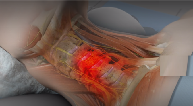
Inactivity and Chronic Pain Risk in Daily Life
The relationship between inactivity and chronic pain risk is stronger than many people realize.
When movement decreases:
- Muscles weaken
- Posture deteriorates
- Joint stress increases
- Core stability reduces
This combination often contributes to chronic discomfort.
Common Pain Areas
People with prolonged sitting habits frequently experience:
- Lower back pain
- Neck pain
- Shoulder pain
- Hip tightness
- Knee discomfort
Research also suggests sedentary behavior contributes to musculoskeletal issues and reduced physical function.

Desk Job Health Issues Prevention Strategies for Long-Term Wellness
Modern workplaces have transformed how people work, but they have also created new health challenges. Many employees spend eight to ten hours daily sitting in front of computers with very little movement throughout the day. Effective desk job health issues prevention methods are no longer optional for maintaining overall health and productivity.
Many individuals assume that discomfort during work is normal. However, recurring stiffness, fatigue and poor posture are often signs that the body is responding negatively to inactivity.
Why Desk Job Health Issues Prevention Matters?
Extended sitting affects multiple body systems simultaneously. Reduced movement decreases circulation and can increase stress on muscles and joints.
Ignoring workplace ergonomics may contribute to:
- Persistent lower back pain
- Neck strain
- Eye fatigue
- Reduced flexibility
- Poor circulation
- Wrist and shoulder discomfort
- Increased tiredness during work hours
Implementing desk job health issues prevention techniques early may help minimize these problems and improve long-term well-being.
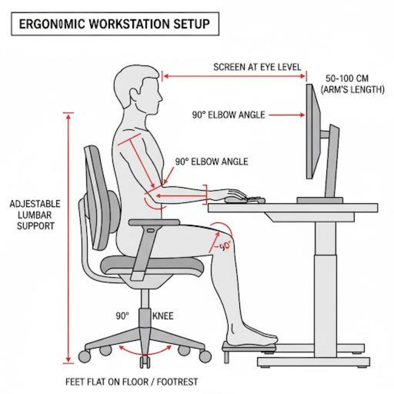
Create an Ergonomic Workspace
Small adjustments to your workstation can significantly improve comfort.
Follow these setup recommendations:
Chair Position
- Feet should remain flat on the floor
- Knees positioned at approximately 90 degrees
- Lower back supported properly
Monitor Height
- Top of screen at eye level
- Screen approximately arm’s length away
Keyboard Placement
- Wrists in neutral alignment
- Elbows relaxed beside the body
Desk Arrangement
- Frequently used items within easy reach
- Avoid repeated twisting motions
Good workspace design remains one of the most effective desk job health issues preventions approaches available.
Movement Break Strategies
Movement does not require an hour-long workout session.
Simple actions include:
- Stand every 30–45 minutes
- Walk while taking phone calls
- Stretch between meetings
- Use stairs when possible
- Perform quick posture resets
Even short movement intervals may help reduce the negative effects of sitting too long health experts often discuss.
Sedentary Lifestyle Health Risks and Their Impact on Heart Health
The sedentary lifestyle health risks affecting cardiovascular health often develop gradually. Many people do not notice symptoms until health conditions become more serious.
Physical movement supports healthy circulation and efficient heart function. Extended inactivity can reduce this natural support system.
How Sitting Affects Circulation?
The body relies on regular muscle movement to help circulate blood efficiently.
When sitting for prolonged periods:
- Blood flow slows
- Muscles remain inactive
- Energy expenditure decreases
- Metabolic activity may decline
Over time, these patterns can influence cardiovascular function.
Hidden Warning Signs
Many people ignore subtle indicators because they develop slowly.
Watch for:
- Reduced stamina
- Shortness of breath with activity
- Persistent fatigue
- Leg discomfort after prolonged sitting
- Reduced energy levels
The sedentary lifestyle health risks linked with prolonged inactivity frequently become more noticeable with age.
Activity Recommendations
Health experts commonly suggest:
- Engage in a minimum of 150 minutes of moderate exercise every week
- Muscle-strengthening activities twice weekly
- Regular movement throughout the day
Small actions often create meaningful long-term changes.
Examples include:
- Walking after meals
- Standing during meetings
- Taking brief movement breaks
- Parking farther away
- Stretching before and after work
Mental Health and the Effects of Sitting Too Long Health Experts Continue to Study
Physical health is not the only concern associated with prolonged inactivity. The effects of sitting too long health researchers examine also include psychological well-being.
Movement and mental wellness are closely connected.
Regular physical activity may support:
- Mood regulation
- Stress reduction
- Energy levels
- Sleep quality
- Cognitive performance
How Inactivity Influences Mood
Long periods of inactivity may affect emotional well-being in several ways.
Potential contributing factors include:
- Reduced social interaction
- Increased fatigue
- Disrupted sleep patterns
- Lower daily energy expenditure
People with highly sedentary schedules frequently report feeling mentally exhausted even after physically inactive days.
Productivity and Focus Challenges
Many individuals notice:
- Difficulty concentrating
- Brain fog
- Reduced motivation
- Mental fatigue
- Lower productivity
Introducing movement throughout the day often helps improve attention and energy levels.
Daily Movement Habits to Reduce Inactivity and Chronic Pain Risk
Preventing long-term health problems does not require dramatic lifestyle changes. Consistency often matters more than intensity. Simple movement habits performed regularly can reduce inactivity and chronic pain risk while supporting overall physical health.
Many people assume they need gym memberships or intense workout routines to improve their health. Incorporating movement throughout the day frequently produces meaningful benefits.
Build Small Habits That Become Routine
Healthy movement habits can fit naturally into daily life.
Try implementing these strategies:
Morning movement routine
- Stretch for 5–10 minutes after waking
- Walk briefly before beginning work
- Perform light mobility exercises
During work hours
- Stand every 30–45 minutes
- Walk during phone calls
- Stretch shoulders and hips
- Change sitting positions regularly
After work
- Take an evening walk
- Perform simple exercises
- Reduce prolonged screen time
These habits may help reduce inactivity and chronic pain risk over time.
Walking: One of the Simplest Solutions
Walking remains one of the easiest and most effective activities for many individuals.
Potential benefits include:
- Increased circulation
- Improved mobility
- Better energy levels
- Reduced stiffness
- Support for cardiovascular health
- Improved mood
Walking also helps counteract the effects of sitting too long health professionals continue to monitor.
Movement Checklist for Busy Individuals
Use this simple daily checklist:
- Walk for 20–30 minutes daily
- Stretch every hour during work
- Use stairs when possible
- Stand during calls or meetings
- Practice proper posture
- Stay hydrated
- Limit unnecessary screen time
Small improvements performed consistently often produce sustainable long-term changes.
Acting Before Symptoms Become Serious
Many health concerns related to inactivity develop slowly. People often adapt to discomfort and assume symptoms are temporary.
Ignoring early warning signs can allow small issues to become persistent challenges.
Signs that may require professional assessment include:
- Recurring back pain
- Neck stiffness
- Frequent headaches
- Reduced flexibility
- Numbness or tingling
- Ongoing fatigue
- Reduced mobility
Early intervention may help identify movement limitations and provide strategies that improve overall function.

Conclusion
The hidden effects of inactivity extend beyond occasional stiffness or fatigue. The long-term sedentary lifestyle health risks associated with prolonged sitting may affect physical health, mental well-being, mobility and overall quality of life.
Fortunately, meaningful improvements do not always require major lifestyle changes. Regularly standing, maintaining proper posture, moving throughout the day and following ergonomic work practices can significantly support long-term health.
If you have been experiencing discomfort related to prolonged sitting or workplace habits, consider seeking professional guidance. Early action can help protect your long-term health and improve daily quality of life. Book your assessment today and take the first step toward a healthier and more active future.
FAQs
1. What are the major sedentary lifestyle health risks?
Common sedentary lifestyle health risks include poor posture, muscle weakness, reduced heart health, weight gain and long-term physical discomfort.
2. Can exercise offset sitting all day?
Exercise helps, but it may not fully prevent the effects of sitting too long, health experts warn. Regular movement breaks are also important.
3. How can I reduce inactivity at work?
Use desk job health issues prevention strategies such as standing every 30–45 minutes, stretching, walking during calls and maintaining good posture to reduce inactivity and chronic pain risk.