Category Archives: Recent Posts

Microdiscectomy vs. Traditional Discectomy: Which Spine Surgery Is Right for You?

Imagine waking up one morning with excruciating pain shooting down your leg, making it nearly impossible to stand or walk. For millions of people suffering from herniated discs, this scenario becomes an unwelcome reality that significantly impacts their quality of life. When conservative treatments like physical therapy, medications, and injections fail to provide relief, spine surgery often becomes the next consideration.

Two primary surgical approaches dominate the landscape of herniated disc treatment: traditional discectomy and microdiscectomy. Both procedures aim to relieve pressure on compressed nerves by removing problematic disc material, yet they differ significantly in technique, recovery time, and overall patient experience. Understanding these differences becomes crucial when facing one of life’s most important healthcare decisions.

The choice between microdiscectomy vs discectomy isn’t always straightforward. Factors such as the size and location of your herniated disc, your overall health status, surgeon expertise, and personal preferences all play vital roles in determining the most appropriate treatment path. This comprehensive guide will walk you through everything you need to know about both procedures, helping you make an informed decision alongside your healthcare team.

We’ll explore the fundamental differences between these spine surgery options, examine their respective benefits and risks, discuss recovery expectations, and provide practical guidance for choosing the right approach for your unique situation. By the end of this article, you’ll have the knowledge necessary to engage in meaningful discussions with your spine specialist about your treatment options.

Understanding Herniated Discs and When Surgery Becomes Necessary

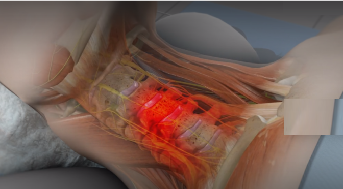
Your spine consists of vertebrae separated by intervertebral discs that act as cushions, absorbing shock and allowing flexible movement. These discs are made up of a jelly-like center called the nucleus pulposus, wrapped by a strong outer layer known as the annulus fibrosus. When the outer layer weakens or tears, the inner material can protrude or “herniate,” potentially pressing against nearby nerves and causing significant pain, numbness, or weakness.

Herniated disc treatment typically begins with conservative approaches. Most patients find relief through a combination of rest, anti-inflammatory medications, physical therapy, and sometimes epidural steroid injections. However, when these non-surgical treatments fail to provide adequate relief after 6-12 weeks, or when patients experience severe neurological symptoms like progressive weakness or loss of bowel/bladder control, surgical intervention becomes necessary.

The decision to pursue spine surgery shouldn’t be taken lightly. Surgeons typically recommend surgical treatment when:

  • Conservative treatments haven’t provided relief after a sufficient trial period
  • The pain greatly affects daily functioning and overall quality of life
  • Neurological symptoms are worsening or severe
  • Imaging studies clearly show nerve compression that correlates with symptoms

Both discectomy and microdiscectomy procedures aim to decompress affected nerves by removing the herniated disc material that’s causing pressure. The fundamental goal remains the same: eliminate the source of nerve compression to restore normal function and alleviate pain. However, the surgical approach and techniques used can vary significantly between these two options.

Understanding your specific condition, including the size, location, and severity of your herniated disc, helps determine which surgical approach might be most appropriate. Your spine specialist will consider these factors alongside your overall health, activity level, and treatment goals when recommending the best course of action.

Traditional Discectomy: The Established Approach

Traditional discectomy has served as the gold standard for treating herniated discs for decades. This open surgical procedure involves making a larger incision, typically 2-3 inches long, to provide surgeons with direct visualization and access to the affected disc space. The surgeon removes a portion of the vertebral bone (laminotomy or laminectomy) to reach the herniated disc material and carefully extract the problematic tissue pressing against the nerve.

During a traditional discectomy, patients receive general anesthesia and are positioned face-down on the operating table. The surgeon makes an incision over the affected spinal level, carefully separates the back muscles from the spine, and removes enough bone to access the disc space. Using specialized instruments and direct visualization, the surgeon identifies and removes the herniated disc fragments while taking care to protect surrounding nerves and tissues.

This spine surgery approach offers several advantages, particularly for complex cases. The larger incision provides excellent visualization of the surgical site, allowing surgeons to thoroughly examine the area and remove all problematic disc material. Traditional discectomy works especially well for large herniations, cases involving multiple disc levels, or situations where significant bone removal is necessary to adequately decompress the nerves.

Recovery from traditional discectomy typically involves a hospital stay of 1-2 days, followed by several weeks of activity restrictions. Patients usually experience immediate relief from leg pain, though some temporary back discomfort at the incision site is normal. Physical therapy often begins within a few weeks to help restore strength and mobility.

While traditional discectomy has proven effective over many years, it does involve more tissue disruption compared to minimally invasive alternatives. The larger incision and more extensive muscle dissection can result in increased post-operative pain, longer recovery times, and potentially higher risks of complications such as infection or excessive scar tissue formation.

Microdiscectomy: The Minimally Invasive Revolution

Microdiscectomy represents a significant advancement in spine surgery techniques, utilizing minimally invasive approaches to achieve the same therapeutic goals as traditional discectomy with potentially fewer complications and faster recovery times. This procedure employs a much smaller incision, typically 1-1.5 inches long, combined with specialized instruments and microscopic or endoscopic visualization to remove herniated disc material.

The microdiscectomy procedure begins similarly to traditional surgery, with the patient under general anesthesia and positioned face-down. However, the surgeon makes a much smaller incision and uses a tubular retractor system to create a narrow working channel directly to the affected disc space. This approach minimizes disruption to surrounding muscles and tissues while still providing adequate access to remove the problematic disc material.

Advanced visualization technology plays a crucial role in microdiscectomy success. Surgeons use either an operating microscope or endoscopic camera system to magnify the surgical site, allowing for precise identification and removal of herniated disc fragments. This enhanced visualization often enables more thorough removal of loose disc material while minimizing trauma to healthy tissues.

One of the most significant advantages of microdiscectomy is the reduced tissue trauma compared to traditional approaches. The smaller incision and muscle-sparing techniques typically result in less post-operative pain, reduced blood loss, and faster healing times. Many patients undergoing microdiscectomy can return home the same day or after an overnight hospital stay.

Spine surgery options like microdiscectomy have revolutionized treatment outcomes for many patients. The minimally invasive nature of this procedure often allows for quicker return to normal activities, with many patients resuming desk work within 1-2 weeks and more strenuous activities within 6-8 weeks, depending on their specific situation and healing progress.

However, microdiscectomy may not be suitable for all cases. Very large herniations, complex anatomy, or situations requiring extensive bone removal might still necessitate traditional open approaches to ensure complete treatment and optimal outcomes.

Microdiscectomy vs Traditional Discectomy: A Comprehensive Comparison

When comparing microdiscectomy vs discectomy, several key factors distinguish these spine surgery options and influence treatment decisions. Understanding these differences helps patients and surgeons collaborate in choosing the most appropriate approach for each unique situation.

Surgical trauma and tissue disruption represent perhaps the most significant difference between these procedures. Microdiscectomy involves minimal muscle dissection and tissue disruption, while traditional discectomy requires more extensive exposure and muscle retraction. This difference directly impacts post-operative pain levels, with microdiscectomy patients typically experiencing less discomfort and requiring fewer pain medications.

Recovery time and return to activities vary considerably between these approaches. Microdiscectomy patients often return to light activities within days and resume normal work within 1-2 weeks. Traditional discectomy recovery typically takes longer, with patients needing 2-4 weeks before returning to desk work and 6-12 weeks for more strenuous activities.

Hospital stay requirements also differ significantly. Many microdiscectomy procedures are performed on an outpatient basis or require only overnight observation. Traditional discectomy usually involves a 1-2 day hospital stay, increasing overall treatment costs and potential exposure to hospital-associated complications.

Complication rates tend to favor minimally invasive approaches. Microdiscectomy is associated with lower risks of infection, excessive bleeding, and scar tissue formation. However, both procedures carry similar risks of nerve injury, recurrent herniation, and anesthesia-related complications.

Long-term outcomes for both procedures are generally excellent, with success rates exceeding 85-90% for appropriate candidates. The choice between microdiscectomy vs traditional discectomy often comes down to individual patient factors rather than significant differences in long-term effectiveness.

Cost considerations may favor microdiscectomy due to shorter hospital stays, reduced need for pain medications, and faster return to work. However, the specialized equipment and training required for minimally invasive techniques may offset some of these savings in certain healthcare systems.

Choosing the Right Spine Surgery Option for Your Situation

Selecting between microdiscectomy and traditional discectomy requires careful consideration of multiple factors unique to your specific situation. This decision should always be made in partnership with an experienced spine specialist who can evaluate your individual circumstances and recommend the most appropriate spine surgery approach.

Disc characteristics play a crucial role in surgical planning. Large, complex herniations or those involving multiple levels may require the enhanced visualization and access provided by traditional discectomy. Conversely, smaller, well-contained herniations often respond excellently to microdiscectomy techniques.

Patient anatomy and previous surgical history significantly influence procedure selection. Patients with previous spine surgery, complex spinal anatomy, or significant scar tissue may benefit from the direct visualization offered by traditional approaches. First-time surgical candidates with straightforward anatomy typically make excellent microdiscectomy candidates.

Surgeon expertise and preference cannot be overlooked when choosing between these spine surgery options. While most spine surgeons are trained in both techniques, individual surgeons may have greater experience or comfort levels with specific approaches. Choosing a surgeon with extensive experience in your recommended procedure often matters more than the specific technique used.

Recovery timeline expectations should align with your personal and professional obligations. If you need to return to work quickly or have limited time for recovery, microdiscectomy may offer advantages. However, if thorough treatment is the primary concern regardless of recovery time, traditional discectomy might be preferred for complex cases.

Risk tolerance and personal preferences vary among patients. Some individuals prefer the most minimally invasive option available, while others prioritize the most thorough treatment approach regardless of invasiveness. Discussing these preferences with your surgeon helps ensure your treatment plan aligns with your values and expectations.

Insurance coverage and financial considerations may influence your decision, though most insurance plans cover both procedures when medically necessary. Understanding your specific coverage and any potential out-of-pocket costs helps inform your treatment choice.

The most important factor in choosing between microdiscectomy vs discectomy is finding an experienced spine specialist who can thoroughly evaluate your condition and recommend the most appropriate treatment approach for your unique situation.

Making a Well-Informed Choice About Your Herniated Disc Treatment

The choice between microdiscectomy and traditional discectomy represents an important decision in your herniated disc treatment journey. Both spine surgery options offer excellent outcomes when performed by experienced surgeons on appropriate candidates. Microdiscectomy provides the advantages of minimal tissue disruption, faster recovery, and reduced post-operative discomfort, making it an attractive option for many patients with suitable disc herniations.

However, traditional discectomy remains the preferred approach for complex cases requiring extensive visualization or bone removal. The key to optimal outcomes lies not necessarily in choosing the “best” technique, but rather in selecting the most appropriate approach for your specific condition and circumstances.

We encourage you to engage in thorough discussions with qualified spine specialists, seek second opinions when appropriate, and carefully consider all factors influencing your treatment decision. Remember that successful spine surgery depends on proper patient selection, surgeon expertise, and realistic expectations about recovery and outcomes.

Your journey toward pain relief and restored function begins with taking that first step to consult with a spine specialist who can guide you through these important decisions and help you reclaim your quality of life.

Is Your Foot Pain Linked to Poor Posture or Nerve Issues?

Understanding Foot Pain: An Overview
Foot pain can be a debilitating condition that affects your quality of life. The foot is a complex
structure composed of bones, joints, ligaments, muscles, and nerves, all working in harmony to
support your body and facilitate movement. When you experience foot pain, it can be challenging to
pinpoint the exact cause due to the intricate nature of the foot’s anatomy. Pain may be localized in
one area, such as the heel, arch, or ball of the foot, or it may be more generalized, affecting the
whole foot. Understanding the potential sources of foot pain is the first step in addressing the issue
effectively.

The causes of foot pain are varied and can include injuries, overuse, and medical conditions such as
arthritis or diabetes. Injuries like sprains, fractures, and tendonitis are common and often result
from accidents or excessive physical activity. Overuse injuries typically occur from repetitive stress
on the foot, such as running or walking long distances without adequate rest. Medical conditions can
lead to inflammation or nerve damage, contributing to foot pain. It’s important to consider all
potential sources when evaluating foot pain, as addressing the root cause is essential for effective
treatment.

Moreover, lifestyle factors and daily habits can significantly influence foot health. Wearing ill-fitting
shoes, standing for extended periods, and neglecting foot care can exacerbate issues and lead to
chronic pain. The foot’s structural integrity is crucial for maintaining balance and proper posture,
which in turn influences overall well-being. By gaining a comprehensive understanding of foot pain
and its origins, you can take proactive steps to alleviate discomfort and prevent future issues.
Recognizing the complexity of foot pain is essential for identifying the underlying causes and
exploring effective treatment options.

The Connection Between Poor Posture and Foot Pain
Poor posture is often overlooked as a contributor to foot pain, yet it plays a significant role in how
your body distributes weight and absorbs shock. When you maintain poor posture, your body’s
alignment is disrupted, leading to uneven pressure on your feet. This imbalance can cause stress on
certain areas of the foot, resulting in pain and discomfort. For example, slouching or leaning forward
can shift your center of gravity, placing additional strain on the balls of your feet. Over time, this
added pressure can lead to conditions such as metatarsalgia or plantar fasciitis.

The connection between posture and foot pain becomes more apparent when considering the
body’s kinetic chain. The kinetic chain is the concept that different parts of the body are
interconnected and affect one another. Poor posture can cause a ripple effect throughout the body,
impacting not just the feet but also the knees, hips, and spine. When one part of the body is
misaligned, it forces other parts to compensate, often leading to discomfort and pain in areas that
may seem unrelated. Understanding this connection is crucial for addressing foot pain holistically
and ensuring long-term relief.

Addressing poor posture involves more than just standing up straight. It requires an awareness of
how your body moves and functions throughout the day. Correcting posture involves strengthening
core muscles, developing body awareness, and making conscious adjustments to your stance and
gait. By improving posture, you can alleviate the undue stress placed on your feet and promote
better overall body mechanics. This not only helps reduce existing foot pain but also prevents future
issues from arising. Taking the time to assess and improve your posture can have a profound impact
on your foot health and overall well-being.

Common Postural Issues That Affect Foot Health
Several common postural issues can directly affect foot health, leading to pain and discomfort. One
such issue is overpronation, where the foot rolls inward excessively while walking or running. This
condition can cause undue stress on the arches and lead to conditions like flat feet, plantar fasciitis,
and shin splints. Overpronation is often exacerbated by poor posture, as it can alter the alignment of
the entire body. Similarly, underpronation, or supination, where the foot rolls outward, can also
result in added pressure on the outer edges of the foot, leading to imbalances and pain.

Another postural issue that impacts foot health is anterior pelvic tilt. This condition occurs when the
pelvis tilts forward, often due to muscle imbalances in the hips and lower back. Anterior pelvic tilt
can cause the lower back to arch excessively, shifting the body’s weight distribution toward the front
of the foot. This added pressure on the forefoot can lead to discomfort and exacerbate existing foot
conditions. Correcting anterior pelvic tilt involves strengthening the core and glute muscles while
stretching the hip flexors and lower back muscles to restore proper alignment.

Forward head posture is another common issue that can indirectly affect foot health. When the
head is positioned forward of the shoulders, it disrupts the body’s alignment, causing compensatory
changes in posture. This forward position can lead to a cascade of adjustments throughout the body,
ultimately affecting the feet. The increased strain on the neck and shoulders can cause tension along
the spine, impacting the lower back and hips, which in turn affects the feet. Addressing forward
head posture involves correcting neck alignment and strengthening the upper back and neck
muscles to support proper posture.

Nerve Issues and Their Impact on Foot Pain
Nerve issues are another significant factor in the development of foot pain. When nerves become
compressed, irritated, or damaged, they can send pain signals to the brain, resulting in discomfort in
the feet. One common nerve-related condition is peripheral neuropathy, which affects the
peripheral nerves responsible for transmitting signals between the central nervous system and the
rest of the body. Peripheral neuropathy can be caused by diabetes, infections, or traumatic injuries,
and it often presents as tingling, numbness, or burning sensations in the feet.

Another nerve-related condition that impacts foot pain is tarsal tunnel syndrome, which is similar to
carpal tunnel syndrome in the wrist. Tarsal tunnel syndrome occurs when the tibial nerve, which
runs along the inside of the ankle, becomes compressed. This compression can lead to pain, tingling,
and numbness in the sole of the foot and the inner ankle. The condition is often caused by repetitive
stress, injury, or anatomical abnormalities such as flat feet. Treating tarsal tunnel syndrome often
involves reducing inflammation, using orthotic supports, and sometimes surgical intervention.

Morton’s neuroma is another nerve issue that can cause foot pain. It involves the thickening of
tissue around one of the nerves leading to the toes, often between the third and fourth toes. This
thickening can compress the nerve, causing sharp, burning pain, and a sensation of a pebble in the
shoe. High-heeled or tight shoes can exacerbate the condition, so wearing appropriate footwear is
essential for relief. Treatment for Morton’s neuroma may include corticosteroid injections, physical
therapy, or surgery in more severe cases. Understanding nerve-related foot pain is crucial for
identifying the appropriate treatment and finding relief.

Symptoms of Nerve-Related Foot Pain
Nerve-related foot pain often presents with a distinct set of symptoms that can help differentiate it
from other types of foot pain. One of the most common symptoms is a burning or tingling sensation,
often described as pins and needles, which can occur in the toes, ball of the foot, or the entire foot.
This sensation may come and go or be constant, depending on the severity and underlying cause of
the nerve issue. Recognizing these symptoms is essential for diagnosis and treatment, as nerve-
related pain requires specific interventions to address the underlying cause.

Numbness is another common symptom of nerve-related foot pain. Affected individuals may
experience a loss of sensation in the feet, which can make it difficult to perceive temperature
changes or feel pressure. This numbness can impact balance and coordination, increasing the risk of
falls and injuries. Additionally, muscle weakness may accompany numbness, further affecting
mobility. These symptoms can be particularly concerning for individuals with conditions like
diabetes, where nerve damage can lead to serious complications if left untreated.

Sharp, shooting pain is also characteristic of nerve-related foot pain. This pain may radiate along the
path of the affected nerve and can be triggered by specific movements or pressure on the foot. In
some cases, the pain may be exacerbated by certain activities, such as walking or standing for
prolonged periods. Identifying the specific symptoms and patterns of nerve-related foot pain is
crucial for determining the appropriate course of treatment. Early intervention can help prevent
further nerve damage and improve the overall quality of life for those affected.

How to Differentiate Between Postural and Nerve-Related Foot Pain
Differentiating between postural and nerve-related foot pain can be challenging, as both can present
with overlapping symptoms. However, understanding the nuances of each type of pain can aid in
accurate diagnosis and treatment. Postural foot pain is often associated with specific activities or

positions, such as standing for long periods or wearing improper footwear. It may be accompanied
by other postural symptoms, such as lower back pain or shoulder discomfort. In contrast, nerve-
related pain is typically characterized by tingling, numbness, or burning sensations that may persist
regardless of activity or position.

A thorough physical examination and medical history are essential for distinguishing between
postural and nerve-related foot pain. A healthcare professional may assess your posture, gait, and
alignment to identify any imbalances contributing to postural pain. They may also perform
neurological tests to evaluate nerve function and identify any areas of nerve compression or
damage. Imaging studies, such as X-rays or MRIs, may be used to rule out other conditions and gain
a better understanding of the underlying issues.

In some cases, electromyography (EMG) or nerve conduction studies may be recommended to
assess nerve health and function. These tests can help identify the specific nerves affected and the
extent of any damage. By combining clinical evaluation with diagnostic tests, healthcare
professionals can accurately differentiate between postural and nerve-related foot pain. This
differentiation is crucial for developing an effective treatment plan tailored to the individual’s needs
and addressing the root cause of the pain.

Exercises to Improve Posture and Alleviate Foot Pain
Incorporating targeted exercises into your routine can significantly improve posture and alleviate
foot pain. Strengthening exercises for the core, hips, and lower back can help correct postural
imbalances and reduce stress on the feet. For example, planks and bridges are effective exercises for
building core stability and promoting proper alignment. These exercises engage the abdominal and
back muscles, supporting the spine and pelvis in a neutral position. Consistent practice can lead to
improved posture and reduced foot pain over time.

Stretching exercises are also crucial for maintaining flexibility and preventing muscle tightness that
can contribute to postural issues. Stretching the calf muscles, hamstrings, and hip flexors can relieve
tension and improve range of motion, allowing for better alignment and weight distribution.
Incorporating yoga or Pilates into your routine can provide a comprehensive approach to stretching
and strengthening, promoting balance and flexibility throughout the body. These practices
emphasize mindfulness and body awareness, helping you maintain correct posture in daily activities.

Foot-specific exercises can further alleviate pain and improve foot health. Simple exercises like toe
curls and heel raises can strengthen the muscles and ligaments in the feet, providing better support
and stability. Additionally, using a foam roller or massage ball can help release tension and improve
circulation in the feet. Regularly performing these exercises can enhance foot function, reduce pain,
and prevent future issues. By adopting a holistic approach to exercise, you can address both postural
and foot-specific concerns, promoting overall well-being and reducing the risk of foot pain.

When to Seek Professional Help for Foot Pain
While self-care measures and exercises can be effective for managing mild foot pain, there are times
when professional help is necessary. If you experience severe pain, swelling, or an inability to bear
weight on the affected foot, it’s important to seek medical attention promptly. These symptoms may
indicate a more serious condition, such as a fracture or severe nerve damage, requiring professional
evaluation and treatment. Early intervention can prevent further complications and support a
quicker recovery.

Persistent or worsening foot pain despite self-care measures also warrants professional evaluation.
A healthcare provider can conduct a thorough assessment to determine the underlying cause of the
pain and develop an appropriate treatment plan. They may recommend physical therapy, orthotic
supports, or medications to address specific issues. In some cases, surgical intervention may be
necessary to correct structural abnormalities or relieve nerve compression. Seeking professional
help ensures that you receive the most effective treatment for your specific condition.

Additionally, if you have underlying medical conditions such as diabetes, arthritis, or neuropathy, it’s
important to regularly consult with a healthcare professional to monitor your foot health. These
conditions can increase the risk of complications, and early detection of any changes in foot health is
crucial for preventing serious issues. Regular check-ups with a podiatrist or specialist can help
manage your condition and maintain optimal foot health. By being proactive and seeking
professional help when needed, you can effectively manage foot pain and improve your overall
quality of life.

Preventative Measures for Maintaining Foot Health
Maintaining foot health requires a proactive approach that includes adopting healthy lifestyle habits
and taking preventative measures. One of the most important steps is wearing appropriate footwear
that provides adequate support and cushioning. Choose shoes with a proper fit, arch support, and a
wide toe box to prevent crowding and pressure on the toes. Avoid high heels and shoes with narrow
toe boxes, as they can exacerbate foot pain and contribute to conditions like bunions and
hammertoes. Investing in quality footwear can significantly reduce the risk of foot pain and promote
overall foot health.

Regular foot care is also essential for preventing issues and maintaining healthy feet. This includes
washing and drying your feet thoroughly, trimming toenails properly, and inspecting your feet
regularly for any signs of changes or abnormalities. Moisturizing your feet can prevent dry, cracked
skin, while exfoliating can remove dead skin cells and promote circulation. If you notice any unusual
symptoms, such as redness, swelling, or persistent pain, it’s important to address them promptly to
prevent more serious issues.

Incorporating foot and ankle exercises into your routine can strengthen the muscles and improve
flexibility, reducing the risk of injuries and strains. Balance exercises, such as standing on one foot or

using a balance board, can enhance stability and coordination, supporting overall foot health.
Maintaining a healthy weight is also crucial, as excess weight can place additional stress on the feet
and lead to pain and discomfort. By adopting these preventative measures, you can support foot
health, reduce the risk of pain, and enjoy a more active and comfortable lifestyle.

Conclusion: Finding the Right Approach to Foot Pain Relief
Foot pain can be a complex and multifaceted issue, with causes ranging from poor posture to nerve-
related conditions. Understanding the interplay between these factors is crucial for finding effective
relief and addressing the root causes of discomfort. By recognizing the impact of posture on foot
health and the potential for nerve issues to contribute to pain, you can take a more informed and
targeted approach to treatment. Whether through exercises, lifestyle changes, or professional
intervention, addressing foot pain requires a comprehensive and individualized strategy.

Taking proactive steps to improve posture, engage in regular exercise, and maintain proper foot care
can significantly reduce the risk of foot pain and enhance overall well-being. Seeking professional
help when necessary ensures that you receive the appropriate diagnosis and treatment for your
specific condition. By prioritizing foot health and being mindful of the factors that contribute to pain,
you can reclaim your stride and enjoy a more active and pain-free lifestyle.

Ultimately, the key to finding relief from foot pain lies in understanding your body’s unique needs
and addressing the underlying causes of discomfort. By adopting a holistic approach that considers
posture, nerve health, and overall lifestyle, you can achieve lasting relief and maintain optimal foot health. Don’t let foot pain hold you back any longer—take charge of your well-being and step
confidently into a future free from discomfort and limitations.

Why Hip and Pelvic Pain Often Go Hand in Hand

Understanding Hip and Pelvic Pain

Hip and pelvic pain can be a confounding experience, often leaving individuals feeling trapped in a cycle of discomfort and confusion. The hips and pelvis serve as crucial support structures for the body, playing a vital role in maintaining balance and facilitating movement. When pain arises in these areas, it can significantly disrupt daily activities, making even the simplest tasks challenging. Understanding the anatomy and function of the hips and pelvis is the first step towards unraveling the mystery behind the pain that often links them together.

The hips are ball-and-socket joints where the femur (thigh bone) connects to the pelvis. This configuration allows for a wide range of motion, essential for activities like walking, running, and bending. The pelvis, on the other hand, is a basin-shaped structure composed of several bones including the ilium, ischium, and pubis. It supports the weight of the upper body and connects the spine to the lower limbs. The complex interplay between these structures means that an issue in one can easily affect the other, leading to concurrent pain.

Many people underestimate the interconnectedness of the hips and pelvis, often viewing them as separate entities. However, the reality is that they work in tandem, supported by a network of muscles, ligaments, and tendons. This intricate relationship means that stress or injury in one area can create a ripple effect, causing discomfort in the other. Recognizing this connection is vital for those seeking to understand and address their hip and pelvic pain effectively.

Common Causes of Hip and Pelvic Pain

Hip and pelvic pain can stem from a myriad of causes, some of which are more common than others. One prevalent cause is osteoarthritis, a degenerative joint disease that affects the cartilage cushioning the hip joint. As the cartilage wears down, it leads to pain and stiffness, which can extend to the pelvic region. Osteoarthritis is particularly common in older adults, though it can affect younger individuals as well, especially those with a family history of the condition.

Another common culprit is tendinitis, which occurs when the tendons connecting muscles to bones become inflamed. This inflammation can result from repetitive motions or overuse, often seen in athletes or individuals with physically demanding jobs. Tendinitis in the hip can cause pain that radiates to the pelvis, as the muscles and tendons work to stabilize the joint. Similarly, bursitis, the inflammation of the small fluid-filled sacs that cushion the joints, can lead to similar discomfort.

In addition to these musculoskeletal issues, hip and pelvic pain can also be caused by injuries such as fractures or dislocations. Trauma from falls, accidents, or sports injuries can impact both the hip and pelvic regions, resulting in acute pain. In some cases, underlying medical conditions like hernias, gynecological issues, or even kidney stones can manifest as hip and pelvic pain, making diagnosis and treatment a complex process. Understanding the potential causes is essential for addressing the root of the problem and finding relief.

The Connection Between Hip and Pelvic Pain

The connection between hip and pelvic pain is a topic that often surprises those unfamiliar with the intricacies of human anatomy. At the heart of this relationship is the shared structural and functional roles that bind the hips and pelvis together. Both areas are integral to the support and movement of the lower body, meaning that dysfunction in one can have a direct impact on the other. This interconnectedness is crucial for understanding why pain in one area is frequently accompanied by discomfort in the other.

Muscular imbalances are a common factor that links hip and pelvic pain. When certain muscles around the hips and pelvis become overactive or underactive, it can lead to changes in posture and movement patterns. For instance, tight hip flexors can pull on the pelvis, creating an anterior tilt that strains the lower back and pelvic region. Conversely, weak gluteal muscles can lead to instability in the hip joint, causing compensatory movements that stress the pelvis.

Additionally, the nerves that supply the hips and pelvis can contribute to their shared pain experience. The sciatic nerve, for example, runs from the lower back through the hips and down each leg. Compression or irritation of this nerve can result in sciatica, a condition characterized by pain that radiates from the lower back to the hips and pelvis. This nerve-related pain highlights the interconnected nature of these regions and underscores the importance of addressing both areas when seeking relief.

Symptoms to Watch For

Recognizing the symptoms of hip and pelvic pain is a critical step towards effective diagnosis and treatment. One of the most common symptoms is a persistent aching or stiffness in the hip joint or pelvic region. This discomfort may worsen with physical activity, prolonged sitting, or standing, and can feel particularly intense in the morning or after periods of rest. Paying attention to the pattern and intensity of the pain can provide valuable insights into its underlying cause.

In addition to general discomfort, individuals may also experience more specific symptoms such as swelling, warmth, or redness around the affected area. These signs suggest inflammation, which is often associated with conditions like tendinitis or bursitis. In some cases, the pain may be accompanied by a reduced range of motion, making it difficult to perform everyday activities like bending, walking, or climbing stairs. Such limitations can significantly impact one’s quality of life, highlighting the need for timely intervention.

Another important symptom to watch for is referred pain, which occurs when discomfort is felt in a location distant from its source. For instance, pain originating in the hip may be perceived in the groin, thigh, or even the knee. Similarly, pelvic pain can manifest as lower back discomfort. Understanding these atypical presentations is essential for accurate diagnosis and ensuring that treatment targets the true source of the pain rather than just its symptoms.

Diagnosing Hip and Pelvic Pain

Accurate diagnosis is paramount when it comes to addressing hip and pelvic pain, as it enables tailored treatment plans that target the specific underlying cause. The diagnostic process typically begins with a comprehensive medical history and physical examination. During this initial assessment, healthcare providers may inquire about the onset, duration, and nature of the pain, as well as any previous injuries or medical conditions that could be contributing factors.

Imaging studies are often employed to gain a clearer picture of the structures involved. X-rays can reveal changes in bone architecture, such as fractures or arthritis-related degeneration. For a more detailed view, magnetic resonance imaging (MRI) or computed tomography (CT) scans may be utilized to assess the soft tissues, including muscles, tendons, and ligaments. These advanced imaging techniques are invaluable in identifying conditions like tendinitis, bursitis, or hernias that may not be visible on standard X-rays.

In some cases, diagnostic injections may be used to pinpoint the source of pain. By injecting a local anesthetic into specific areas, doctors can determine if temporary pain relief occurs, thereby confirming the involvement of that structure in the patient’s symptoms. This targeted approach helps distinguish between multiple potential sources of pain and ensures that treatment strategies are appropriately directed. Ultimately, a thorough diagnostic process is essential for developing a treatment plan that effectively addresses the root cause of hip and pelvic pain.

Treatment Options for Relief

Once the cause of hip and pelvic pain has been identified, a range of treatment options can be explored to provide relief and restore function. One of the most common approaches is physical therapy, which focuses on strengthening the muscles around the hip and pelvis to improve stability and reduce strain. Customized exercise programs tailored to the individual’s needs can enhance flexibility, correct muscular imbalances, and promote better posture and movement patterns.

In addition to physical therapy, medication may be prescribed to manage pain and inflammation. Nonsteroidal anti-inflammatory drugs (NSAIDs) such as ibuprofen or naproxen are often recommended for their ability to reduce swelling and alleviate discomfort. For more severe pain, doctors may consider corticosteroid injections to deliver targeted relief directly to the affected area. These injections can provide significant pain reduction, allowing individuals to participate more effectively in rehabilitation exercises.

In cases where conservative treatments are ineffective, surgical intervention may be necessary. Surgical options vary depending on the specific condition, ranging from minimally invasive procedures to more extensive surgeries. For example, arthroscopic surgery can address issues like labral tears or impingement in the hip joint, while hip replacement surgery may be considered for severe arthritis. The decision to pursue surgery is typically made after a thorough evaluation of the patient’s condition and consideration of potential risks and benefits.

Preventative Measures to Avoid Pain

Prevention plays a vital role in minimizing the risk of developing hip and pelvic pain, and adopting certain lifestyle changes can be highly effective. One of the most important preventative measures is maintaining a healthy weight, as excess body weight places additional stress on the hips and pelvis, increasing the likelihood of joint degeneration and injury. A balanced diet and regular exercise can help manage weight and promote overall musculoskeletal health.

Engaging in regular physical activity is another key factor in preventing hip and pelvic pain. Activities that promote strength, flexibility, and cardiovascular fitness can enhance joint stability and reduce the risk of injury. Low-impact exercises such as swimming, cycling, or walking are particularly beneficial, as they provide cardiovascular benefits without placing excessive strain on the joints. Incorporating strength training exercises that target the core, hips, and lower extremities can further bolster stability and support.

Proper body mechanics and ergonomic considerations are also crucial for preventing pain. Paying attention to posture during daily activities, such as sitting, standing, or lifting, can reduce stress on the hips and pelvis. Using ergonomic furniture and equipment, taking frequent breaks during prolonged activities, and practicing good lifting techniques can all contribute to minimizing the risk of strain or injury. By adopting these preventative measures, individuals can significantly reduce their risk of developing hip and pelvic pain and promote long-term joint health.

The Role of Physical Therapy

Physical therapy is a cornerstone in the treatment and prevention of hip and pelvic pain, offering a non-invasive approach that can yield significant benefits. A physical therapist works closely with patients to develop individualized exercise programs that target specific weaknesses and imbalances. These programs are designed to improve flexibility, strength, and stability, ultimately enhancing joint function and reducing pain.

One of the primary goals of physical therapy is to increase the range of motion in the hip and pelvic joints. Through a combination of stretching exercises and manual therapy techniques, therapists can help release tight muscles and improve joint mobility. This increased flexibility not only alleviates existing discomfort but also helps prevent future pain by promoting more efficient movement patterns.

Another critical aspect of physical therapy is education. Therapists provide valuable guidance on body mechanics, posture, and ergonomic adjustments that can be applied to daily activities. By teaching patients how to move safely and efficiently, therapists empower them to take an active role in managing their condition and preventing recurrence. This educational component, combined with targeted exercises, makes physical therapy an essential tool in addressing hip and pelvic pain effectively.

When to Seek Medical Attention

While many cases of hip and pelvic pain can be managed with conservative measures, there are certain situations where seeking medical attention becomes imperative. If the pain is severe, persistent, or accompanied by other concerning symptoms, such as fever, unexplained weight loss, or changes in bowel or bladder function, it is crucial to consult a healthcare professional. These symptoms may indicate an underlying condition that requires prompt evaluation and treatment.

Additionally, if pain is the result of a traumatic injury, such as a fall or accident, immediate medical attention is necessary to rule out fractures or dislocations. Delaying treatment in such cases can lead to complications and prolonged recovery. Similarly, if conservative treatments like rest, ice, or over-the-counter medications fail to provide relief after a reasonable period, it is advisable to seek further evaluation to determine the cause and explore alternative treatment options.

Ultimately, listening to your body and seeking timely medical attention can prevent minor issues from escalating into more significant problems. Early intervention not only facilitates more effective treatment but also improves the chances of a full recovery. By prioritizing health and being proactive in seeking care when needed, individuals can better manage their hip and pelvic pain and maintain an active, fulfilling lifestyle.

Conclusion: Managing Hip and Pelvic Pain Effectively

Managing hip and pelvic pain requires a multifaceted approach that addresses both the underlying causes and the symptoms. By understanding the intricate relationship between these two areas, individuals can take proactive steps to alleviate discomfort and improve their quality of life. Whether through lifestyle modifications, physical therapy, or medical interventions, there are numerous strategies available to manage and prevent pain effectively.

Educating oneself about the anatomy and function of the hips and pelvis is a crucial first step in this journey. Recognizing the common causes and symptoms of pain allows for more informed discussions with healthcare providers and empowers individuals to make decisions that align with their health goals. With the right knowledge and resources, it is possible to navigate the complexities of hip and pelvic pain successfully.

Ultimately, the key to managing hip and pelvic pain lies in a combination of proactive prevention, timely intervention, and a commitment to maintaining overall health and well-being. By embracing a holistic approach and working closely with healthcare professionals, individuals can reclaim their comfort and mobility, paving the way for a more active and fulfilling lifestyle. Through understanding and action, the puzzle of hip and pelvic pain can be unraveled, leading to lasting relief and improved quality of life.