Thoracic Outlet Syndrome vs. Carpal Tunnel: Key Differences

Have you ever felt tingling, numbness, or weakness in your hand and wondered whether it’s just fatigue—or something more serious? Many people assume nerve-related symptoms in the arm or hand automatically mean carpal tunnel syndrome. However, another condition called Thoracic Outlet Syndrome can cause very similar symptoms, making diagnosis confusing and sometimes delayed.

Both conditions affect nerves and can significantly impact your daily life. They can make simple activities like typing, driving, or even holding a phone uncomfortable or painful. Without proper diagnosis and treatment, these symptoms may worsen over time and reduce mobility, strength, and overall quality of life.

Understanding the differences between these conditions is essential for getting the right treatment. Although they share overlapping symptoms, they originate in completely different parts of the body and require different treatment approaches. An incorrect diagnosis may result in ineffective treatment and extended discomfort.

In this comprehensive guide, you will learn the key differences between these two conditions, including symptoms, causes, risk factors, diagnostic methods, and treatment options. By the end, you will have the knowledge needed to recognize warning signs early and seek the appropriate care for faster relief and long-term recovery.

Understanding Carpal Tunnel Syndrome

Carpal tunnel syndrome is one of the most common nerve compression disorders, affecting millions of people worldwide. It happens when the median nerve is compressed while traveling through the carpal tunnel in the wrist.

This tunnel is a narrow passage made of bones and ligaments. When inflammation, swelling, or structural changes occur, pressure builds up and compresses the nerve.

Anatomy and Mechanism

The median nerve controls sensation and movement in several parts of the hand. When compressed, it affects:

  • Thumb
  • Index finger
  • Middle finger
  • Part of the ring finger

This compression disrupts nerve signalling, leading to sensory and motor symptoms.

Key anatomical features involved:

  • Median nerve
  • Carpal tunnel ligament
  • Wrist bones
  • Tendons surrounding the nerve

According to the Mayo Clinic, prolonged pressure on the median nerve leads to progressive symptoms if left untreated.

Common Symptoms of Carpal Tunnel Syndrome

Symptoms often develop gradually and worsen over time. Many patients first notice symptoms at night or during repetitive activities.

Most common symptoms include:

  • Tingling or numbness in fingers
  • Hand weakness
  • Difficulty gripping objects
  • Pain radiating up the arm
  • Frequent dropping of objects

Symptom pattern clues:

  • Symptoms worsen at night
  • Shaking the hand provides temporary relief
  • Symptoms affect thumb, index, and middle fingers

If untreated, muscle weakness and permanent nerve damage may occur.

Causes and Risk Factors

Carpal tunnel syndrome develops due to increased pressure within the wrist tunnel.

Common causes include:

  • Repetitive hand movements
  • Typing or computer work
  • Wrist injuries
  • Arthritis
  • Diabetes
  • Pregnancy-related swelling

High-risk professions:

  • Office workers
  • Assembly line workers
  • Drivers
  • Musicians
  • Healthcare professionals

Early intervention can prevent progression and permanent nerve damage.

What Is Thoracic Outlet Syndrome?

This condition affects the nerves or blood vessels located between the collarbone and the first rib. This area is known as the thoracic outlet, and compression here can disrupt nerve and vascular function.

Unlike carpal tunnel syndrome, which affects the wrist, this condition originates near the neck and shoulder region.

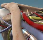
Anatomy of the Thoracic Outlet

The thoracic outlet contains critical structures including:

  • Brachial plexus nerves
  • Subclavian artery
  • Subclavian vein
  • Muscles and bones

Compression in this area affects nerve signals traveling from the neck to the arm.

Types include:

  1. Neurogenic (most common)
  2. Venous
  3. Arterial

According to the Cleveland Clinic, neurogenic type accounts for over 90% of cases.

Symptoms Originating from the Neck and Shoulder

Symptoms often begin in the shoulder and travel downward into the arm.

Common symptoms include:

  • Neck pain
  • Shoulder tightness
  • Arm fatigue
  • Tingling in fingers
  • Weak grip strength

Unique distinguishing signs:

  • Symptoms worsen when raising arms
  • Shoulder discomfort precedes hand symptoms
  • Pain may involve entire arm

These symptoms can mimic other nerve disorders, which makes diagnosis challenging.

Causes and Risk Factors

This condition can develop due to structural abnormalities or repetitive strain.

Common causes include:

  • Poor posture
  • Neck injuries
  • Repetitive overhead activities
  • Trauma
  • Anatomical abnormalities

High-risk individuals include:

  • Athletes
  • Office workers with poor posture
  • People with neck injuries
  • Weightlifters

Early diagnosis improves recovery outcomes.

Key Differences Between the Two Conditions

Recognizing the differences is crucial for ensuring proper treatment and effective recovery.

Location of Nerve Compression

Carpal tunnel syndrome:

  • Compression occurs in wrist
  • Affects median nerve only

Thoracic outlet-related compression:

  • Occurs near neck and shoulder
  • Affects brachial plexus nerves

This difference explains the variation in symptom patterns.

Symptom Distribution

Carpal tunnel syndrome symptoms typically affect specific fingers.

Thoracic outlet-related symptoms may affect the entire arm.

Comparison:

FeatureCarpal TunnelThoracic Outlet Related
OriginWristNeck/Shoulder
Fingers affectedSpecificEntire arm possible
Neck painRareCommon
Shoulder painRareCommon

Activity Triggers

Carpal tunnel syndrome worsens with wrist activity.

Thoracic outlet-related compression worsens with arm elevation.

Examples:

  • Typing worsens wrist-related compression
  • Overhead lifting worsens outlet compression

Understanding triggers helps differentiate between the two.

Diagnosis and Testing Methods

Accurate diagnosis is essential for effective treatment.

Physical Examination

Doctors evaluate:

  • Muscle strength
  • Sensory response
  • Reflexes
  • Range of motion

These tests help identify nerve involvement.

Imaging and Nerve Testing

Common diagnostic tools include:

  • MRI
  • X-ray
  • Nerve conduction studies
  • Ultrasound

These tests locate compression areas.

Differential Diagnosis

Doctors must rule out:

  • Cervical spine disorders
  • Peripheral neuropathy
  • Other nerve compressions

Accurate diagnosis ensures proper treatment.

Treatment Options and Recovery

Treatment depends on severity and cause.

Conservative Treatment

Most patients improve without surgery.

Non-surgical treatments include:

  • Physical therapy
  • Posture correction
  • Anti-inflammatory medications
  • Activity modification

These treatments reduce nerve compression.

Physical Therapy and Rehabilitation

Therapy focuses on improving alignment and reducing pressure.

Goals include:

  • Strengthening muscles
  • Improving posture
  • Reducing inflammation
  • Restoring nerve function

Consistent therapy improves outcomes.

Surgical Treatment Options

Surgery is recommended if conservative treatment fails.

Procedures may include:

  • Nerve decompression
  • Removal of compressive structures

Surgery success rates are high when properly indicated.

Prevention Strategies for Long-Term Relief

Preventing nerve compression requires lifestyle adjustments.

Posture Improvement

Good posture reduces nerve pressure.

Tips include:

  • Keep shoulders relaxed
  • Avoid slouching
  • Maintain neutral wrist position

Proper posture prevents recurrence.

Ergonomic Adjustments

Workplace ergonomics plays a crucial role.

Important adjustments include:

  • Proper chair height
  • Wrist support
  • Monitor positioning

Ergonomics reduces nerve stress.

Exercise and Strengthening

Regular exercise improves muscle balance.

Helpful exercises include:

  • Stretching
  • Strengthening routines
  • Mobility exercises

Exercise promotes nerve health.

When to See a Specialist for Thoracic Outlet Syndrome

Early medical evaluation prevents long-term complications. Many patients ignore symptoms until weakness or chronic pain develops, which makes treatment more complex and recovery slower.

You should consult a specialist if symptoms persist beyond a few weeks or worsen over time. Early diagnosis helps reduce the risk of permanent nerve damage and enhances treatment outcomes.

Seek medical care if you experience:

  • Persistent numbness or tingling
  • Arm weakness
  • Shoulder pain
  • Reduced grip strength
  • Symptoms interfering with daily activities

A specialist can perform diagnostic tests and recommend the most effective treatment plan based on your condition.

Conclusion

Nerve compression disorders can significantly affect daily function, comfort, and quality of life. Understanding the differences between carpal tunnel syndrome and Thoracic Outlet Syndrome is essential for accurate diagnosis and effective treatment. While both conditions share similar symptoms, their causes, locations, and treatment approaches differ.

Early diagnosis and proper treatment can prevent long-term complications and restore normal function. If you experience persistent symptoms such as numbness, weakness, or pain, seeking medical evaluation is the best step toward recovery. Book your consultation today and take the first step toward lasting relief and improved nerve health.

How long does healing take after laminectomy surgery?

If you are planning spine surgery or recently underwent one, you may be wondering: How long does it take to recover from a Laminectomy? This is one of the most important questions patients ask because recovery directly affects mobility, work, and overall quality of life. While this procedure is highly effective in relieving nerve pressure and improving symptoms, the healing process takes time and varies from person to person.

This surgical procedure is performed to relieve pressure on spinal nerves caused by conditions such as spinal stenosis, herniated discs, or bone overgrowth. By removing part of the vertebra, surgeons create more space in the spinal canal, allowing compressed nerves to function normally again. Although the surgery can significantly reduce pain, numbness, and weakness, recovery involves multiple phases that require patience and proper care.

Many patients expect immediate relief, but nerve healing is gradual. Some symptoms improve quickly, while others take weeks or months to fully resolve. Recovery also depends on factors such as age, overall health, physical condition, and whether additional spinal procedures were performed.

In this comprehensive guide, you will learn the complete recovery timeline, what happens during each healing phase, factors that influence recovery speed, and what you can do to accelerate healing safely. Understanding the process helps you set realistic expectations and achieve the best possible outcome.

What Is a Laminectomy and How Does It Affect Recovery?

A Laminectomy is a spinal decompression procedure that removes part or all of the lamina, which is the bony structure covering the back of the spinal canal. This creates more space for the spinal cord and nerves, relieving compression that causes pain, numbness, and weakness. The surgery is commonly performed in the lower back (lumbar spine) but can also be done in the neck or mid-back depending on the location of nerve compression.

This procedure directly addresses the structural cause of nerve compression rather than just managing symptoms. When nerves are compressed for extended periods, they become inflamed and dysfunctional. Removing the compressive structure allows nerves to recover, but the healing process continues long after the surgery is completed. The body must repair tissues, reduce inflammation, and restore normal nerve function, which explains why recovery takes time.

Recovery also involves healing of muscles, ligaments, and soft tissues that were moved or affected during surgery. Even though modern surgical techniques minimize tissue damage, the body still requires several weeks to repair surgical areas. During this period, patients may experience soreness, stiffness, and fatigue as part of the natural healing process.

According to the Cleveland Clinic, decompression surgery like this procedure significantly improves mobility and reduces nerve-related pain in most patients. However, the timeline varies depending on the severity of nerve compression and the patient’s overall health.

The recovery process can be divided into multiple phases, each with specific healing milestones. Understanding these phases helps patients remain patient and committed to rehabilitation, which is essential for long-term success.

Laminectomy Recovery Timeline: What Happens at Each Stage

Recovery occurs in stages, and each stage represents different healing processes within the body. While some improvements occur quickly, full recovery requires gradual nerve healing and tissue repair.

Immediate Recovery: First 24 to 72 Hours

This is the acute healing phase when the body begins repairing surgical tissues. Patients are closely monitored for complications, and pain management is a priority during this period. Most patients are encouraged to stand and walk within the first day after surgery to promote circulation and prevent complications such as blood clots.

During this phase, patients may experience soreness, stiffness, and fatigue. These symptoms are normal and gradually improve as inflammation decreases. Walking short distances helps improve blood flow, which accelerates healing and reduces stiffness.

Early Recovery Phase: First 2 to 6 Weeks

During this phase, surgical wounds heal, and inflammation continues to decrease. Patients generally experience gradual improvements in pain relief and mobility over time.

Nerve symptoms such as tingling or numbness may improve slowly because nerves heal at a slower rate than other tissues.

Patients are encouraged to increase activity gradually while avoiding heavy lifting or twisting movements. Walking is one of the most effective ways to improve recovery because it strengthens muscles and promotes circulation without placing excessive strain on the spine.

Intermediate Recovery Phase: 6 Weeks to 3 Months

By this stage, most patients experience significant improvement in symptoms and function. Muscles become stronger, and mobility improves considerably. Physical therapy often begins during this period to restore flexibility and strengthen spinal support muscles.

Nerve healing continues during this phase. Some patients notice complete symptom relief, while others experience gradual improvement over several months. The recovery speed depends on how long the nerve was compressed before surgery.

Long-Term Recovery Phase: 3 Months to 1 Year

Full recovery may take several months to a year, especially if nerve compression was severe. During this phase, nerves continue to regenerate and regain normal function. Most patients return to normal activities and experience improved quality of life.

This phase focuses on strengthening the spine and preventing future problems. Maintaining proper posture, staying active, and following medical advice ensures long-term success.

Factors That Affect Laminectomy Recovery Time

Several factors influence how quickly patients recover from spinal surgery. Understanding these factors helps patients set realistic expectations and take steps to improve healing outcomes.

Age and Overall Health

Younger patients generally recover faster because their bodies heal more efficiently. Older patients may take longer due to reduced tissue regeneration and pre-existing health conditions. Chronic illnesses such as diabetes can slow healing by affecting circulation and tissue repair.

Good cardiovascular health improves oxygen delivery to healing tissues, which accelerates recovery. Patients who maintain a healthy lifestyle typically experience smoother and faster healing compared to those with poor health habits.

Severity and Duration of Nerve Compression

The length of time nerves was compressed before surgery significantly affects recovery speed. Nerves that were compressed for shorter periods typically recover faster. Long-term compression may cause nerve damage that takes longer to heal.

In some cases, nerve function continues to improve for months after surgery. This gradual improvement reflects the slow regeneration process of nerve tissue.

Physical Condition and Muscle Strength

Patients with strong muscles and good physical conditioning often recover faster. Strong muscles support the spine and reduce strain during recovery. Weak muscles may require longer rehabilitation to restore stability and function.

Physical therapy helps rebuild strength and improve mobility, which accelerates recovery and prevents complications.

How to Speed Up Laminectomy Recovery Safely

Proper care and rehabilitation play a critical role in recovery speed and long-term success. Following medical recommendations helps prevent complications and improves healing outcomes.

Essential Recovery Strategies

Patients can improve healing and reduce recovery time by following these recommendations:

  • Follow all post-surgical instructions carefully
  • Attend physical therapy sessions consistently
  • Avoid lifting heavy objects during early recovery
  • Maintain proper posture while sitting and standing
  • Stay physically active with gentle exercises like walking

These actions promote circulation, strengthen muscles, and prevent stiffness, all of which support faster recovery.

Lifestyle Habits That Improve Healing

Certain lifestyle choices significantly influence recovery outcomes:

  • Eating a balanced diet rich in nutrients
  • Staying hydrated
  • Getting adequate rest
  • Avoiding smoking

Smoking slows healing by reducing blood flow to tissues. Proper nutrition provides essential nutrients required for tissue repair.

When Can You Return to Normal Activities After a Laminectomy?

Returning to normal activities depends on the healing stage, physical condition, and type of activity. Each activity places different levels of stress on the spine, which influences when it can be safely resumed.

Most patients can begin light activities such as walking within a few days after surgery. Walking helps strengthen muscles, enhances circulation, and reduces stiffness. Gradual activity progression helps rebuild strength without causing strain.

Patients with desk jobs typically return to work within four to six weeks. These jobs place minimal physical stress on the spine, allowing earlier return compared to physically demanding jobs. Patients with physically demanding occupations may require several months before safely returning to work.

Exercise and recreational activities should resume gradually under medical supervision. High-impact activities such as running or heavy lifting require complete healing and strong spinal support muscles. Returning too early increases the risk of complications or reinjury.

Listening to your body and following medical advice ensures safe recovery. Gradual progression prevents strain and supports long-term success.

Signs of Healthy Healing and Warning Signs After a Laminectomy

Understanding normal healing signs helps patients track progress and identify potential complications early.

Signs of Normal Healing

These signs indicate healthy recovery progress:

  • Gradual reduction in pain
  • Improved mobility
  • Increased strength
  • Reduced nerve symptoms

These improvements reflect tissue healing and nerve recovery.

Warning Signs That Require Medical Attention

Contact your doctor immediately if you experience:

  • Severe or worsening pain
  • Fever or signs of infection
  • Persistent weakness
  • Loss of bladder or bowel control

Early medical intervention prevents serious complications.

Long-Term Outlook After a Laminectomy

Most patients experience significant improvement in symptoms and quality of life after recovery. The procedure effectively relieves nerve compression, allowing patients to regain mobility and function. Long-term success depends on maintaining spinal health through exercise, posture, and healthy lifestyle habits.

Physical therapy strengthens muscles and improves spinal stability, reducing the risk of future problems. Maintaining a healthy weight also reduces stress on the spine and prevents degeneration.

Many patients return to normal activities and enjoy improved mobility and reduced pain. Following medical recommendations ensures lasting benefits and prevents complications.

Conclusion

Recovering from a Laminectomy is a gradual process that involves multiple healing phases, from immediate tissue repair to long-term nerve recovery. While many patients experience symptom relief within weeks, full recovery may take several months depending on individual factors. Following medical advice, staying active, and maintaining a healthy lifestyle significantly improve healing outcomes.

Early rehabilitation and proper care help restore mobility, reduce pain, and improve quality of life. Understanding the recovery timeline helps patients stay informed and confident throughout the healing process. Book your spine consultation today to ensure proper recovery and regain your active, pain-free life.

Exercises That Help Relieve Thoracic Outlet Syndrome Pain

Living with nerve pain in your neck, shoulder, and arm can make even simple daily tasks feel exhausting. Many patients recovering from spinal procedures like Laminoplasty or dealing with nerve compression conditions such as thoracic outlet syndrome experience stiffness, weakness, and limited mobility. These symptoms can interfere with work, sleep, and overall quality of life, making proper rehabilitation essential for long-term recovery and comfort.

Exercise plays a critical role in relieving nerve pressure, restoring movement, and strengthening supportive muscles. Without proper rehabilitation, muscles can weaken, posture can worsen, and nerve compression symptoms may persist or return. Targeted exercises improve circulation, reduce inflammation, and help restore normal nerve function, which accelerates healing and prevents complications.

Thoracic outlet syndrome develops when nerves or blood vessels become compressed numbness, and weakness in the arm and shoulder. Fortunately, specific exercises can relieve pressure, improve posture, and restore strength in affected areas.

In this guide, you will learn which exercises are most effective, how they work, when to start them, and how they support recovery and nerve health. You will also learn how rehabilitation supports long-term spinal stability and prevents future problems.

Understanding Thoracic Outlet Syndrome and Nerve Compression

Thoracic outlet syndrome develops when nerves or blood vessels in the thoracic outlet become compressed. This narrow space between the collarbone and first rib contains critical nerves responsible for arm movement and sensation. When compression occurs, nerve signals become disrupted, causing pain, weakness, and mobility limitations.

This condition often develops gradually due to poor posture, repetitive movements, or muscle imbalances. Modern lifestyles involving prolonged computer use, poor ergonomics, and limited physical activity contribute significantly to its development. Over time, tight muscles and weak stabilizing muscles create structural imbalance, increasing nerve compression risk.

According to the Cleveland Clinic, nerve compression conditions affecting the neck and shoulder can significantly impair mobility and quality of life if not properly treated. Exercise therapy is one of the most effective non-surgical treatments to relieve compression and restore function.

Without proper rehabilitation, symptoms may worsen and lead to chronic nerve dysfunction. Targeted exercises help restore muscle balance, improve posture, and reduce pressure on nerves. This process promotes healing and prevents long-term complications.

How Exercises Help Relieve Nerve Compression and Improve Recovery

Exercise is one of the most effective treatments for relieving nerve compression because it addresses the underlying causes rather than just symptoms. Proper exercises strengthen muscles, improve posture, and increase flexibility, all of which reduce pressure on nerves.

When muscles become tight or weak, they alter the alignment of bones and joints. This structural imbalance creates pressure points where nerves become compressed. Exercises restore muscle balance and proper alignment, reducing compression and improving nerve function.

Benefits of Targeted Rehabilitation Exercises

Exercises provide several important benefits for recovery and nerve health:

  • Improve posture and spinal alignment
  • Strengthen muscles supporting the neck and shoulders
  • Increase flexibility and reduce muscle tightness
  • Improve blood circulation and oxygen delivery
  • Reduce inflammation around nerves
  • Restore mobility and functional movement

Improved circulation helps deliver nutrients and oxygen necessary for tissue healing. This process accelerates recovery and improves nerve regeneration.

According to the Mayo Clinic, therapeutic exercises improve recovery outcomes and reduce long-term complications in patients with nerve compression conditions.

Best Posture Correction Exercises for Thoracic Outlet Syndrome

Posture plays a critical role in nerve compression. Poor posture places excessive strain on neck and shoulder muscles, increasing compression risk. Correcting posture reduces pressure and restores proper alignment.

Chin Tuck Exercise for Neck Alignment

This exercise strengthens muscles that support proper neck posture.

Steps:

  1. Sit or stand upright
  2. Gently pull your chin backward
  3. Hold for 5 seconds
  4. Repeat 10 times

This exercise strengthens deep neck muscles and improves spinal alignment.

Shoulder Blade Squeeze Exercise

This exercise helps strengthen the upper back muscles and enhances posture.

Steps:

  • Sit upright with arms relaxed
  • Squeeze shoulder blades together
  • Hold for 5 seconds
  • Repeat 10 to 15 times

This movement strengthens muscles that stabilize the shoulder.

Wall Posture Exercise

This exercise retrains proper spinal alignment.

Steps:

  • Stand with your back against a wall
  • Keep shoulders and head touching the wall
  • Hold for 10 seconds
  • Repeat several times

This exercise improves posture awareness and alignment.

Stretching Exercises That Reduce Nerve Compression

Stretching exercises improve flexibility and reduce muscle tightness that contributes to nerve compression. Tight muscles restrict movement and increase pressure on nerves.

Neck Stretch for Muscle Relaxation

This stretch relieves tension in neck muscles.

Steps:

  • Tilt your head toward your shoulder
  • Hold for 15 seconds
  • Repeat on both sides

This stretch improves flexibility and reduces muscle tension.

Chest Stretch to Improve Shoulder Mobility

This stretch opens the chest and improves posture.

Steps:

  • Stand in a doorway
  • Place arms on door frame
  • Lean forward gently
  • Hold for 20 seconds

This stretch reduces forward shoulder posture.

Strengthening Exercises After Laminoplasty and Thoracic Outlet Syndrome

Recovery exercises are especially important after Laminoplasty because muscles and nerves require rehabilitation to restore strength and function. Strengthening exercises stabilize the spine and reduce nerve compression risk.

Resistance Band Row Exercise for Laminoplasty Recovery

This exercise strengthens upper back muscles that support posture and spinal alignment.

Steps:

  • Hold resistance band with both hands
  • Pull band toward your body
  • Squeeze shoulder blades together
  • Repeat 10 to 15 times

This exercise improves spinal stability and reduces nerve compression.

Strengthening exercises help restore muscle balance and improve spinal support. Strong muscles protect nerves and prevent compression recurrence.

When to Start Exercises After Laminoplasty

Starting exercises at the right time is essential for safe recovery. Too much activity too soon can delay healing, while too little activity can cause stiffness and weakness. Proper timing ensures safe and effective recovery.

Doctors typically recommend starting gentle movements within days or weeks after Laminoplasty depending on individual recovery progress. Early movement improves circulation, reduces stiffness, and accelerates healing. However, exercises should always be performed under medical supervision to prevent complications.

Rehabilitation programs usually begin with gentle stretching and posture correction exercises. As healing advances, strengthening exercises are slowly incorporated. This gradual progression ensures safe recovery while restoring muscle strength and mobility.

Patients who follow structured rehabilitation programs typically experience faster recovery and better long-term outcomes. Consistency is key to restoring normal function and preventing future problems.

Long-Term Exercise Strategies to Prevent Nerve Compression

Long-term exercise is essential for maintaining spinal health and preventing recurrence of nerve compression. Even after recovery, maintaining muscle strength and flexibility protects nerves and improves overall function.

Regular exercise improves posture, strengthens muscles, and maintains proper spinal alignment. These benefits reduce stress on nerves and prevent compression. Maintaining good posture during daily activities also prevents muscle imbalances that contribute to nerve problems.

Aerobic exercise improves circulation and supports tissue health. Improved circulation delivers nutrients and oxygen to tissues, promoting long-term healing and preventing degeneration.

Consistency is essential for long-term success. Patients who maintain regular exercise routines experience fewer complications and better overall spinal health.

Signs Exercises Are Helping Recovery

Recognizing positive recovery signs helps patients stay motivated and confident in their rehabilitation process.

Positive Recovery Indicators

These signs indicate effective rehabilitation:

  • Reduced pain and stiffness
  • Improved mobility
  • Increased strength
  • Better posture
  • Improved endurance

These improvements reflect nerve healing and muscle strengthening.

Conclusion

Exercise plays a critical role in relieving nerve compression, restoring mobility, and improving long-term spinal health. Proper rehabilitation strengthens muscles, improves posture, and reduces pressure on nerves, allowing the body to heal naturally. Patients recovering from Laminoplasty or managing nerve compression conditions benefit significantly from structured exercise programs designed to restore strength and flexibility.

Consistent rehabilitation improves recovery speed, reduces pain, and prevents future complications. Working with a specialist ensures exercises are safe and effective for your specific condition. Book your spine consultation today to receive a personalized exercise plan and achieve lasting relief and recovery.

Living With Spinal Stenosis: Pain Relief and Mobility Improvement Tips

Living with Spinal Stenosis can quietly change how you move, rest, and interact with the world. What may start as mild stiffness or leg fatigue often progresses into persistent discomfort, limited walking tolerance, or pain that interrupts daily routines. Many people find themselves planning their day around where they can sit, rest, or lean forward for relief.

Have you noticed that standing or walking feels harder than it used to? Do symptoms ease when you sit down or bend slightly forward? These patterns are common, yet many individuals are unsure why they occur or what they can safely do about them.

The condition is frequently associated with aging, but that doesn’t mean pain or reduced mobility are inevitable. While structural changes in the spine may not be reversible, symptoms are often manageable with the right approach. Education, targeted movement, and lifestyle adjustments can dramatically improve comfort and independence.

In this article, you’ll learn how spinal narrowing affects nerves, why symptoms fluctuate, and which strategies support pain relief and mobility. We’ll explore movement-based solutions, strength training, daily habit adjustments, and when professional care may be appropriate. The goal is not just symptom control, but long-term function and confidence in your body.

Understanding Spinal Stenosis and How It Influences Your Everyday Activities

What Is Happening Inside the Spine

Spinal Stenosis refers to a gradual narrowing of spaces within the spine, most commonly in the lumbar or cervical regions. This narrowing reduces the room available for nerves, which can lead to irritation during certain movements or positions.

Degenerative changes are the most common contributors. Over time, spinal discs may lose height, joints can thicken, and bone spurs may develop. Ligaments that once flexed easily may stiffen and occupy more space. Individually, these changes are often manageable. Together, they can significantly affect nerve comfort.

According to the National Institute of Arthritis and Musculoskeletal and Skin Diseases, these age-related changes are a leading reason adults experience nerve-related leg or arm symptoms later in life.

How Symptoms Affect Daily Activities

The functional impact often extends beyond pain alone. Many individuals notice reduced endurance and confidence with movement.

Common daily challenges include:

  • Difficulty standing for extended periods
  • Needing frequent breaks while walking
  • Heaviness or weakness in the legs
  • Trouble maintaining balance on uneven surfaces

These limitations can slowly reduce activity levels, which may contribute to muscle weakness and further discomfort if not addressed.

Why Pain Changes Throughout the Day

Posture, Position, and Nerve Space

Symptoms often fluctuate depending on posture. Upright standing or spinal extension can decrease space around nerves, increasing discomfort. In contrast, flexed positions often create temporary relief by opening the spinal canal slightly.

This is why many people feel better when sitting, leaning forward, or using a shopping cart for support. Recognizing these positional patterns allows you to modify activities rather than avoid them altogether.

Understanding Flare-Ups

Flare-ups are not random. They often follow specific stressors, such as:

  • Prolonged standing without breaks
  • Sudden increases in physical activity
  • Poor posture over extended periods
  • Muscle fatigue from overuse

Tracking triggers helps you plan movement more strategically and reduce symptom escalation.

Pain Relief Strategies That Support Long-Term Comfort

Activity Modification Instead of Avoidance

Managing Spinal Stenosis effectively does not mean stopping movement. In fact, complete inactivity often worsens stiffness, weakness, and pain sensitivity. The key is modifying how and when you move.

Helpful approaches include:

  • Breaking tasks into shorter segments
  • Alternating between sitting and standing
  • Choosing supportive footwear and surfaces

Research published in The Spine Journal indicates that individuals who remain moderately active often report better function than those who severely limit activity.

Heat, Cold, and Recovery Tools

Simple recovery strategies can reduce discomfort when used consistently.

Heat therapy supports muscle relaxation and circulation, making it useful before activity. Cold therapy reduces inflammation and nerve sensitivity, often helping after movement.

Using these tools appropriately can improve tolerance for daily tasks without relying solely on medication.

Improving Mobility Through Safe, Purposeful Movement

Flexion-Based Exercises for Symptom Relief

Certain movements naturally reduce nerve pressure by increasing space within the spine. These exercises are often more comfortable and easier to tolerate.

Examples include:

  • Knee-to-chest movements
  • Seated forward bends
  • Posterior pelvic tilts

Movements should be slow, controlled, and stopped before pain increases. Consistency matters more than intensity.

Walking Without Overdoing It

Walking is among the most effective activities for preserving mobility. However, symptom-limited walking is essential.

Strategies to improve tolerance include:

  • Walking shorter distances multiple times per day
  • Using a slight forward lean if needed
  • Gradually increasing duration rather than speed

Over time, these adjustments can improve endurance without triggering flare-ups.

Strengthening the Body to Reduce Spinal Stress

Core Strength and Stability

Weak core muscles place extra strain on the spine during everyday movement. Strengthening these muscles improves posture, balance, and movement efficiency.

Effective exercises include:

  • Abdominal bracing
  • Modified planks
  • Seated stability exercises

Harvard Health Publishing reports that core-focused strengthening improves functional outcomes in people with degenerative spinal conditions.

Lower Body Strength for Mobility

Strong hips and legs help distribute load away from the spine. This reduces fatigue and improves balance.

Recommended movements include:

  • Sit-to-stand exercises
  • Step-ups using support
  • Resistance band exercises

Improved lower-body strength supports safer walking and daily activity.

Lifestyle Adjustments That Reduce Daily Strain

Ergonomics at Home and Work

Environmental setup plays a major role in symptom management for Spinal Stenosis. Poor ergonomics increase unnecessary stress on the spine throughout the day.

Supportive changes include:

  • Chairs with lumbar support
  • Elevated work surfaces
  • Regular posture check-ins

The Occupational Safety and Health Administration highlights ergonomics as a key factor in reducing musculoskeletal strain.

Sleep Positions and Recovery

Quality sleep supports pain management and tissue recovery. Certain positions reduce pressure on sensitive structures.

Helpful positions include:

  • Side sleeping with knees slightly bent
  • Back sleeping with a pillow under the knees

Avoid positions that force the spine into excessive extension.

Managing Daily Activities Safely

Lifting and Bending Techniques

Improper lifting can worsen symptoms quickly. Safe movement mechanics reduce strain on the spine.

Key principles include:

  • Bending at the hips and knees
  • Keeping objects close to the body
  • Avoiding twisting while carrying weight

Learning these techniques helps protect mobility during everyday tasks.

Managing Fatigue and Energy Levels

Fatigue often contributes to poor posture and increased pain. Planning activities strategically helps maintain comfort.

Helpful tips include:

  • Scheduling demanding tasks earlier in the day
  • Taking brief, planned rest breaks
  • Avoiding long periods in one position

Small adjustments can prevent symptom escalation.

When Professional Care Becomes Important

Conservative Treatment Options

If symptoms continue to interfere with daily life, professional care may provide additional support. Conservative approaches focus on restoring movement and reducing nerve irritation.

These may include:

  • Physical therapy
  • Manual therapy
  • Guided exercise programs

Early intervention often prevents further functional decline.

Warning Signs That Require Medical Evaluation

Seek immediate medical attention if you experience:

  • Progressive weakness
  • Loss of bladder or bowel control
  • Severe pain that does not improve with rest

These symptoms may indicate advanced nerve involvement requiring urgent care.

Conclusion: Protecting Mobility and Independence

Living well with Spinal Stenosis is possible with the right knowledge and strategy. While structural changes may remain, pain and mobility limitations do not have to define your daily life.

By understanding how posture, movement, strength, and habits affect symptoms, you can make informed choices that support comfort and function. Consistent movement, targeted strengthening, and thoughtful lifestyle adjustments can significantly improve quality of life over time.

If pain or mobility limitations are holding you back, professional guidance can help you build a personalized plan for safe, effective progress. Book a consultation today to take the next step toward improved mobility, reduced pain, and greater confidence in movement.

How to Relieve Sciatica Naturally and Prevent Future Flare-Ups

If you’ve ever experienced sharp, radiating pain that starts in your lower back and travels down your leg, you’re familiar with how disruptive Sciatica can be. For some people, it feels like burning or electric shock–like pain. For others, it’s numbness, tingling, or weakness that makes everyday movement difficult.

What makes this condition especially frustrating is its tendency to return. Many people experience temporary relief, only to have symptoms flare up again weeks or months later. This cycle often happens because treatment focuses only on pain relief, not the underlying cause.

Sciatica is not a diagnosis by itself. It’s a symptom of nerve irritation, usually caused by mechanical stress, muscle imbalance, or spinal dysfunction. Understanding this distinction is critical if you want long-term relief instead of short-term fixes.

The encouraging news is that most cases improve with conservative, natural care. According to the Mayo Clinic, the majority of patients recover without surgery by improving movement, posture, and lifestyle habits.

In this article, you’ll learn how to relieve discomfort naturally, reduce nerve irritation, and prevent future flare-ups. We’ll break down causes, effective strategies, and prevention techniques so you can take control of your recovery safely and confidently.

Understanding Sciatica: Causes, Symptoms, and Risk Factors

What Happens When the Sciatic Nerve Is Irritated

The sciatic nerve is the largest nerve in the human body. It originates in the lower spine and branches through the hips, buttocks, and down each leg. Sciatica occurs when this nerve becomes compressed or inflamed, sending pain signals along its pathway.

The most common sources of irritation include:

  • Herniated or bulging discs pressing on nerve roots
  • Spinal stenosis narrowing the spinal canal
  • Tight or inflamed muscles, particularly the piriformis
  • Degenerative disc changes
  • Poor movement mechanics

According to the National Institute of Neurological Disorders and Stroke, disc-related compression is one of the leading contributors to nerve-related leg pain.

Common Symptoms to Watch For

Symptoms of Sciatica vary depending on the location and severity of nerve involvement.

Typical signs include:

  • Pain radiating from the lower back into the leg
  • Tingling or “pins and needles” sensations
  • Muscle weakness in the leg or foot
  • Pain that worsens with sitting or bending

These symptoms may appear gradually or suddenly and can range from mild to debilitating.

Why Rest Alone Doesn’t Fix Sciatica

The Problem With Prolonged Inactivity

One of the most common mistakes people make when dealing with Sciatica is excessive rest. While short-term rest may reduce acute pain, prolonged inactivity often worsens the condition.

When you stop moving:

  • Muscles weaken and tighten
  • Joint mobility decreases
  • Blood flow to healing tissues slows

Research published in The Spine Journal shows that patients who remain moderately active recover faster than those who avoid movement altogether.

The Role of Gentle, Controlled Movement

Movement stimulates circulation, reduces inflammation, and helps restore proper mechanics. Walking, light stretching, and mobility exercises encourage healing without overstressing the nerve.

Low-impact activities that support recovery include:

  • Walking on level ground
  • Swimming or water therapy
  • Gentle yoga or mobility routines

The key is consistency and moderation, not intensity.

Natural Pain Relief Techniques That Support Healing

Heat and Cold Therapy Explained

Temperature therapy is a simple yet effective way to manage Sciatica symptoms.

Cold therapy helps by:

  • Reducing inflammation
  • Numbing sharp pain
  • Calming irritated nerve tissue

Heat therapy helps by:

  • Relaxing tight muscles
  • Improving circulation
  • Reducing stiffness

Apply either for 15–20 minutes, depending on which feels more relieving. Many people alternate both methods for optimal results.

Anti-Inflammatory Nutrition

Chronic inflammation can worsen nerve irritation. Adjusting your diet may help reduce flare-ups.

Foods that support nerve health include:

  • Fatty fish rich in omega-3s
  • Leafy green vegetables
  • Berries and antioxidant-rich fruits
  • Nuts and seeds

Limiting processed foods, refined sugars, and excessive alcohol can further reduce inflammatory stress on the body.

Stretching Strategies to Relieve Nerve Compression

Piriformis and Hip Mobility

Tight hip muscles are a major contributor to Sciatica. The piriformis muscle, located deep in the buttock, lies close to the sciatic nerve and can compress it when tight or inflamed.

Effective stretches include:

  • Seated figure-four stretch
  • Supine knee-to-opposite-shoulder stretch
  • Gentle hip rotation stretches

Hold stretches for 20–30 seconds and repeat 2–3 times per session.

Hamstring and Lower Back Flexibility

Restricted hamstrings pull on the pelvis, increasing strain on the lower spine. Improving flexibility reduces nerve tension.

Helpful movements include:

  • Lying hamstring stretches with a strap
  • Pelvic tilts
  • Cat-cow spinal mobility

Avoid bouncing or forcing range of motion. Stretching should feel relieving, not painful.

Strengthening the Body to Prevent Sciatica Flare-Ups

Why Core Strength Matters

Weak core muscles place excessive stress on the spine, increasing the likelihood of nerve irritation. Strengthening the deep abdominal and spinal stabilizers improves load distribution and posture.

Effective exercises include:

  • Modified planks
  • Dead bugs
  • Bird dogs

According to Harvard Health Publishing, core stabilization significantly reduces recurrence of lower back nerve pain.

Glute and Hip Strength

Strong glute muscles reduce strain on the lower back during walking, lifting, and standing.

Key strengthening movements include:

  • Glute bridges
  • Step-ups
  • Resistance band side walks

Balanced strength improves movement efficiency and protects the sciatic nerve during daily activity.

Posture, Ergonomics, and Daily Habits

How Sitting Affects Sciatica

Prolonged sitting increases disc pressure and tightens hip muscles, both of which can aggravate Sciatica.

Improve sitting posture by:

  • Keeping feet flat on the floor
  • Supporting the lower back
  • Positioning screens at eye level

Stand up and move every 30–45 minutes to reduce nerve stress.

Sleep Positions That Reduce Pressure

Sleep posture plays a critical role in overnight recovery.

Best positions include:

  • Side-lying with a pillow between the knees
  • Back-lying with a pillow under the knees

Avoid stomach sleeping, which increases spinal compression.

Preventing Future Sciatica Episodes

Lifting and Movement Mechanics

Improper lifting is a common trigger for Sciatica flare-ups.

Protect your spine by:

  • Bending at the hips and knees
  • Keeping objects close to your body
  • Avoiding twisting while lifting

Learning proper mechanics significantly reduces injury risk.

Stress and Muscle Tension

Stress increases muscle tension, which can worsen nerve compression.

Helpful stress-management strategies include:

  • Deep breathing exercises
  • Mindfulness or meditation
  • Gentle stretching before bed

Reducing stress supports both nervous system and muscle health.

When to Seek Professional Care

Conservative Treatment Options

If symptoms persist, professional care may be necessary. Conservative approaches often include:

  • Physical therapy
  • Chiropractic care
  • Manual therapy

These treatments focus on restoring mobility, alignment, and nerve function without invasive procedures.

Warning Signs That Need Immediate Attention

Seek urgent medical care if you experience:

  • Loss of bladder or bowel control
  • Progressive leg weakness
  • Severe, unrelenting pain

These symptoms may indicate serious nerve compression requiring immediate evaluation.

Conclusion: Take a Proactive Approach to Recovery

Relieving pain naturally requires more than quick fixes. Sciatica improves when you address movement, strength, posture, and daily habits together.

By staying active, stretching tight muscles, strengthening weak areas, and improving ergonomics, you can significantly reduce discomfort and prevent future flare-ups.

If pain continues or interferes with your quality of life, professional evaluation can help identify the root cause and guide personalized care. Schedule your consultation today and begin your journey toward lasting relief and confident movement.

Common and Rare Lumbar Puncture Side Effects You Should Know

A lumbar puncture, also known as a spinal tap, is a medical procedure performed to collect cerebrospinal fluid (CSF) for diagnostic or therapeutic purposes. The procedure involves placing a needle in the lower back to access the spinal canal. Often used to diagnose conditions such as meningitis, multiple sclerosis, or haemorrhages, a lumbar puncture can provide invaluable insights into a patient’s neurological health.

The process of a lumbar puncture, while generally safe, involves certain risks that every patient should be aware of. Understanding the potential side effects, both common and rare, is crucial for anyone undergoing this procedure. As a healthcare professional, I always emphasize the importance of informed consent and ensure that patients fully understand what to expect before, during, and after the procedure.

In this article, we will dive into the common and rare side effects of a lumbar puncture, factors that may influence these side effects, and how to manage them. Additionally, we will outline when medical attention is necessary and provide insights into the recovery process.

Common Lumbar Puncture Side Effects

After a lumbar puncture, patients often experience headaches, which are among the most frequently reported side effects. These headaches, known as post-dural puncture headaches, occur due to a leakage of cerebrospinal fluid from the puncture site, leading to a drop in CSF pressure. Typically, these headaches can manifest within a few hours to a couple of days post-procedure and may be accompanied by nausea or neck stiffness. Rest, adequate hydration, and over-the-counter pain medications usually alleviate these symptoms.

Another common side effect is back pain at the site of the puncture. This discomfort is usually mild to moderate and subsides within a few days. It is important to follow post-procedure care instructions, such as avoiding strenuous activities and maintaining proper posture, to minimize discomfort and promote healing.

Some patients may also experience temporary numbness or tingling in the legs or lower back. This is generally due to irritation of the nerves in the area and should resolve on its own. However, if these sensations persist or worsen, it is important to consult a healthcare provider for further evaluation.

Rare Lumbar Puncture Side Effects

While rare, some patients may experience more severe side effects following a lumbar puncture. One such side effect is a cerebrospinal fluid leak, which can lead to a persistent headache that does not improve with typical measures. In such cases, a procedure called a blood patch may be required to seal the leak and alleviate symptoms.

Infection at the puncture site is another rare complication. Although strict sterile techniques are employed during the procedure to minimize this risk, it is essential to monitor for any signs of infection, such as redness, swelling, or discharge, and seek medical attention promptly if they occur.

In very rare instances, bleeding within the spinal canal can happen, particularly in patients with bleeding disorders or those on anticoagulant therapy. Symptoms may include severe back pain, weakness, or numbness in the legs. Timely medical care is important in these cases to reduce the risk of further complications.

Factors That Influence Side Effects

Several factors can influence the likelihood and severity of side effects following a lumbar puncture. One major factor is the patient’s overall health and medical history. Individuals with conditions such as bleeding disorders, low platelet counts, or those on blood-thinning medications may be at a higher risk for complications and should discuss these risks with their healthcare provider prior to the procedure.

The technique used during the lumbar puncture can also impact the incidence of side effects. Experienced practitioners who use proper techniques and equipment can minimize trauma to the tissues and reduce the risk of complications. Patient positioning during the procedure is another critical factor, as proper alignment can aid in smooth needle insertion and decrease the risk of nerve irritation.

Finally, following post-procedure instructions is vital in mitigating side effects. Patients should be advised to rest, stay hydrated, and avoid heavy lifting or strenuous activities for a specified period after the lumbar puncture. Adhering to these guidelines can greatly influence recovery and minimize potential side effects.

Managing and Mitigating Side Effects

Managing the side effects of a lumbar puncture effectively involves a combination of self-care measures and, in some cases, medical interventions. For common headaches, resting in a horizontal position and increasing fluid intake can help alleviate symptoms. Caffeinated beverages may also provide relief by constricting blood vessels and reducing headache severity.

For back pain or discomfort at the puncture site, applying a warm compress can soothe the area and reduce muscle tension. Over-the-counter pain medications like acetaminophen or ibuprofen can also be used, but it’s important to consult a healthcare provider before taking any medication, especially if you have existing medical conditions.

In instances where side effects persist or worsen, it may be necessary to seek medical attention. A healthcare provider can evaluate the situation and recommend additional treatments, such as a blood patch for severe headaches or antibiotics for potential infections. Proactive communication with your healthcare team is essential to address any concerns promptly.

When to Seek Medical Attention

While most side effects of a lumbar puncture are mild and self-limiting, certain symptoms warrant immediate medical attention. Severe or prolonged headaches that do not respond to home remedies or medications should not be ignored, as they may indicate a cerebrospinal fluid leak requiring intervention.

Signs of infection, such as fever, increased pain, redness, or discharge at the puncture site, should also prompt a visit to the doctor. Early detection and treatment of infections are critical to prevent further complications.

Additionally, if you experience neurological changes, such as weakness, numbness, or difficulty walking, it is crucial to seek medical evaluation immediately. These symptoms could indicate more serious complications and require prompt attention to ensure appropriate care.

Recovery Process After a Lumbar Puncture

The recovery process following a lumbar puncture is usually straightforward but requires adherence to specific guidelines to ensure optimal healing. Most patients can resume normal activities within a day or two, but it’s advisable to take it easy for the first 24 to 48 hours to allow the body to recover.

Hydration plays a key role in recovery, as it helps replenish lost fluids and maintain cerebrospinal fluid pressure. Patients are encouraged to drink plenty of water and other non-caffeinated fluids to support recovery and prevent headaches.

Monitoring for any unusual symptoms during the recovery period is also important. Keeping a journal of any side effects or changes in symptoms can be helpful when discussing your recovery with your healthcare provider. This proactive approach can aid in identifying any potential issues early on.

Conclusion: The Importance of Awareness

Understanding the potential side effects of a lumbar puncture is crucial for patients and healthcare providers alike. Being informed allows for better preparation, management, and recovery, ensuring that the procedure’s benefits outweigh any temporary discomfort or complications. By staying informed and proactive, patients can navigate the lumbar puncture process with confidence and ease.

As we conclude, I encourage you to share this information with anyone who may be undergoing a lumbar puncture or considering the procedure. Spreading awareness about both common and rare lumbar puncture side effects can empower individuals to make informed decisions about their health.

If you have further questions or need personalized advice, don’t hesitate to reach out to your healthcare provider. Remember, your health and comfort are of utmost importance, and open communication with your medical team is key to a successful recovery.

For more information or to discuss any concerns you may have about lumbar puncture side effects, please consult your healthcare provider. Staying informed and prepared is the best way to ensure a smooth and successful recovery.

How Spondylolisthesis Affects Spinal Stability and Daily Movement

Have you ever experienced persistent lower back pain, stiffness, or a strange sense that your back isn’t quite “aligned”? You might be dealing with spondylolisthesis, a condition that affects spinal stability and can influence almost every aspect of your daily movement.

Spondylolisthesis occurs when a vertebra, one of the small bones in your spine, slips forward over the vertebra below it. This misalignment may seem minor at first, but it can affect surrounding muscles, ligaments, and nerves. The result? Pain, limited mobility, and difficulty performing routine tasks like bending, lifting, walking, or even standing for long periods.

In this article, we’ll explore how spondylolisthesis impacts spinal stability and daily movement. You’ll learn about its causes, types, symptoms, diagnosis, treatment options, and practical tips for maintaining an active lifestyle while managing discomfort.

By understanding this condition, you’ll gain insights into how to protect your spine, reduce pain, and improve your quality of life. Whether you’ve recently been diagnosed or want to take preventive steps to protect your back, this guide provides actionable advice that can make a real difference.

We’ll also discuss exercises, posture adjustments, lifestyle changes, and treatment options that can help you stay mobile and reduce the risk of further spinal instability. By the end, you’ll know how to approach spondylolisthesis with confidence and take steps to maintain a healthy, active life.

Understanding Spondylolisthesis

What Is Spondylolisthesis?

At its core, spondylolisthesis is the forward slippage of one vertebra over the one beneath it. This misalignment can happen anywhere along the spine but is most commonly found in the lower back (lumbar spine). The degree of slippage determines the severity of symptoms and the impact on daily movement.

Even a small shift can reduce spinal stability, leading to stress on ligaments, muscles, and discs. Over time, this instability can worsen, causing chronic pain or difficulty performing daily activities.

Key points:

  • Vertebral slippage can be mild (Grade I) or severe (Grade IV)
  • The spine loses structural support as the vertebra moves forward
  • Nerves passing through the spinal canal may become compressed, causing radiating pain

Types of Spondylolisthesis

Understanding the type helps guide treatment:

  1. Congenital Spondylolisthesis: Present from birth due to abnormal spinal formation. Often diagnosed in childhood or adolescence.
  2. Isthmic Spondylolisthesis: Occurs when a small fracture (often stress-related) weakens the vertebra. Common in athletes or individuals with repetitive stress.
  3. Degenerative Spondylolisthesis: Most common in adults over 50. Caused by age-related wear and tear of discs and joints, leading to vertebral slippage.
  4. Traumatic Spondylolisthesis: Resulting from acute injuries, like falls or car accidents.
  5. Pathologic Spondylolisthesis: Caused by diseases weakening the bone, including osteoporosis, tumours, or infections.

Causes and Risk Factors

Several factors contribute to the development of spondylolisthesis:

  • Age-related degeneration: Discs and facet joints wear down with age, increasing vertebral instability.
  • Repetitive stress or athletic activity: Gymnastics, weightlifting, football, or similar high-impact sports can strain the spine.
  • Genetics: Congenital defects in vertebrae increase susceptibility.
  • Spinal injury: Trauma can weaken vertebrae, causing slippage.
  • Lifestyle factors: Poor posture, obesity, and weak core muscles increase pressure on the spine.

Symptoms and Signs of Spondylolisthesis

Common Symptoms

Not everyone experiences obvious signs of spondylolisthesis, especially in mild cases. When symptoms do appear, they often include:

  • Lower back pain that worsens with activity or prolonged standing
  • Stiffness or tightness in the hamstrings, making bending difficult
  • Muscle spasms in the lower back
  • Sciatica-like pain, radiating into the buttocks or legs
  • Numbness, tingling, or weakness in the legs in severe cases

Pain may increase after activities such as lifting, bending, or exercising. Some people notice that pain relieves when lying down, which is a helpful diagnostic clue.

How It Affects Daily Movement

Spondylolisthesis can subtly interfere with everyday life:

  • Bending and lifting: Even picking up groceries can be painful
  • Walking or standing: Prolonged movement may cause discomfort
  • Balance issues: Severe slippage can slightly alter posture, affecting stability
  • Exercise limitations: High-impact activities may worsen symptoms

For example, someone with degenerative spondylolisthesis may struggle to stand for long periods, leading to fatigue, reduced physical activity, and decreased quality of life. Recognizing symptoms early allows interventions that prevent further instability.

Impact on Spinal Stability

How Vertebral Slippage Weakens the Spine

The forward movement of a vertebra changes the spine’s natural alignment. This misalignment:

  • Weakens ligaments and surrounding muscles, which are essential for support
  • Increases mechanical stress on neighbouring vertebrae
  • May compress nerves, causing pain or numbness
  • Leads to compensatory postures, straining other spinal regions

Even mild instability can alter gait, posture, and the body’s ability to perform simple tasks safely.

Long-Term Effects on Spinal Function

Without management, spondylolisthesis can cause:

  • Chronic back pain due to ongoing stress on spinal structures
  • Degenerative disc disease as other discs absorb extra load
  • Postural changes, including forward lean or swayback
  • Loss of flexibility, making bending, twisting, or lifting difficult

Maintaining Spinal Stability

Improving stability is achievable through consistent interventions:

  • Core-strengthening exercises like planks or bridges
  • Posture correction with ergonomic chairs and mindful alignment
  • Keeping a healthy weight to ease pressure on your lower back

Daily routines incorporating these strategies can prevent further vertebral slippage and maintain functional movement.

Diagnosing Spondylolisthesis

Medical Evaluation

Diagnosis begins with a thorough evaluation:

  • A physical check-up that looks at posture, movement, and nerve function
  • Reflex and strength testing to identify nerve involvement
  • Assessing pain patterns to distinguish from other causes

Imaging Tests

To confirm spondylolisthesis, doctors often order:

  • X-rays: Reveal vertebral slippage and alignment
  • MRI or CT scans: Show nerve compression, disc health, and soft tissue involvement

Grading the Severity

Grading spondylolisthesis helps guide treatment:

  • Grade I: 0–25% slippage (mild)
  • Grade II: 26–50% (moderate)
  • Grade III: 51–75% (severe)
  • Grade IV: 76–100% (very severe)

Treatment decisions are based on grade, symptoms, and impact on daily life.

Treatment and Management

Non-Surgical Options

Most mild to moderate cases improve with non-surgical, conservative treatment.

  • Physical therapy: Strengthens core and back muscles for support
  • Pain management: Over-the-counter anti-inflammatories or prescription medications
  • Activity modification: Avoid heavy lifting or high-impact sports temporarily
  • Bracing: Stabilizes the spine during physical activity or recovery

Example:
A patient with Grade II spondylolisthesis may perform guided core exercises and adjust daily tasks to reduce bending, which often relieves pain without surgery.

Surgical Options

Surgery is considered when conservative measures fail:

  • Spinal fusion: Joining two vertebrae to provide spinal stability
  • Laminectomy: Relieves pressure on compressed nerves

Post-surgery rehabilitation includes months of physical therapy, careful lifting, and gradual return to activities.

Lifestyle Adjustments for Daily Movement

Practical steps for managing movement include:

  • Maintain healthy weight to reduce spinal load
  • Try gentle, low-impact activities like walking, swimming, or cycling.
  • Use ergonomic adjustments when sitting, standing, or lifting
  • Stretch daily to maintain flexibility in the back and hamstrings

Even small changes, like using a lumbar-support pillow or taking short breaks when standing, can significantly reduce pain and prevent worsening.

Preventing Complications and Supporting Mobility

Early Intervention

Early recognition prevents worsening:

  • Regular check-ups if you have persistent back pain
  • Prompt referral to physical therapy for strengthening exercises
  • Avoid repetitive strain or high-impact activities until cleared

Daily Habits for Spinal Health

Sit with proper lumbar support

  • Lift objects using knee-bending techniques
  • Avoid prolonged sitting or standing without breaks
  • Incorporate core-strengthening exercises regularly

Pain Management Tips

  • Use heat or cold packs for temporary relief
  • Practice mindful movement when bending or twisting
  • Engage in gentle yoga or Pilates for flexibility and stability

Conclusion

Spondylolisthesis can impact spinal stability and daily movement, but with proper care, it is manageable. From early diagnosis to strengthening exercises, lifestyle adjustments, and medical interventions, there are multiple strategies to reduce pain, maintain mobility, and protect your spine.

Conservative treatments like physical therapy, core strengthening, posture correction, and low-impact exercise often help patients regain comfort and confidence in their daily activities. For severe cases, surgical options can restore stability and relieve nerve pressure. If you are experiencing symptoms of spondylolisthesis or want a personalized plan to protect your spine, don’t wait. Schedule a consultation with a spine specialist today to take proactive steps toward a healthier back and an active, pain-free life.

How Facet Joint Syndrome Causes Chronic Back Pain and How to Manage It

Managing the healthcare revenue cycle requires a comprehensive understanding of various medical conditions, their diagnosis, and treatment options. Facet Joint Syndrome represents one of the most common yet frequently misdiagnosed causes of chronic back pain, creating challenges for both clinical management and accurate medical billing. At Access Healthcare, we partner with healthcare organizations to navigate these complex conditions, ensuring proper documentation and coding to prevent denials and optimize reimbursements.

Facet Joint Syndrome affects millions of Americans, accounting for approximately 15-40% of chronic lower back pain cases. When these small, stabilizing joints in the spine become inflamed or damaged, they can cause persistent, debilitating pain that significantly impacts a patient’s quality of life. For healthcare providers, accurate diagnosis and documentation of Facet Joint Syndrome are essential for proper coding, billing, and ultimately, timely reimbursement.

Are you struggling with revenue leakage due to improper documentation of spinal conditions like Facet Joint Syndrome? This comprehensive guide explores the anatomy of facet joints, the causes and symptoms of Facet Joint Syndrome, diagnostic approaches, and treatment options. We’ll also highlight the importance of accurate documentation and coding for optimal revenue cycle management of this common spinal condition.

Understanding Facet Joints: Anatomy and Function

Facet joints play a crucial role in spine health and stability, and understanding their anatomy and function is essential for proper diagnosis, treatment planning, and accurate medical coding. At Access Healthcare, we help healthcare organizations ensure comprehensive documentation of these anatomical structures to prevent claim denials and improve reimbursements.

The Anatomical Structure of Facet Joints

Facet joints, also known as zygapophyseal joints, are small synovial joints located between adjacent vertebrae in the spine. Each vertebra has four facet joints:

  • Two superior facets that connect with the vertebra above
  • Two inferior facets that connect with the vertebra below

These joints are lined with articular cartilage and encapsulated by a synovial membrane that produces lubricating fluid, allowing for smooth movement between vertebrae. The orientation of facet joints varies throughout the spine:

  • In the cervical spine (neck), facet joints are oriented more horizontally, facilitating greater range of motion
  • In the thoracic spine (mid-back), they are positioned more vertically, limiting movement
  • In the lumbar spine (lower back), they have a more sagittal orientation, allowing for flexion and extension while restricting rotation

The Functional Role of Facet Joints

Facet joints serve several critical functions in spinal health:

  • Stability: They provide structural support and prevent excessive movement, particularly backward motion of the spine
  • Motion Guidance: They guide and limit the direction of spinal movement
  • Weight-Bearing: They share the load of body weight with the intervertebral discs, especially during extension movements
  • Proprioception: They contain nerve endings that provide information about body position and movement

As we age, these joints are subject to considerable wear and tear, which can lead to the development of Facet Joint Syndrome. Understanding the normal anatomy and function of these joints is crucial for proper diagnosis and documentation, which directly impacts the revenue cycle by ensuring accurate coding and appropriate reimbursement.

At Access Healthcare, we apply disruptive automation technologies to ensure precise documentation of facet joint anatomy and pathology, ultimately delivering revenue cycle excellence for healthcare organizations.

Facet Joint Syndrome: Causes and Risk Factors

Facet Joint Syndrome develops through various mechanisms, and understanding these causes is essential for accurate diagnosis, appropriate treatment planning, and proper medical coding. At Access Healthcare, we partner with healthcare organizations to ensure comprehensive documentation of these etiological factors, ultimately improving reimbursements and preventing denials.

Common Causes of Facet Joint Syndrome

Facet Joint Syndrome typically results from degenerative changes and other factors that affect joint integrity:

  • Osteoarthritis: The most common cause, involving the breakdown of cartilage within the facet joints. This degenerative process leads to inflammation, joint space narrowing, and bone spur formation.
  • Traumatic Injury: Sudden trauma from accidents, falls, or sports injuries can damage facet joints, leading to acute inflammation that may develop into chronic pain if not properly managed.
  • Spinal Stenosis: Narrowing of the spinal canal often affects facet joints, either as a cause or consequence of facet joint pathology.
  • Degenerative Disc Disease: As intervertebral discs thin and lose height, increased pressure is placed on facet joints, accelerating their degeneration.
  • Spondylolisthesis: Forward slippage of a vertebra can alter biomechanical forces on facet joints, contributing to their deterioration and symptom development.

Risk Factors That Increase Susceptibility

Several factors can increase the likelihood of developing Facet Joint Syndrome:

  • Age: The primary risk factor, as joint degeneration naturally increases with age. This condition most commonly affects adults older than 50 years.
  • Occupation: Jobs requiring repetitive movements, heavy lifting, or prolonged sitting/standing place additional stress on facet joints.
  • Previous Spinal Surgery: Prior procedures can alter spinal biomechanics, potentially increasing stress on facet joints.
  • Obesity: Excess weight places additional stress on weight-bearing joints, including facet joints.
  • Sedentary Lifestyle: Lack of physical activity weakens supporting muscles, increasing the load on facet joints.
  • Genetic Predisposition: Family history of degenerative joint conditions may increase susceptibility.

Understanding these causes and risk factors is crucial for healthcare providers to document accurately and code appropriately. At Access Healthcare, we help healthcare organizations navigate this complex maze of administrative and clinical processes, ensuring proper documentation of Facet Joint Syndrome causes for optimal revenue cycle management.

Clinical Presentation and Diagnosis of Facet Joint Syndrome

Accurate diagnosis of Facet Joint Syndrome requires a comprehensive understanding of its clinical presentation and appropriate diagnostic approaches. At Access Healthcare, we understand that precise documentation of symptoms and diagnostic findings is essential for proper medical coding and billing, ultimately preventing denials and ensuring timely reimbursements.

Recognizing Symptoms of Facet Joint Syndrome

Facet Joint Syndrome presents with characteristic symptoms that help distinguish it from other causes of back pain:

  • Localized Pain: Typically felt on one or both sides of the spine rather than in the midline. Pain is often more severe in the morning and after inactivity.
  • Pain Patterns: Often radiates in predictable patterns depending on the affected level:
    • Pain from the cervical facet joints may travel to the shoulders or upper back and, less commonly, down the arms.
    • Thoracic facet joint pain may wrap around the ribcage
    • Lumbar facet joint pain commonly radiates to the buttocks and posterior thighs, rarely extending below the knee
  • Mechanical Symptoms: Pain typically worsens with extension (bending backward) and rotation of the spine, while flexion (bending forward) often provides relief.
  • Stiffness: Many patients report significant stiffness, particularly after periods of immobility.
  • Pseudo-Radicular Symptoms: Unlike true radiculopathy from disc herniation, facet joint pain rarely causes neurological symptoms like numbness, tingling, or weakness.
  • Weather Sensitivity: Some patients report increased pain with changes in weather, similar to other arthritic conditions.

Diagnostic Approaches and Imaging

Diagnosing Facet Joint Syndrome involves a combination of clinical assessment and specialized tests:

  • Physical Examination: Specific maneuvers that stress the facet joints, such as extension and rotation, typically reproduce the pain.
  • Imaging Studies:
    • X-rays may show degenerative changes like joint space narrowing and osteophyte (bone spur) formation
    • MRI provides detailed images of soft tissues and can reveal inflammation, joint effusion, and cartilage damage
    • CT scans offer excellent visualization of bone details and can show facet hypertrophy and sclerosis
  • Diagnostic Blocks: The gold standard for diagnosis involves fluoroscopically guided injections:
    • Medial branch blocks target the nerves innervating the facet joints.
    • Intra-articular injections place anesthetic directly into the joint
    • A positive response (significant pain relief) confirms the facet joints as the pain source

Proper documentation of these diagnostic findings is crucial for accurate medical coding and appropriate reimbursement. Incomplete or ambiguous documentation can lead to claim denials and revenue leakage.

At Access Healthcare, we apply disruptive automation technologies to ensure comprehensive documentation of diagnostic processes, helping healthcare organizations improve reimbursements and prevent denials related to Facet Joint Syndrome cases.

Conservative Treatment Approaches for Facet Joint Syndrome

For most patients with Facet Joint Syndrome, non-surgical management is the first-line treatment approach. Understanding these conservative options is essential for healthcare providers both from a clinical perspective and for accurate documentation and coding. At Access Healthcare, we help healthcare organizations navigate these treatment protocols to ensure proper reimbursement and prevent claim denials.

Physical Therapy and Exercise Programs

A comprehensive rehabilitation program is central to managing Facet Joint Syndrome:

  • Core Strengthening: Exercises that target the deep abdominal and back muscles provide better support for the spine, reducing stress on facet joints
  • Flexibility Training: Gentle stretching programs help maintain range of motion and reduce muscle tension around affected joints
  • Postural Education: Training in proper body mechanics for daily activities helps reduce joint stress
  • Manual Therapy: Techniques performed by physical therapists, including joint mobilization and soft tissue manipulation, can provide significant relief

Evidence suggests that combining structured physical therapy with other conservative interventions leads to pain reduction and functional improvement in up to 70% of patients with facet joint syndrome.

Medication Management

Various medications can help manage the pain and inflammation associated with Facet Joint Syndrome:

  • Non-steroidal Anti-inflammatory Drugs (NSAIDs): Reduce inflammation and pain at the joint level
  • Muscle Relaxants: Address secondary muscle spasm that often accompanies facet joint pain
  • Analgesics: Provide pain relief without addressing the underlying inflammation
  • Short-term Opioids: Reserved for severe cases that don’t respond to other medications (requiring careful monitoring)

Interventional Procedures

When physical therapy and medications provide insufficient relief, minimally invasive interventional procedures may be considered:

  • Facet Joint Injections: Corticosteroid and anesthetic medications injected directly into the affected facet joints can reduce inflammation and provide pain relief for months in some patients
  • Medial Branch Blocks: Diagnostic injections that can also provide temporary therapeutic benefit
  • Radiofrequency Ablation: Following successful diagnostic blocks, this procedure uses heat generated by radio waves to disrupt the sensory nerves supplying the facet joints, potentially providing relief for 6-12 months or longer
  • Regenerative Treatments: Emerging options include platelet-rich plasma (PRP) and stem cell injections aimed at promoting tissue healing

Accurate documentation of these conservative treatments is essential for proper medical coding and billing. At Access Healthcare, we partner with healthcare organizations to ensure comprehensive documentation of treatment plans, helping to improve reimbursements and prevent denials related to Facet Joint Syndrome management.

Surgical Interventions and Long-Term Management

When conservative treatments fail to provide adequate relief for Facet Joint Syndrome, surgical interventions may be considered. Understanding these options is crucial for healthcare providers both clinically and from a revenue cycle perspective. At Access Healthcare, we help healthcare organizations navigate the complex coding and billing requirements associated with these procedures to prevent denials and optimize reimbursements.

Surgical Options for Facet Joint Syndrome

Surgery is typically reserved for cases with severe, debilitating pain that hasn’t responded to comprehensive conservative management:

  • Facet Joint Fusion: A minimally invasive procedure that fuses the affected facet joints, eliminating movement and thereby reducing pain. Various techniques and implants are available, including:
    • Traditional screw fixation
    • Specialized facet fusion implants
    • Biological fusion techniques
  • Partial Facetectomy: Surgical removal of a portion of the facet joint to relieve pressure and pain, often performed as part of a broader decompression procedure
  • Medial Branch Neurotomy: While technically an interventional procedure, surgical approaches to permanently disrupt the nerves supplying the facet joints may be considered for long-term management
  • Spinal Fusion: In cases with significant instability or when Facet Joint Syndrome occurs alongside other degenerative conditions, spinal fusion may be necessary to stabilize the affected segments

These surgical interventions require precise documentation of medical necessity, procedural details, and post-operative care to ensure appropriate coding and billing. Insufficient documentation increases the risk of claim denials and delayed reimbursement.

Long-Term Management Strategies

Facet Joint Syndrome is often a chronic condition requiring ongoing management:

  • Maintenance Physical Therapy: Periodic “tune-ups” with a physical therapist to reinforce proper body mechanics and exercise techniques
  • Regular Exercise Program: Consistent core strengthening and flexibility exercises help maintain spine health
  • Weight Management: Maintaining healthy weight reduces stress on facet joints
  • Ergonomic Modifications: Adjustments to work and home environments to minimize spine stress
  • Repeat Interventions: Procedures like radiofrequency ablation may need to be repeated periodically when symptoms return
  • Complementary Approaches: Some patients benefit from acupuncture, massage therapy, or mind-body practices like yoga and tai chi

Developing a comprehensive long-term management plan is essential for patients with Facet Joint Syndrome, and accurately documenting this plan is crucial for proper medical coding and billing.

At Access Healthcare, we apply disruptive automation technologies to ensure comprehensive documentation of both surgical and long-term management approaches, helping healthcare organizations improve reimbursements and prevent denials related to Facet Joint Syndrome care.

Conclusion: Optimizing Management and Revenue Cycle for Facet Joint Syndrome

Facet Joint Syndrome represents a significant challenge not only for patient care but also for healthcare revenue cycle management. With proper understanding of this condition—its causes, clinical presentation, diagnostic approaches, and treatment options—healthcare organizations can provide optimal care while ensuring appropriate reimbursement.

At Access Healthcare, we partner with healthcare organizations to improve and accelerate reimbursements, prevent denials, arrest revenue leakage, and improve the patient experience. Our comprehensive approach to revenue cycle management ensures accurate coding and documentation for complex conditions like Facet Joint Syndrome, ultimately leading to better financial outcomes for healthcare providers.

If you want to stop revenue leakage and optimize your revenue cycle for spine-related conditions like Facet Joint Syndrome, you need experts. Join 80+ health systems and hospitals who trust Access Healthcare to be their experts in revenue cycle management.

Let us help you streamline your revenue cycle processes and achieve better financial outcomes for your organization. With our expertise and disruptive automation technologies, we can help you navigate the complex maze of administrative and clinical processes associated with Facet Joint Syndrome and other challenging medical conditions.

Sciatica Pain: Causes, Symptoms, and Effective Treatment Options

Imagine waking up one morning, ready to start your day, only to be struck by a sharp, shooting pain that radiates from your lower back down through your leg. This debilitating sensation makes even the simplest tasks—like getting dressed or walking to the kitchen—feel insurmountable. If this scenario sounds familiar, you’re likely experiencing sciatica, a condition that affects millions of people worldwide.

Sciatica isn’t just another backache; it’s a complex neurological condition that can significantly impact your quality of life. The pain can range from a mild ache to an excruciating, burning sensation that leaves you searching desperately for relief. Whether you’re a healthcare professional seeking comprehensive information or someone personally battling this condition, understanding the intricacies of sciatica is crucial for effective management and recovery.

In this comprehensive guide, we’ll explore the underlying causes of sciatica, help you recognize its distinctive symptoms, and most importantly, discuss the wide range of effective treatment options available today. From conservative approaches that you can implement at home to advanced medical interventions, we’ll provide you with the knowledge needed to make informed decisions about your care. By the end of this article, you’ll have a clear roadmap for addressing sciatica pain and reclaiming your mobility and comfort.

Understanding What Sciatica Really Is

Sciatica causes pain that starts in the lower back and moves through the hips and buttocks, often traveling down one leg. This condition typically affects only one side of your body, though bilateral sciatica can occur in rare cases. As the longest and largest nerve in the human body, the sciatic nerve is especially vulnerable to compression and irritation.

Sciatica is considered a clinical symptom rather than a standalone diagnosis. It describes the characteristic pain pattern that occurs when the sciatic nerve becomes compressed, irritated, or inflamed. This compression can happen at various points along the nerve’s pathway, most commonly in the lower lumbar spine where the nerve roots exit the spinal column.

Understanding the anatomy involved is crucial for comprehending why sciatica can be so debilitating. The sciatic nerve is formed by nerve roots from L4 to S3 in the spine, creating a nerve bundle as thick as your thumb. When this massive nerve becomes compromised, the resulting pain can be intense and far-reaching, affecting not just your back but your entire leg and foot.

The pain associated with sciatica is often described as electric, burning, or stabbing. Unlike general back pain, sciatica has a distinctive quality that patients often recognize immediately. The pain follows a specific pathway, typically starting in the lower back or buttock and traveling down the back or side of the leg, sometimes reaching all the way to the foot and toes.

Uncovering the Root Causes of Sciatica

Several underlying conditions can lead to sciatica, with herniated discs being the most common culprit. When the soft, gel-like center of a spinal disc pushes through a crack in the tougher exterior casing, it can press against nearby nerve roots, triggering the characteristic pain of sciatica. This herniation often occurs due to age-related wear and tear, sudden movements, or heavy lifting with poor form.

Spinal stenosis represents another significant cause of sciatica. This condition involves the narrowing of the spinal canal, which can compress the sciatic nerve roots. Spinal stenosis typically develops gradually over time, often affecting older adults as bone spurs, thickened ligaments, or enlarged joints encroach upon the space available for nerves.

Piriformis syndrome, though less common, can also trigger sciatica-like symptoms. The piriformis muscle, located deep in the buttock region, can sometimes spasm or tighten, compressing the sciatic nerve that runs nearby or, in some cases, directly through the muscle. This condition is often seen in athletes or individuals who engage in repetitive activities involving the hips and legs.

Spondylolisthesis, a condition where one vertebra slips forward over another, can create instability in the spine and potentially compress nerve roots. This slippage can be congenital, develop due to stress fractures, or result from degenerative changes in the spine. When the displaced vertebra puts pressure on the sciatic nerve roots, sciatica symptoms can develop.

Other less common causes include spinal tumors, infections, or trauma to the spine. Pregnancy can also trigger sciatica as the growing uterus puts pressure on the sciatic nerve, and hormonal changes can affect ligament stability. Understanding these various causes is essential for developing targeted treatment strategies that address the root problem rather than just managing symptoms.

Recognizing the Distinctive Symptoms of Sciatica

The defining symptom of sciatica is radiating pain that extends from the lower back through the buttock and into the lower limb. However, clinical presentation may vary significantly between patients. Some individuals experience a constant, dull ache, while others suffer from sharp, shooting pains that can be triggered by movement, coughing, or sneezing.

Numbness and tingling are common companions to sciatica pain. These sensations typically follow the same pathway as the pain, affecting the buttock, leg, and sometimes extending into the foot and toes. The numbness can range from mild to severe, and some patients report feeling as though their leg has “fallen asleep” or feeling disconnected from their limb.

Muscle weakness in the affected leg is another significant symptom of sciatica. This weakness can manifest as difficulty lifting the foot (foot drop), trouble standing on tiptoes, or general weakness when walking or climbing stairs. The weakness occurs because the compressed nerve cannot properly transmit signals to the muscles it innervates.

The pain pattern of sciatica is typically unilateral, meaning it affects one side of the body. The pain often worsens with prolonged sitting, bending forward, coughing, or sneezing—activities that increase pressure on the spinal discs and nerve roots. Conversely, many patients find relief when walking or lying down with their legs elevated.

Some individuals with sciatica experience what’s known as “neurogenic claudication,” where walking or standing for extended periods triggers leg pain, numbness, or weakness. This symptom is particularly common in cases where spinal stenosis is the underlying cause. The pain typically improves when sitting down or leaning forward, such as when pushing a shopping cart.

Comprehensive Treatment Options for Sciatica Relief

Conservative treatment approaches form the foundation of sciatica management and are often highly effective. Rest, while important initially, should be limited to avoid muscle deconditioning. Most healthcare professionals recommend staying active within comfortable limits rather than prolonged bed rest, which can actually worsen symptoms and delay recovery.

Physical therapy plays a crucial role in treating sciatica and preventing future episodes. A skilled physical therapist can design a personalized exercise program that includes stretching to improve flexibility, strengthening exercises to support the spine, and techniques to improve posture and body mechanics. Specific exercises like the McKenzie method or neural mobilization techniques can be particularly beneficial for certain types of sciatica.

Medication management offers multiple options for controlling sciatica pain and inflammation. Over-the-counter anti-inflammatory drugs (NSAIDs) like ibuprofen or naproxen can help reduce inflammation around the compressed nerve. For more severe pain, prescription medications such as muscle relaxants, anticonvulsants like gabapentin, or short-term use of oral corticosteroids may be recommended.

Heat and cold therapy can provide significant relief for many patients with sciatica. Ice packs applied for 15-20 minutes several times daily can help reduce inflammation, particularly in the acute phase. Heat therapy, through heating pads or warm baths, can help relax muscles and improve blood flow to the affected area. Alternating between heat and cold therapy often provides optimal symptom relief for patients.

For cases that don’t respond to conservative treatment, more advanced interventions may be necessary. Epidural steroid injections can deliver powerful anti-inflammatory medication directly to the area around the compressed nerve roots. These injections can provide significant pain relief and allow patients to participate more effectively in physical therapy and rehabilitation programs.

Surgical intervention is typically reserved for severe cases of sciatica that don’t respond to conservative treatment or when there’s evidence of progressive neurological deterioration. Procedures like microdiscectomy, laminectomy, or spinal fusion may be recommended depending on the underlying cause. Modern minimally invasive techniques have significantly reduced recovery times and improved outcomes for surgical patients.

Prevention Strategies and Long-term Management

Preventing sciatica recurrence requires a comprehensive approach that addresses the underlying risk factors and promotes spinal health. Maintaining good posture throughout daily activities is fundamental. This includes using proper ergonomics at work, sleeping in positions that maintain spinal alignment, and being mindful of body mechanics during lifting and bending activities.

Regular exercise is perhaps the most important factor in preventing sciatica. A well-rounded fitness program should include cardiovascular exercise, strength training focusing on core muscles, and flexibility work. Strong core muscles provide crucial support for the spine, reducing the risk of disc herniation and other conditions that can lead to sciatica.

Weight management plays a significant role in spinal health and sciatica prevention. Excess weight, particularly around the midsection, places additional stress on the lower back and can contribute to disc degeneration and nerve compression. Maintaining a healthy weight through proper diet and regular exercise can significantly reduce the risk of developing sciatica.

Stress management shouldn’t be overlooked in sciatica prevention and treatment. Chronic stress can contribute to muscle tension and inflammation, potentially exacerbating sciatica symptoms. Techniques such as meditation, yoga, deep breathing exercises, or counseling can help manage stress levels and improve overall well-being.

Creating an ergonomic environment at home and work is essential for long-term spinal health. This includes using supportive chairs, adjusting computer monitor heights, taking regular breaks from prolonged sitting, and ensuring that frequently used items are within easy reach to avoid repetitive bending or twisting motions.

Taking Control of Your Sciatica Journey

Sciatica can be a challenging condition that significantly impacts your daily life, but with proper understanding and treatment, most people can achieve substantial relief and return to their normal activities. The key to successful management lies in early intervention, accurate diagnosis, and a comprehensive treatment approach that addresses both symptoms and underlying causes.

Remember that sciatica treatment is not one-size-fits-all. What works for one person may not be effective for another, which is why working with healthcare professionals who can tailor treatment plans to your specific needs is so important. Whether your management includes conservative therapies, advanced interventions, or a multimodal approach, adherence to the treatment plan and consistent communication with your healthcare providers are critical.

The journey to overcoming sciatica requires patience, persistence, and often lifestyle modifications. However, with the right approach and professional guidance, you can not only find relief from your current symptoms but also take steps to prevent future episodes. Don’t let sciatica control your life—take the first step toward recovery by scheduling a consultation with a healthcare professional who specializes in spine care.

Ready to take control of your sciatica pain? Contact our experienced healthcare team today to schedule a comprehensive evaluation and develop a personalized treatment plan that addresses your specific needs and goals.

Living with Failed Back Syndrome: Pain Management and Treatment Strategies

Failed Back Syndrome (FBS) is a complex condition that often arises after spinal surgery, which doesn’t achieve the intended outcomes, leaving the patient with persistent or even worsening pain. This syndrome is frustrating both for the individuals experiencing it and for the healthcare providers attempting to treat it. Understanding FBS is the first step in managing it effectively.

Failed Back Syndrome isn’t a single disorder but rather a collection of symptoms. These symptoms can include chronic pain, muscle spasms, and neurological problems. Understanding the syndrome involves recognizing that it can result from various factors, including nerve damage, scar tissue formation, or even misdiagnosis before surgery. By identifying these underlying causes, we can better tailor our approach to treatment.

Moreover, FBS is not just about physical pain; it can also significantly affect the emotional and psychological well-being of those who suffer from it. Chronic pain is often associated with anxiety, depression, and diminished quality of life. Therefore, addressing these aspects is crucial for a holistic approach to managing the syndrome.

Common Causes of Failed Back Syndrome

The causes of Failed Back Syndrome are as varied as the individuals who experience it. One prevalent cause is the formation of scar tissue around nerve roots after surgery, which can lead to ongoing pain and discomfort. This scarring can sometimes compress nerves, causing symptoms that are similar to the original problem the surgery aimed to address.

Another common cause is inadequate or incorrect diagnosis before the initial surgery. If the original source of pain is not accurately identified, the surgery may not target the correct area, leading to continued pain. Surgery can also sometimes exacerbate the problem, causing new issues to arise.

Finally, the body’s natural healing processes can sometimes work against the goals of the surgery. For example, bone regrowth or fusion issues can occur after spinal surgery, leading to further complications. Understanding these causes is essential for developing effective treatment plans that address the root of the problem rather than just the symptoms.

Symptoms and Diagnosis of Failed Back Syndrome

Symptoms of Failed Back Syndrome can vary widely from person to person, but they often include persistent back or leg pain, muscle spasms, and in some cases, numbness or tingling in the extremities. These symptoms may be debilitating and can significantly interfere with daily functioning and overall quality of life.

Diagnosing FBS typically involves a comprehensive evaluation, including a detailed medical history and physical examination. Imaging studies such as MRI or CT scans are often used to evaluate the spine’s condition and identify any underlying issues, such as nerve compression or scar tissue.

In some cases, additional diagnostic procedures, like nerve conduction studies, may be necessary to pinpoint the exact source of pain. This thorough diagnostic process is crucial, as it helps to rule out other potential causes of pain and ensures that the treatment plan is appropriately tailored to the individual’s needs.

Pain Management Strategies for Failed Back Syndrome

Managing pain in Failed Back Syndrome often requires a multifaceted approach. One common strategy is the use of medications, such as non-steroidal anti-inflammatory drugs (NSAIDs), to alleviate pain and reduce inflammation. In some cases, stronger pain medications or muscle relaxants may be prescribed to manage more severe symptoms.

Another effective pain management strategy is the use of nerve blocks or epidural steroid injections. These procedures can provide temporary relief by targeting specific nerves or areas of inflammation. While they don’t offer a permanent solution, they can be invaluable for managing acute pain episodes and improving quality of life.

Additionally, complementary therapies like acupuncture, massage therapy, and chiropractic care may be beneficial for some individuals. These therapies can help to relieve muscle tension, improve circulation, and promote relaxation, contributing to overall pain management. It’s important to work with a healthcare provider to develop a personalized pain management plan that incorporates these various strategies.

Treatment Options for Failed Back Syndrome

Treatment options for Failed Back Syndrome are diverse and should be tailored to each individual’s needs. One option is revision surgery, which involves correcting any issues from the initial procedure or addressing new problems that have arisen. However, this approach carries risks and is not suitable for everyone.

Non-surgical treatments are often preferred due to their lower risk profiles. These can include physical therapy, which helps to strengthen muscles, improve mobility, and reduce pain. Physical therapy is often combined with other treatments like medication or injections for optimal results.

Another promising treatment is spinal cord stimulation, which involves implanting a device that sends electrical signals to the spinal cord to interrupt pain signals. This can be an effective option for individuals who have not found relief through other methods. Again, consulting with a healthcare provider is essential to determine the most appropriate treatment plan.

The Role of Physical Therapy in Recovery

Physical therapy plays a pivotal role in the recovery process for individuals with Failed Back Syndrome. It focuses on improving mobility, strength, and flexibility, which are crucial for pain management and overall function. A well-designed physical therapy program can help reduce pain, enhance quality of life, and prevent further injury.

Therapists often employ a combination of exercises, manual therapy, and modalities like heat or cold therapy to achieve these goals. Exercise programs are usually tailored to the individual’s needs and abilities, focusing on strengthening the core muscles, which support the spine. Improved core strength can reduce pressure on the spine and alleviate pain.

In addition to physical benefits, therapy can also have positive psychological effects. Engaging in regular physical activity can boost mood, reduce anxiety, and promote a sense of control over one’s health. This holistic approach can make a significant difference in the lives of those living with Failed Back Syndrome.

Psychological Impact of Failed Back Syndrome

The psychological impact of Failed Back Syndrome should not be underestimated. Chronic pain can lead to a range of emotional challenges, including anxiety, depression, and feelings of helplessness. These psychological effects can exacerbate physical symptoms, creating a cycle of pain and distress.

Addressing the emotional aspects of FBS is crucial for comprehensive treatment. Cognitive-behavioral therapy (CBT) is one effective approach, helping individuals develop coping strategies, reframe negative thought patterns, and manage stress. CBT can be a valuable part of a multidisciplinary treatment plan, working alongside physical therapy and medical interventions.

Support groups and counseling can also provide emotional support and foster a sense of community among individuals with similar experiences. Exchanging experiences and learning from others can be empowering and may reduce feelings of isolation. Overall, addressing the psychological impact of Failed Back Syndrome is integral to enhancing overall well-being and quality of life.

Lifestyle Changes to Manage Symptoms

Implementing lifestyle changes can be a powerful way to manage symptoms of Failed Back Syndrome. One important change is maintaining a healthy weight, as excess weight can place additional strain on the spine and exacerbate pain. A balanced diet rich in anti-inflammatory foods can support weight management and overall health.

Regular physical activity is also essential, even outside of physical therapy sessions. Activities like walking, swimming, or low-impact aerobics can improve cardiovascular health, strengthen muscles, and enhance flexibility. Consistent exercise can help manage pain and prevent further deterioration of the spine.

Additionally, adopting stress management techniques such as meditation, yoga, or deep breathing exercises can be beneficial. These practices can reduce stress, promote relaxation, and improve mental well-being, which are crucial for managing chronic pain. By making these lifestyle changes, individuals with FBS can take an active role in their health and symptom management.

Navigating Support Systems and Resources

Navigating support systems and resources is a crucial aspect of living with Failed Back Syndrome. Many organizations offer resources and support for individuals with chronic pain, including educational materials, support groups, and advocacy efforts. These resources can provide valuable information and foster connections with others facing similar challenges.

Healthcare providers can also be an important part of the support system, offering guidance, treatment options, and referrals to specialists as needed. Open communication with healthcare providers is essential for developing and adjusting treatment plans as needed. Patients should feel empowered to ask questions and advocate for their needs.

Family and friends can also play a vital role in providing emotional support and practical assistance. Involving loved ones in the treatment process can create a supportive environment and enhance overall well-being. By utilizing these support systems and resources, individuals with FBS can better manage their condition and improve their quality of life.

Conclusion: Living Well with Failed Back Syndrome

Living with Failed Back Syndrome is undoubtedly challenging, but with the right strategies and support, it is possible to manage symptoms effectively and maintain a good quality of life. Understanding the condition, exploring treatment options, and making lifestyle changes are critical steps in this journey.

It’s important to remember that every individual’s experience with FBS is unique, and treatment plans should be tailored to each person’s specific needs. Working closely with healthcare providers, staying informed, and actively participating in one’s own care are essential components of successful management.

Ultimately, while Failed Back Syndrome can be a lifelong condition, it doesn’t have to define one’s life. With determination, support, and the right resources, individuals can lead fulfilling and active lives despite the challenges they face. If you’re living with Failed Back Syndrome, I encourage you to reach out to healthcare professionals, support groups, and other resources to find the guidance and assistance you need to navigate this condition effectively.

If you’re experiencing symptoms of Failed Back Syndrome, don’t hesitate to seek professional medical advice. Early intervention and a comprehensive treatment plan can make a significant difference in managing your symptoms and improving your quality of life. Reach out to your healthcare provider today to explore your options and take the first step towards effective pain management and recovery.

Fibromyalgia vs Other Chronic Pain Conditions: What Makes It Different?

Fibromyalgia is a complex and often misunderstood condition characterized by widespread musculoskeletal pain, fatigue, and tenderness in localized areas. It affects millions of people worldwide, and its exact cause remains elusive. While researchers continue to explore its origins, it is widely accepted that fibromyalgia involves a variety of genetic, environmental, and psychological factors. This multifaceted nature makes it challenging to diagnose and treat effectively.

One of the hallmark features of fibromyalgia is its pervasive and chronic pain. Unlike other conditions where pain is localized or stems from specific injuries, fibromyalgia pain is widespread, often described as a constant dull ache affecting several areas of the body. This persistent pain can vary in intensity and is often accompanied by stiffness, especially in the morning. These symptoms can significantly impact daily life, making it crucial to understand and manage the condition.

In addition to pain, fibromyalgia is frequently associated with a range of other symptoms, including fatigue, sleep disturbances, and cognitive difficulties, often referred to as “fibro fog.” These symptoms can be debilitating, affecting a person’s ability to function at work, maintain social relationships, and perform everyday activities. Understanding these aspects of fibromyalgia is essential for both patients and healthcare providers to develop effective management strategies.

Common Symptoms of Fibromyalgia

The symptoms of fibromyalgia can vary widely from person to person, but certain signs are commonly reported. The most prevalent symptom is widespread pain, which is often described as a constant dull ache that lasts for at least three months. This pain must occur on both sides of the body and above and below the waist to meet the diagnostic criteria for fibromyalgia.

Another common symptom is fatigue. People with fibromyalgia often wake up tired, even after a long period of sleep. Sleep is frequently disrupted by pain, and many patients report sleep disorders such as restless legs syndrome and sleep apnea. Fatigue can be so profound that it affects daily functioning and quality of life, leading to frustration and a sense of helplessness.

Cognitive difficulties, often referred to as “fibro fog,” are also a significant concern for those with fibromyalgia. This can include problems with focus, memory, and concentration, making it challenging to complete tasks or hold conversations. These cognitive issues can be particularly distressing for individuals who are otherwise highly functional and can exacerbate feelings of isolation and depression.

How Fibromyalgia Differs from Other Chronic Pain Conditions

Fibromyalgia is often compared to other chronic pain conditions, but several key differences set it apart. One of the primary distinctions is the nature and distribution of pain. While conditions like arthritis or localized injuries have specific areas of pain, fibromyalgia’s pain is widespread and affects multiple sites across the body. This can make it difficult for patients to pinpoint the origin of their discomfort.

Another differentiating factor is the presence of multiple symptoms beyond pain. While chronic pain conditions often focus on pain management, fibromyalgia requires a more holistic approach due to its array of symptoms, including fatigue, sleep disturbances, and cognitive challenges. This complexity can complicate treatment and requires a comprehensive strategy tailored to the individual’s needs.

Furthermore, the diagnostic process for fibromyalgia is distinct. Unlike other conditions that may have clear diagnostic tests, fibromyalgia is diagnosed primarily through clinical evaluation. This involves a careful assessment of symptoms, medical history, and the exclusion of other potential causes of the symptoms. This reliance on clinical judgment necessitates a strong patient-provider relationship and a thorough understanding of the condition.

The Role of Diagnosis in Fibromyalgia

Diagnosing fibromyalgia can be a lengthy and challenging process due to the absence of specific laboratory tests or imaging studies that can confirm the condition. Diagnosis is primarily based on the patient’s reported symptoms and a physical examination. Healthcare providers often use diagnostic criteria established by the American College of Rheumatology, which includes the presence of widespread pain and other symptoms such as fatigue and sleep disturbances.

The process of obtaining a diagnosis can be an emotional journey for patients, often filled with frustration and uncertainty. Many individuals with fibromyalgia experience feelings of disbelief or dismissal from others, including healthcare professionals. This can lead to a delay in diagnosis and treatment, exacerbating symptoms and negatively impacting quality of life.

Once a diagnosis is confirmed, it opens the door to targeted treatment and management strategies. It allows patients to understand their condition better and take proactive steps in managing their symptoms. Additionally, a formal diagnosis can provide validation and support, helping patients connect with support groups and resources specifically designed for those living with fibromyalgia.

Treatment Options for Fibromyalgia

Managing fibromyalgia involves a combination of pharmacological and non-pharmacological approaches. Medications such as pain relievers, antidepressants, and anti-seizure drugs can help reduce pain and improve sleep. However, medication alone is often not sufficient to manage all aspects of the condition.

Non-pharmacological treatments play a crucial role in fibromyalgia management. Physical therapy, exercise, and lifestyle changes can help improve function and reduce pain. Regular aerobic exercise, in particular, has been shown to be beneficial, as it helps increase endorphins and improve mood. Additionally, cognitive-behavioral therapy (CBT) can assist patients in developing coping strategies and managing the psychological impact of the condition.

Complementary therapies such as acupuncture, chiropractic treatment, and massage may also provide relief for some individuals. These therapies can help reduce stress, improve relaxation, and promote a sense of well-being. A personalized treatment plan that incorporates these various approaches is often the most effective way to manage fibromyalgia symptoms.

Living with Fibromyalgia: Coping Strategies

Living with fibromyalgia requires a proactive approach to managing symptoms and maintaining quality of life. Developing effective coping strategies is essential for navigating the challenges posed by this condition. One of the key strategies is to prioritize self-care and listen to one’s body. This includes getting adequate rest, eating a balanced diet, and engaging in regular physical activity that is tailored to the individual’s capabilities.

Stress management is another critical component of coping with fibromyalgia. Stress can worsen symptoms, making effective stress management essential. Techniques such as mindfulness meditation, deep-breathing exercises, and yoga can help reduce stress levels and improve overall well-being. These practices can also enhance mental clarity and reduce the impact of “fibro fog.”

Support from family, friends, and support groups can provide emotional comfort and practical assistance. Connecting with others who understand the challenges of living with fibromyalgia can be empowering and help reduce feelings of isolation. Sharing experiences and strategies can lead to new insights and approaches to managing the condition.

The Impact of Fibromyalgia on Daily Life

Fibromyalgia can significantly impact an individual’s daily life, affecting their ability to work, maintain relationships, and engage in leisure activities. The chronic pain and fatigue associated with the condition can make even simple tasks feel overwhelming. This can lead to feelings of frustration and a sense of loss, as individuals may have to adjust their expectations and lifestyle.

One of the most challenging aspects of fibromyalgia is the unpredictability of symptoms. Flare-ups can arise unpredictably, causing disruptions to plan and routines. This can make it difficult for individuals to commit to activities or maintain steady employment. Understanding and accommodating these fluctuations is essential for both patients and those around them.

Despite these challenges, many individuals with fibromyalgia find ways to adapt and lead fulfilling lives. This often involves redefining priorities and setting realistic goals. With the right support and management strategies, it is possible to minimize the impact of fibromyalgia and maintain a good quality of life.

Myths and Misconceptions about Fibromyalgia

Despite increased awareness, fibromyalgia remains a condition surrounded by myths and misconceptions. One common myth is that fibromyalgia is “all in the head” or not a real condition. This misconception can lead to stigma and misunderstanding, making it difficult for individuals to seek help and receive appropriate care.

Another misconception is that fibromyalgia only affects women. While it is true that the condition is more prevalent in women, men can also develop fibromyalgia. This myth can contribute to underdiagnosis and a lack of support for male patients who may be experiencing symptoms.

There is also a belief that fibromyalgia is untreatable or that individuals with the condition will never improve. While fibromyalgia is a chronic condition, many people can manage their symptoms effectively with the right combination of treatments and lifestyle changes. Dispelling these myths is crucial for improving understanding and support for those living with fibromyalgia.

Support and Resources for Fibromyalgia Patients

Finding support and resources is an integral part of managing fibromyalgia. There are numerous organizations and online communities dedicated to providing information, support, and advocacy for individuals with fibromyalgia. These resources can offer valuable insights into the condition and provide a platform for connecting with others who share similar experiences.

Support groups, whether in-person or online, can be a lifeline for individuals with fibromyalgia. They offer a safe space to share experiences, exchange coping strategies, and receive emotional support. Participating in a support group can help reduce feelings of isolation and empower individuals to take control of their condition.

Educational resources, such as books, articles, and webinars, can provide valuable information on managing fibromyalgia. Staying informed about the latest research and treatment options can help individuals make informed decisions about their care. Engaging with these resources can also foster a sense of empowerment and optimism.

Conclusion: Embracing a Holistic Approach to Managing Fibromyalgia

In conclusion, fibromyalgia is a complex condition that requires a comprehensive and individualized approach to management. By understanding its unique characteristics and differentiating it from other chronic pain conditions, we can better support those living with fibromyalgia. Embracing a holistic approach that includes medication, lifestyle changes, and complementary therapies can help individuals manage their symptoms and improve their quality of life.

Living with fibromyalgia can be challenging, but it is possible to lead a fulfilling life with the right strategies and support. By prioritizing self-care, managing stress, and seeking support from others, individuals with fibromyalgia can navigate the challenges of their condition and maintain a positive outlook. For those seeking to learn more or connect with others, I encourage you to explore the resources available and reach out for support. Together, we can work towards a better understanding and management of fibromyalgia.

If you or someone you know is living with fibromyalgia, consider joining a local support group or an online community to connect with others who understand what you’re going through. Sharing experiences and learning from others can be a powerful tool in managing fibromyalgia. Please remember that you are not alone in this journey, and support is available.

How Foraminal Stenosis Leads to Nerve Pain and Mobility Issues

Foraminal stenosis is a condition that affects the spinal column, specifically the foramina—small openings where nerve roots exit the spine. These openings can become narrowed due to various factors, leading to compression of the nerves. As someone who has studied this condition extensively, I can attest to the fact that this narrowing can cause significant discomfort and impact daily life.

The spine is a crucial part of our body, serving as a conduit for nerve signals that travel between the brain and the rest of the body. When the foramina become stenotic, or narrowed, it can hinder this essential communication. This disruption can manifest as nerve pain, tingling sensations, and even muscle weakness, severely affecting one’s quality of life.

Understanding the mechanics of foraminal stenosis is the first step toward managing it effectively. By comprehending how the narrowing occurs and the symptoms it presents, we can better navigate the challenges it poses. This knowledge empowers us to seek appropriate medical advice and consider lifestyle adjustments that may alleviate symptoms.

Causes of Foraminal Stenosis

Several factors contribute to the development of foraminal stenosis, with age being a primary element. As we age, the spine undergoes degenerative changes, including the wear and tear of intervertebral discs and the growth of bone spurs. These changes can lead to a narrowing of the foramina, compressing the nerves that pass through them.

In addition to the natural aging process, other causes may include arthritis, herniated discs, and spinal injuries. Arthritis can lead to inflammation and the growth of bone spurs, while herniated discs can protrude into the foraminal space, both resulting in nerve compression. Spinal injuries, whether from accidents or repetitive strain, can also contribute to stenosis by altering the alignment and structure of the vertebrae.

Understanding these causes is vital in both preventing and managing foraminal stenosis. By identifying risk factors early, we can adopt preventive measures such as maintaining a healthy weight, practicing good posture, and engaging in regular physical activity. These proactive steps can help preserve spinal health and reduce the likelihood of developing this condition.

Symptoms and Signs of Foraminal Stenosis

Recognizing the symptoms of foraminal stenosis is crucial for timely diagnosis and treatment. The condition often presents with nerve pain, which can vary in intensity and location depending on which part of the spine is affected. Common symptoms include sharp or burning pain in the arms, legs, or back, as well as tingling or numbness.

In some cases, individuals may experience muscle weakness in the affected areas. This weakness can hinder daily activities, making tasks like lifting objects, walking, or even standing challenging. It’s important to note that symptoms may worsen with certain movements or positions, such as bending or twisting the spine.

Early identification of these signs can lead to more effective management of the condition. If you or someone you know is experiencing these symptoms, it may be helpful to seek medical advice. A healthcare professional can conduct a thorough evaluation to determine the cause of the symptoms and recommend appropriate treatment options.

How Foraminal Stenosis Affects Nerve Function

The impact of foraminal stenosis on nerve function is profound, as it directly interferes with the transmission of signals between the brain and the body. When the foramina narrow, the nerves passing through them can become compressed, leading to impaired nerve function. This compression can result in a range of symptoms that affect various bodily functions.

Nerve compression can cause pain, tingling, and numbness in the areas supplied by the affected nerves. This can lead to difficulties in performing everyday tasks and affect one’s overall quality of life. The pain and discomfort can be debilitating, making it challenging to focus on work, enjoy hobbies, or even relax.

By understanding the effects of foraminal stenosis on nerve function, we can appreciate the importance of seeking treatment. Addressing the condition early can help prevent further deterioration and improve quality of life. Treatment options may include physical therapy, medication, or in some cases, surgery, depending on the severity of the condition.

The Impact of Foraminal Stenosis on Mobility

Mobility is a critical aspect of our daily lives, and foraminal stenosis can significantly impact it. The condition can lead to muscle weakness, making it difficult to walk, stand, or perform physical activities. This limitation can affect one’s independence and ability to engage in social and recreational activities.

In addition to muscle weakness, the pain associated with foraminal stenosis can further hinder mobility. Pain during movement can lead to a sedentary lifestyle, which can have additional health implications, such as weight gain and reduced cardiovascular fitness. This can create a vicious cycle, where reduced mobility leads to further health problems.

It is essential to address mobility issues caused by foraminal stenosis through appropriate interventions. Physical therapy can help strengthen muscles, improve flexibility, and enhance mobility. Additionally, pain management strategies, such as medications or alternative therapies, can provide relief and enable individuals to maintain an active lifestyle.

Diagnosis of Foraminal Stenosis

Diagnosing foraminal stenosis involves a comprehensive evaluation by a healthcare professional. The process typically begins with a detailed medical history and physical examination to assess symptoms and identify potential causes. During the examination, the healthcare provider may perform specific tests to evaluate nerve function and mobility.

Imaging studies, such as X-rays, MRI scans, or CT scans, play a crucial role in confirming a diagnosis of foraminal stenosis. These imaging techniques provide detailed views of the spine, allowing healthcare providers to visualize the foramina and assess the degree of narrowing. By identifying the precise location and extent of the stenosis, a tailored treatment plan can be developed.

Early diagnosis is key to effective management of foraminal stenosis. If you suspect you may have this condition, it is important to seek medical attention promptly. Timely intervention can help prevent further complications and improve overall outcomes.

Treatment Options for Foraminal Stenosis

Several treatment options are available for managing foraminal stenosis, ranging from conservative approaches to surgical intervention. The selection of treatment depends on the condition’s severity as well as the individual’s specific needs and preferences.

Conservative treatments, such as physical therapy, medication, and lifestyle modifications, are often the first line of defence. Physical therapy can enhance flexibility, build muscle strength, and alleviate pain. Medications, such as anti-inflammatory drugs or pain relievers, can provide relief from symptoms. Additionally, lifestyle changes, such as maintaining a healthy weight and practicing good posture, can support spine health.

In cases where conservative treatments are not sufficient, surgical intervention may be considered. Surgery aims to relieve nerve compression by widening the foramina and removing any obstructions. While surgery carries risks, it can provide significant relief for individuals with severe foraminal stenosis.

Preventive Measures for Foraminal Stenosis

Prevention is always better than cure, and there are several measures we can take to reduce the risk of developing foraminal stenosis. Maintaining a healthy lifestyle is key, as it supports overall spine health and minimizes the risk of degenerative changes.

Regular exercise is one of the most effective ways to prevent foraminal stenosis. Engaging in activities that promote flexibility, strength, and cardiovascular fitness can help keep the spine in optimal condition. Additionally, practicing good posture and using ergonomic furniture can reduce strain on the spine and prevent injury.

By adopting these preventive measures, we can protect our spine health and reduce the likelihood of developing foraminal stenosis. Incorporating these habits into our daily routine can have long-term benefits and improve our overall well-being.

Living with Foraminal Stenosis: Tips for Management

Living with foraminal stenosis can be challenging, but there are strategies that can help manage the condition and improve quality of life. Adopting a positive mindset and taking proactive steps can make a significant difference in managing symptoms and maintaining an active lifestyle.

One of the most important aspects of managing foraminal stenosis is working closely with healthcare professionals. Regular check-ups and follow-up appointments can help monitor the condition and ensure that treatment plans are effective. Additionally, seeking guidance from physical therapists, occupational therapists, and pain management specialists can provide valuable support.

Incorporating relaxation techniques, such as yoga or meditation, into daily routines can also be beneficial. These practices can help reduce stress, improve flexibility, and promote overall well-being. By exploring various management strategies, individuals with foraminal stenosis can find what works best for them and live a fulfilling life.

Conclusion: The Importance of Early Intervention

In conclusion, foraminal stenosis is a condition that can significantly impact nerve function and mobility. Acting early is key to managing the condition and preventing additional problems. By understanding the causes, symptoms, and treatment options, we can take proactive steps to protect our spine health and improve our quality of life.

If you suspect you may have foraminal stenosis, I encourage you to seek medical advice promptly. Early diagnosis and treatment can make a significant difference in managing the condition and ensuring a positive outcome. Remember, you are not alone, and there are resources and support available to help you navigate this journey. If you’re experiencing symptoms of foraminal stenosis, don’t wait for the pain to worsen. Reach out to a healthcare professional today to explore your options and find a treatment plan that works for you. Acting now can help you achieve a healthier, pain-free future. Let’s prioritize our spine health together!

Thoracic Outlet Syndrome Treatment Options: From Therapy to Surgery

Have you ever experienced persistent pain, numbness, or tingling in your shoulder, arm, or hand? These symptoms might be pointing to a condition called Thoracic Outlet Syndrome (TOS), a complex disorder affecting the nerves, blood vessels, and muscles in the space between your collarbone and first rib. Thoracic Outlet Syndrome can significantly impact your daily life, making simple tasks like typing or reaching overhead challenging and painful. The good news is that effective treatment options exist, ranging from conservative approaches to surgical interventions.

Living with untreated Thoracic Outlet Syndrome can lead to chronic pain, limited mobility, and even serious complications involving blood flow to your arms. Understanding the available treatment paths is crucial for anyone diagnosed with this condition. Whether you’re newly diagnosed or exploring better management options, this comprehensive guide will walk you through all available Thoracic Outlet Syndrome treatment approaches.

In this article, you’ll discover the full spectrum of Thoracic Outlet Syndrome treatments, from physical therapy exercises and lifestyle modifications to medication options and surgical procedures. We’ll examine when each approach might be appropriate and what you can expect from each treatment modality. By the end, you’ll have a clearer understanding of how to address your symptoms and work effectively with healthcare providers to improve your quality of life.

Understanding Thoracic Outlet Syndrome

What Is Thoracic Outlet Syndrome?

Thoracic Outlet Syndrome refers to a group of disorders that occur when blood vessels or nerves in the space between your collarbone and first rib (thoracic outlet) are compressed. Such compression can result in discomfort in the neck and shoulders and numbness in the fingers. The thoracic outlet is a narrow passageway that contains important structures including nerves, arteries, and veins. When these structures are compressed, different types of Thoracic Outlet Syndrome can develop.

Types of Thoracic Outlet Syndrome

Understanding the specific type of Thoracic Outlet Syndrome you have is crucial for proper treatment planning:

  • Neurogenic TOS: The most common form, affecting the brachial plexus nerves that pass from your neck to your arm
  • Venous TOS: Involves compression of the major veins passing through the thoracic outlet
  • Arterial TOS: The rarest form, involving compression of the major arteries in the thoracic outlet

Each type presents different symptoms and may require specialized treatment approaches. A thorough diagnosis by a healthcare professional is essential before beginning any treatment regimen for Thoracic Outlet Syndrome.

Conservative Treatment Approaches

Physical Therapy: The First Line of Defense

For most patients diagnosed with Thoracic Outlet Syndrome, physical therapy represents the cornerstone of treatment. A skilled physical therapist can design a customized program focusing on:

  • Postural correction exercises to improve alignment
  • Strengthening of shoulder and neck muscles
  • Stretching techniques to relieve compression in the thoracic outlet
  • Movement patterns that avoid symptom triggers

Physical therapy for Thoracic Outlet Syndrome typically requires a commitment of several weeks to months, with regular practice of home exercises between formal sessions. Many patients experience significant relief through consistent physical therapy alone, particularly those with neurogenic TOS. The exercises work by creating more space in the thoracic outlet and improving the support structures around this narrow passage.

Lifestyle Modifications and Home Remedies

Simple changes to your daily habits can have a profound impact on Thoracic Outlet Syndrome symptoms:

  • Maintain proper posture while sitting and standing
  • Take frequent breaks from repetitive activities
  • Adjust workstations to be ergonomically correct
  • Apply heat to relax muscles in the affected area
  • Practice gentle self-massage techniques
  • Use proper body mechanics when lifting objects
  • Maintain a healthy weight to reduce strain on the shoulders and neck

These modifications work hand-in-hand with formal therapy to create a comprehensive approach to managing Thoracic Outlet Syndrome. For many patients, these conservative measures provide sufficient relief to avoid more invasive treatments.

Medication Options for Symptom Relief

Several medication approaches may help manage the discomfort associated with Thoracic Outlet Syndrome:

  • Anti-inflammatory medications (NSAIDs) to reduce swelling and pain
  • Muscle relaxants to decrease tension in the shoulder and neck muscles
  • Pain medications for temporary relief during flare-ups
  • Botulinum toxin (Botox) injections to relax specific muscles that may be compressing the thoracic outlet
  • Steroid injections may be administered to decrease inflammation in the affected area.

Medication is typically used as a supportive treatment alongside physical therapy rather than as a standalone solution for Thoracic Outlet Syndrome. Your healthcare provider will determine the most appropriate medication based on your specific symptoms and type of TOS.

Advanced Treatment Options

Specialized Therapy Techniques

When standard physical therapy approaches aren’t providing sufficient relief, more specialized therapeutic techniques may be recommended:

  • Manual therapy focusing on soft tissue mobilization
  • Nerve gliding exercises to improve nerve movement through the thoracic outlet
  • Ultrasound therapy to reduce muscle tightness and improve blood flow
  • Electrical stimulation for pain management and muscle reeducation
  • Kinesio taping techniques to improve posture and provide support

These specialized approaches are particularly valuable for patients with challenging cases of Thoracic Outlet Syndrome who haven’t responded adequately to standard physical therapy but aren’t yet candidates for surgical intervention.

Vascular Interventions for Venous and Arterial TOS

For patients with vascular forms of Thoracic Outlet Syndrome (venous or arterial), specific treatments addressing blood flow may be necessary:

  • Thrombolytic therapy to dissolve blood clots
  • Anticoagulation medications to prevent future clot formation
  • Angioplasty to open narrowed blood vessels
  • Stent placement to keep vessels open

These interventions are typically performed by vascular specialists and may be required before or alongside other treatments for Thoracic Outlet Syndrome. The goal is to restore proper blood flow and prevent serious complications like deep vein thrombosis or arterial insufficiency.

Surgical Treatment Options

When Is Surgery Necessary?

Surgery for Thoracic Outlet Syndrome is typically considered when:

  • Conservative management has failed to relieve symptoms after a period of 3–6 months.
  • Symptoms are severe or worsening despite other interventions
  • There is evidence of significant vascular compression
  • Neurological symptoms are progressive
  • Quality of life is substantially impacted

The decision to pursue surgical treatment for Thoracic Outlet Syndrome is complex and should involve thorough discussion with your healthcare team, weighing the potential benefits against the risks and recovery requirements.

Thoracic Outlet Decompression Surgery

The most common surgical procedure for Thoracic Outlet Syndrome is thoracic outlet decompression, which aims to create more space for the nerves and blood vessels. This may involve:

  • First rib resection (removal of the first rib)
  • Scalenectomy (removal of the anterior scalene muscle)
  • Removal of a cervical rib if present
  • Neurolysis (freeing compressed nerves from surrounding scar tissue)

The specific approach will depend on the type of Thoracic Outlet Syndrome and the anatomical structures involved in each case. These procedures are typically performed by thoracic surgeons or vascular surgeons with specialized training.

Vascular Reconstruction Procedures

For patients with arterial or venous Thoracic Outlet Syndrome with significant vascular damage, additional procedures may be necessary:

  • Bypass grafting to reroute blood around damaged vessels
  • Repair of damaged arteries or veins
  • Removal of blood clots (thrombectomy)
  • Venous or arterial patch angioplasty

These more complex procedures address the vascular complications that can arise from long-standing Thoracic Outlet Syndrome and are essential for restoring proper circulation to the affected arm.

Recovery and Long-Term Management

Post-Surgery Rehabilitation

Recovery from Thoracic Outlet Syndrome surgery typically follows a careful progression:

  • Initial phase: Protecting the surgical site and managing pain
  • Early mobility: Gentle range of motion exercises
  • Progressive strengthening: Rebuilding support for the shoulder and neck
  • Return to activities: Gradual reintroduction of normal activities with modified techniques

A dedicated rehabilitation program is essential for optimizing surgical outcomes for Thoracic Outlet Syndrome. Most patients require 3-6 months of therapy after surgery to achieve maximum benefit.

Preventing Recurrence

Whether treated surgically or conservatively, preventing recurrence of Thoracic Outlet Syndrome symptoms requires ongoing attention to:

  • Maintaining good posture
  • Regular practice of strengthening exercises
  • Ergonomic workspace setup
  • Avoiding activities that trigger symptoms
  • Weight management
  • Stress reduction techniques

These preventive measures become a lifestyle approach for those who have experienced Thoracic Outlet Syndrome, helping to maintain the benefits achieved through treatment.

Conclusion

Thoracic Outlet Syndrome presents unique challenges, but with the right treatment approach, most patients can experience significant improvement in symptoms and quality of life. From conservative physical therapy to advanced surgical techniques, the treatment options for Thoracic Outlet Syndrome offer hope for those affected by this condition.

The key to successful management lies in accurate diagnosis, personalized treatment planning, and commitment to the rehabilitation process. If you’re experiencing symptoms that suggest Thoracic Outlet Syndrome, don’t hesitate to seek evaluation from a healthcare provider experienced with this condition.

Ready to find relief from your Thoracic Outlet Syndrome symptoms? Schedule a consultation with our specialists today to discuss which treatment options might be right for you.

Laminoplasty vs Laminectomy: Understanding the Key Differences

Are you experiencing persistent neck or back pain that radiates down your arms or legs? Your spine specialist might have mentioned surgical options like laminoplasty or laminectomy to address spinal compression issues. Understanding the difference between these procedures is crucial when making decisions about your spinal health. Both laminoplasty and laminectomy are decompression surgeries designed to relieve pressure on the spinal cord and nerves, but they differ significantly in their approach, preservation of spinal structures, and long-term outcomes.

Spinal stenosis, herniated discs, or bony overgrowths can compress your spinal cord and nerves, leading to debilitating symptoms that affect your quality of life. Surgical intervention is indicated when conservative management fails to alleviate symptoms. Laminoplasty and laminectomy represent two different surgical philosophies for addressing these compression issues – one preserves certain spinal structures while creating more space, and the other removes these structures entirely to eliminate pressure.

The choice between laminoplasty and laminectomy isn’t simply a matter of preference; it requires careful consideration of your specific condition, spinal stability, and long-term goals for mobility and function. This comprehensive guide will walk you through the key differences between laminoplasty and laminectomy procedures, including their surgical techniques, ideal candidates, recovery processes, and potential complications. By understanding these differences, you’ll be better equipped to participate in discussions about your treatment options with your healthcare provider and make informed decisions about your spine health.

Understanding Spinal Anatomy and Compression Conditions

The Role of the Lamina in Spinal Health

Before diving into the differences between laminoplasty and laminectomy, it’s important to understand the structure they address: the lamina. The lamina constitutes the posterior “roof” of each vertebra, providing protection to the spinal canal. This bony arch plays a critical role in:

  • Protecting the spinal cord and nerve roots
  • Providing attachment points for muscles and ligaments
  • Plays a role in maintaining the structural stability of the spinal column
  • Facilitating proper spinal alignment and movement

When examining laminoplasty versus laminectomy, the fundamental difference lies in how each procedure handles this important structural component. While laminectomy completely removes the lamina, laminoplasty preserves and repositions it, creating a different biomechanical environment in the spine post-surgery.

Common Conditions Requiring Decompression Surgery

Several spine conditions may necessitate surgical decompression through either laminoplasty or laminectomy:

  • Narrowing of the spinal canal in the neck (cervical) or lower back (lumbar), called spinal stenosis.
  • Degenerative disc disease with nerve compression
  • Herniated discs that press on nerve roots
  • Ossification of the posterior longitudinal ligament (OPLL)
  • Cervical spondylotic myelopathy
  • Congenital spinal stenosis

The severity and location of these conditions, along with other patient factors, help determine whether laminoplasty or laminectomy would be more appropriate. For example, multilevel cervical stenosis often responds well to laminoplasty, while certain lumbar conditions may require laminectomy, sometimes with fusion.

Laminoplasty: The “Open Door” Approach

What Is Laminoplasty and How Is It Performed?

Laminoplasty is often described as an “open door” technique because it creates a hinge on one side of the lamina while making a complete cut on the opposite side. This allows the lamina to be opened like a door, creating more space for the spinal cord without removing the lamina entirely. During a laminoplasty procedure:

  1. The surgeon makes an incision over the affected area of the spine
  2. Muscles and soft tissues are carefully retracted to expose the lamina
  3. A partial cut is made on one side of the lamina to create a hinge
  4. A complete cut is made on the opposite side
  5. The lamina is then “opened” and secured in the new position using small plates or spacers
  6. The incision is closed and the recovery process begins

Laminoplasty is usually done in the neck and less often in the mid-back or lower back. The procedure typically takes several hours and requires general anaesthesia.

Benefits and Advantages of Laminoplasty

Laminoplasty offers several potential advantages over laminectomy for certain patients:

  • Preservation of the lamina and its attachments to surrounding structures
  • Maintenance of more normal spinal biomechanics
  • Reduced risk of post-surgical spinal instability
  • Lower likelihood of requiring spinal fusion
  • Preservation of some degree of motion in the affected segments
  • Potentially decreased risk of developing kyphosis (abnormal forward curvature)
  • Protection against scar tissue formation directly on the dura (covering of the spinal cord)

These advantages make laminoplasty particularly valuable for younger patients, those with multilevel cervical stenosis, and individuals who wish to maintain as much natural spinal motion as possible.

Ideal Candidates for Laminoplasty

Not everyone with spinal compression is an ideal candidate for laminoplasty. The procedure tends to work best for:

  • Patients with cervical stenosis affecting multiple levels
  • Those with compression primarily from the posterior elements of the spine
  • Individuals without significant kyphosis (forward curve) in the affected area
  • Patients without significant instability in the spine
  • Those with relatively well-preserved disc heights
  • Patients who have good bone quality for securing the “open door” with hardware
  • Younger, active individuals who benefit from motion preservation

Your spine surgeon will evaluate your specific condition, including detailed imaging studies, to determine if laminoplasty is appropriate for your situation.

Laminectomy: The Complete Removal Approach

What Is Laminectomy and How Is It Performed?

In contrast to laminoplasty, laminectomy involves the complete removal of the lamina to create more space within the spinal canal. During a laminectomy procedure:

  1. The surgeon makes an incision over the affected area
  2. Muscles and tissues are retracted to expose the spine
  3. The entire lamina is removed from the affected vertebrae
  4. Sometimes portions of the facet joints and thickened ligaments are also removed
  5. In some cases, fusion may be performed simultaneously to provide stability
  6. The incision is closed, and recovery begins

Laminectomy can be performed in any region of the spine—cervical, thoracic, or lumbar—and may address one or multiple levels depending on the extent of compression. The procedure effectively eliminates pressure on neural structures by removing the structural elements causing compression.

Benefits and Advantages of Laminectomy

Laminectomy offers several potential advantages in specific situations:

  • Maximum decompression of neural elements
  • Complete removal of problematic bone spurs or overgrowth
  • Excellent visualization of compressed nerves
  • Ability to address compression from multiple directions
  • Option to combine with fusion when instability is present or anticipated
  • Sometimes performed through minimally invasive approaches
  • Effective for both central canal and foraminal stenosis

These benefits make laminectomy the preferred choice for certain conditions and patient populations where maximum decompression is the primary goal.

Ideal Candidates for Laminectomy

Laminectomy may be more appropriate than laminoplasty for:

  • Patients with severe stenosis requiring maximum decompression
  • Those with significant facet joint hypertrophy contributing to compression
  • Individuals with pre-existing instability requiring concurrent fusion
  • Patients with lumbar stenosis (where laminoplasty is rarely performed)
  • Those with compression primarily affecting nerve roots in the neural foramina
  • Individuals with asymmetric compression
  • Older patients where motion preservation is less critical

The decision between laminectomy and laminoplasty often depends on the specific pathology, location, and individual patient factors.

Comparing Recovery and Outcomes

Recovery Process: Laminoplasty vs Laminectomy

The recovery experiences after laminoplasty and laminectomy show some notable differences:

Laminoplasty Recovery:

  • Hospital stay typically 1-3 days
  • Neck collar or brace often worn for 2-6 weeks to protect the hinge
  • Range of motion exercises usually begin after the initial healing period
  • Return to light activities in 4-6 weeks
  • Full recovery typically takes 3-6 months
  • Physical therapy focuses on proper movement patterns and strengthening

Laminectomy Recovery:

  • Hospital stay of 1-3 days without fusion, longer with fusion
  • Bracing requirements vary based on extent of surgery and if fusion was performed
  • Activity restrictions are typically more stringent if fusion was performed
  • Return to light activities in 4-6 weeks without fusion, longer with fusion
  • Complete recovery takes 3-12 months depending on extent of surgery
  • Physical therapy emphasizes core strengthening and proper body mechanics

Both procedures require careful attention to post-operative instructions and gradual return to activities, but the preservation of the lamina in laminoplasty can sometimes allow for an earlier return to certain movements.

Potential Complications and Long-term Considerations

Both procedures carry potential risks, but with some differences:

Laminoplasty Complications:

  • “Hinge” failure or displacement of hardware
  • C5 nerve root palsy (temporary weakness in the shoulder)
  • Limited range of motion, especially in extension
  • Axial neck pain from muscle detachment and reattachment
  • Potential for incomplete decompression
  • Hardware-related issues

Laminectomy Complications:

  • Development of spinal instability
  • Higher risk of kyphotic deformity (abnormal forward curvature)
  • Post-laminectomy syndrome
  • Epidural fibrosis (scarring around nerve roots)
  • Higher likelihood of requiring future fusion surgery
  • Greater disruption of normal spine biomechanics

Long-term outcomes also show some differences. Studies suggest that laminoplasty may better preserve spinal alignment in multilevel cervical procedures, while laminectomy provides more complete decompression for severe stenosis but with potentially greater impact on stability.

Pain Relief and Functional Improvements

When considering laminoplasty versus laminectomy, patients often want to know which procedure offers better pain relief and functional improvement. Research findings suggest:

  • Both procedures effectively relieve neurological symptoms when appropriately selected
  • Laminectomy may provide more immediate and complete decompression
  • Laminoplasty may result in less post-operative neck pain in some studies
  • Laminectomy with fusion offers excellent stability but at the cost of motion
  • Laminoplasty balances decompression with motion preservation
  • Nerve recovery depends more on pre-operative condition and duration of symptoms than the specific technique
  • For operations involving 4 or more levels, some studies suggest laminectomy may be associated with less axial pain than laminoplasty

The best procedure for pain relief varies by individual, making personalized consultation with your spine surgeon essential.

Making the Right Choice for Your Spine

Questions to Ask Your Surgeon

When discussing laminoplasty versus laminectomy with your surgeon, consider asking these important questions:

  • Given my specific condition, why do you recommend laminoplasty (or laminectomy)?
  • What are the success rates for each procedure for my particular condition?
  • How will either procedure affect the motion in my spine?
  • What is the likelihood I’ll need additional surgery in the future with each option?
  • How does my age and activity level influence which procedure is better for me?
  • What specific recovery restrictions will I face with each procedure?
  • If you’re recommending fusion with laminectomy, why is that necessary in my case?
  • How experienced are you with both laminoplasty and laminectomy procedures?

These questions can help clarify which procedure aligns better with your goals and circumstances.

Factors That Influence the Decision

Several factors influence whether laminoplasty or laminectomy is more appropriate:

  • Location of compression (cervical laminoplasty is more common than lumbar)
  • Number of levels requiring decompression (multilevel often favors laminoplasty)
  • Presence of spinal instability (may necessitate laminectomy with fusion)
  • Existing spinal alignment (kyphosis may contraindicate laminoplasty)
  • Patient age and activity level (younger, active patients may benefit more from laminoplasty)
  • Surgeon experience and preference
  • Primary symptoms (radicular vs. myelopathic)
  • Bone quality and overall health status

Your surgeon will weigh these factors when making recommendations tailored to your unique situation.

Conclusion

Choosing between laminoplasty and laminectomy is a decision that should be made carefully, with full understanding of how each procedure addresses spinal compression differently. While laminoplasty preserves the lamina and potentially offers better maintenance of spinal biomechanics, laminectomy provides maximum decompression that may be necessary in certain conditions. The ideal choice depends on your specific diagnosis, spinal characteristics, surgeon expertise, and personal health goals.

Both procedures have proven effective for appropriate candidates, with success rates typically between 70-90% for relieving neurological symptoms when patients are properly selected. By understanding the key differences between laminoplasty and laminectomy outlined in this guide, you’re better prepared to have meaningful discussions with your healthcare provider about which approach might best address your spinal condition.

Ready to learn more about which decompression procedure is right for you? Schedule a consultation with our spine specialists today to receive personalized guidance based on your unique situation and needs.

Spondylolisthesis Explained: Causes, Grades, and Treatment Options

Managing the healthcare revenue cycle can be complex and challenging, especially when dealing with specific medical conditions that require accurate coding and proper documentation. Spondylolisthesis is one such condition that demands precise understanding for appropriate medical billing and revenue cycle management. At Access Healthcare, we recognize the importance of comprehending these conditions to ensure accurate reimbursements and prevent denials.

Spondylolisthesis occurs when one of the vertebrae in your spine slips forward and out of place, potentially causing significant pain and functional limitations. This condition affects approximately 4-6% of the U.S. population and can occur anywhere along the spine, though it is most common in the lower back (lumbar spine). For healthcare providers, understanding spondylolisthesis is crucial for proper diagnosis coding, treatment planning, and ensuring appropriate reimbursement.

In this comprehensive guide, we’ll explore the causes, classifications, symptoms, grades, and treatment options for spondylolisthesis. Whether you’re a healthcare provider seeking to improve your clinical documentation or a revenue cycle professional aiming to optimize reimbursement processes, this information will help you navigate the complex maze of administrative and clinical processes associated with this spinal condition.

Understanding Spondylolisthesis: Types and Classifications

Spondylolisthesis refers to the displacement of a vertebra from its normal position in the spinal column. This condition is often preceded by spondylolysis, a degenerative condition or stress fracture in the vertebra. Understanding the different types of spondylolisthesis is essential for accurate diagnosis and appropriate treatment planning, which ultimately impacts proper coding and revenue cycle management.

Types of Spondylolisthesis

Spondylolisthesis can be classified into five main groups based on the cause of displacement:

  • Dysplastic Spondylolisthesis: A congenital condition caused by abnormal bone formation of the facet part of the vertebra. This type is present at birth and may progress during childhood and adolescence.
  • Isthmic Spondylolisthesis: Results from a defect in the pars interarticularis part of the vertebra. This type is more common in athletes and gymnasts who suffer from overuse injuries. In most cases, the pars fracture occurs during adolescence and goes unnoticed until adulthood.
  • Degenerative Spondylolisthesis: Occurs due to degeneration or wear and tear of the intervertebral discs and ligaments. Osteoarthritis of the facet joints plays an important role in the development of instability and slippage. This type usually affects people over 60 years of age.
  • Traumatic Spondylolisthesis: Results from acute injury or trauma to the spine, causing fractures that lead to vertebral slippage.
  • Pathologic Spondylolisthesis: Caused by disease processes such as tumors, infections, or other bone disorders that weaken the vertebral structures.

Understanding these classifications is crucial for healthcare organizations to ensure accurate clinical documentation and appropriate medical billing, ultimately improving reimbursements and preventing denials.

Common Causes and Risk Factors of Spondylolisthesis

The causes of spondylolisthesis are multifactorial, and identifying these factors is essential for proper diagnosis, treatment planning, and accurate medical coding. At Access Healthcare, we partner with healthcare organizations to navigate these complexities and ensure comprehensive documentation.

Primary Causes of Spondylolisthesis

  • Degenerative Changes: The most common cause in adults over 60, occurring when the intervertebral discs and facet joints deteriorate with age, leading to instability and slippage.
  • Congenital Abnormalities: Some individuals are born with vertebral defects that predispose them to spondylolisthesis later in life.
  • Stress Fractures: Particularly common in athletes who repeatedly hyperextend their spine, such as gymnasts, football linemen, and weightlifters. These microfractures in the pars interarticularis can eventually lead to slippage.
  • Trauma: Acute injuries from falls, motor vehicle accidents, or sports injuries can cause fractures that result in vertebral displacement.
  • Bone Disorders: Conditions such as osteoporosis, which weakens bone density, can contribute to the development of spondylolisthesis.

Risk Factors That Increase Susceptibility

  • Age: Degenerative spondylolisthesis is more common in adults over 60, while isthmic spondylolisthesis often begins in adolescence but may not cause symptoms until adulthood.
  • Gender: Degenerative spondylolisthesis is more common in women, particularly after menopause, while isthmic spondylolisthesis is more prevalent in males.
  • Genetics: Family history plays a role, with certain genetic factors increasing susceptibility to pars defects.
  • Athletic Activities: Sports that place repetitive stress on the lower back increase the risk, particularly gymnastics, football, and weightlifting.

Understanding these causes and risk factors helps healthcare providers deliver more accurate diagnoses and documentation, which directly impacts the revenue cycle by improving coding accuracy and reducing claim denials.

Grading and Symptoms of Spondylolisthesis

Proper grading of spondylolisthesis is essential for accurate clinical documentation, which directly impacts medical billing and reimbursement processes. At Access Healthcare, we apply disruptive automation technologies to ensure precise documentation of these grades, ultimately delivering revenue cycle excellence.

The Meyerding Classification System

Spondylolisthesis is graded according to the severity of the slippage using the Meyerding classification:

  • Grade 1: Less than 25% slippage of the vertebra
  • Grade 2: 25-50% slippage
  • Grade 3: 50-75% slippage
  • Grade 4: 75-100% slippage

If the slippage is greater than 100% and the vertebra fully slips off the one beneath it, the condition is known as spondyloptosis, the most severe type.

Recognizing Symptoms of Spondylolisthesis

The symptoms of spondylolisthesis vary widely depending on the grade, location, and type of slippage. Some patients may be completely asymptomatic, especially in lower grades, while others experience significant pain and functional limitations. Common symptoms include:

  • Lower Back Pain: Often the primary complaint, typically worsening with activity and improving with rest
  • Stiffness: Characterized by restricted range of motion in the lower lumbar region
  • Muscle Spasms: Tightening of the hamstring muscles and back muscles
  • Radicular Symptoms: Pain, numbness, or weakness in the legs due to nerve compression
  • Postural Changes: Increased lordosis (swayback) or visible step-off in the lower back
  • Neurological Symptoms: In severe cases, loss of bowel or bladder control may occur due to significant nerve compression

Understanding these symptoms and accurately documenting the grade of spondylolisthesis is crucial for proper medical coding, which directly affects reimbursement rates and reduces claim denials. At Access Healthcare, we partner with healthcare organizations to ensure comprehensive and accurate documentation of these clinical findings.

Diagnostic Approaches for Spondylolisthesis

Accurate diagnosis of spondylolisthesis is crucial not only for appropriate treatment but also for proper medical coding and billing. At Access Healthcare, we understand the importance of comprehensive diagnostic documentation in the revenue cycle process to prevent denials and ensure timely reimbursements.

Clinical Evaluation and Physical Examination

The diagnostic process begins with a thorough clinical history and physical examination. Healthcare providers typically assess:

  • Pain patterns and functional limitations
  • Neurological status, including motor strength, sensation, and reflexes
  • Range of motion in the spine
  • Postural changes or visible step-offs in the spine
  • Hamstring tightness, a common finding in patients with spondylolisthesis

Advanced Imaging Modalities

While clinical evaluation is important, imaging studies are essential for confirming the diagnosis, determining the type and grade of spondylolisthesis, and planning appropriate treatment:

  • X-rays: Usually the first imaging study performed, providing views from different angles to assess vertebral alignment and measure the degree of slippage. These are essential for grading spondylolisthesis according to the Meyerding classification.
  • CT (Computed Tomography) Scans: Offer more detailed images of bone structures, helping to identify pars defects, fractures, and degenerative changes that may not be visible on standard X-rays.
  • MRI (Magnetic Resonance Imaging): Provides excellent visualization of soft tissues, including intervertebral discs, ligaments, and neural structures. MRI is particularly useful for assessing nerve compression and disc degeneration associated with spondylolisthesis.
  • Bone Scans: Occasionally used to identify areas of active bone remodeling or stress reactions, particularly in cases where recent pars fractures are suspected.

Proper documentation of these diagnostic findings is essential for accurate medical coding and billing. Missing or incomplete diagnostic information can lead to claim denials and delayed reimbursements, creating revenue leakage for healthcare organizations.

At Access Healthcare, we apply disruptive automation technologies to ensure comprehensive documentation of diagnostic processes, helping healthcare organizations improve reimbursements and prevent denials related to spondylolisthesis cases.

Non-Surgical Treatment Options for Spondylolisthesis

For most patients with spondylolisthesis, non-surgical approaches are the first line of treatment. Understanding these options is crucial for healthcare providers in terms of both patient care and revenue cycle management. At Access Healthcare, we help healthcare organizations document these treatments accurately to ensure proper reimbursement and prevent claim denials.

Physical Therapy and Exercise Programs

A comprehensive physical rehabilitation program plays a central role in managing spondylolisthesis:

  • Core Strengthening Exercises: Focus on strengthening abdominal and back muscles to provide better support for the spine
  • Flexibility Training: Gentle stretching to improve range of motion and reduce muscle tightness, particularly in the hamstrings
  • Posture Correction: Training in proper body mechanics to reduce stress on the affected area
  • Aerobic Conditioning: Low-impact activities to maintain overall fitness without aggravating symptoms

Physical therapy typically involves a combination of approaches including traditional physiotherapy, clinical Pilates, and hydrotherapy, all designed to improve mobility, strength, and function while reducing pain.

Medication Management

Various medications can help manage pain and inflammation associated with spondylolisthesis:

  • Analgesics: To provide pain relief
  • Nonsteroidal anti-inflammatory drugs (NSAIDs), such as ibuprofen and naproxen, are prescribed for inflammation and pain control.
  • Muscle Relaxants: To alleviate muscle spasms and associated discomfort
  • Short-term Opioids: For severe pain that doesn’t respond to other medications (requiring careful monitoring)

Interventional Procedures

For patients who don’t respond adequately to physical therapy and medications, various interventional procedures may be considered:

  • Epidural Steroid Injections: Corticosteroid injections into the epidural space can reduce inflammation around compressed nerves, providing temporary pain relief
  • Facet Joint Injections: For patients whose pain originates primarily from the facet joints
  • Medial Branch Blocks: Diagnostic injections that can help identify the source of pain
  • Radiofrequency Ablation: For longer-term pain relief following successful diagnostic blocks

Activity Modification and Supportive Devices

Modifications to daily activities and the use of supportive devices can help manage symptoms:

  • Bracing: Lumbar support braces may be prescribed to limit motion and provide stability, especially during recovery phases
  • Activity Restriction: Temporary limitation of activities that aggravate symptoms, such as heavy lifting, excessive bending, or high-impact sports
  • Ergonomic Modifications: Changes to work and home environments to reduce spinal stress

Accurate documentation of these non-surgical treatments is essential for proper medical coding and billing. At Access Healthcare, we partner with healthcare organizations to ensure comprehensive documentation of treatment plans, helping to improve reimbursements and prevent denials.

Surgical Interventions for Spondylolisthesis

Surgery may be considered when non-surgical treatments fail to adequately relieve symptoms. Understanding the surgical options for spondylolisthesis is crucial for healthcare providers both clinically and from a revenue cycle perspective. At Access Healthcare, we help healthcare organizations navigate the complex coding and billing requirements associated with these procedures to prevent denials and optimize reimbursements.

Indications for Surgical Management

Surgery for spondylolisthesis is typically considered when:

  • Non-surgical treatments have failed to provide adequate relief after 3-6 months
  • Neurological symptoms are progressive or severe
  • The patient experiences significant functional limitations affecting quality of life
  • There is evidence of progressive vertebral slippage
  • Cauda equina syndrome (a medical emergency involving compression of nerve roots at the end of the spinal cord) develops

Common Surgical Procedures

The surgical approach depends on the type, location, and severity of spondylolisthesis, as well as patient-specific factors:

Decompression Procedures

  • Laminectomy: Removal of the lamina (part of the vertebral arch) to create more space for the compressed nerves
  • Foraminotomy: Enlargement of the neural foramen to relieve pressure on exiting nerve roots

Stabilization Procedures

  • Spinal Fusion: The most common approach for spondylolisthesis, involving fusion of the affected vertebrae to prevent further slippage and provide stability. Various fusion techniques include:
    • Transforaminal Lumbar Interbody Fusion (TLIF)
    • Posterior Lumbar Interbody Fusion (PLIF)
    • Instrumented Posterolateral Fusion
    • Anterior Lumbar Interbody Fusion (ALIF)
    • Extracavitary Lateral Interbody Fusion (XLIF)
    • Oblique Lateral Interbody Fusion (OLIF)
  • Pars Repair: In selected cases of isthmic spondylolisthesis (particularly in younger patients without significant degenerative changes), direct repair of the pars defect may be performed as a motion-preserving alternative to fusion

Post-Surgical Care and Rehabilitation

Following surgery, a structured rehabilitation program is essential for optimal outcomes:

  • Initial period of activity restriction to allow proper healing
  • Gradual return to activities under physical therapy guidance
  • Core strengthening and stabilization exercises
  • Education on proper body mechanics and ergonomics

Accurate documentation of surgical procedures, including specific techniques, implants used, and post-operative care, is crucial for proper medical coding and billing. Missing or incomplete documentation can lead to claim denials and reimbursement delays.

At Access Healthcare, we apply disruptive automation technologies to ensure comprehensive documentation of surgical interventions, helping healthcare organizations improve reimbursements and prevent denials related to spondylolisthesis surgeries.

Conclusion: Optimizing Management and Revenue Cycle for Spondylolisthesis

Spondylolisthesis presents challenges not only for patient care but also for healthcare revenue cycle management. With proper understanding of this condition—its causes, classifications, diagnostic approaches, and treatment options—healthcare organizations can provide optimal care while ensuring appropriate reimbursement.

At Access Healthcare, we partner with healthcare organizations to improve and accelerate reimbursements, prevent denials, arrest revenue leakage, and improve the patient experience. Our comprehensive approach to revenue cycle management ensures accurate coding and documentation for complex conditions like spondylolisthesis, ultimately leading to better financial outcomes for healthcare providers.

If you want to stop revenue leakage and optimize your revenue cycle for complex spinal conditions like spondylolisthesis, you need experts. Join 80+ health systems and hospitals who trust Access Healthcare to be their experts in revenue cycle management. Let us help you streamline your revenue cycle processes and achieve better financial outcomes for your organization. With our expertise and disruptive automation technologies, we can help you navigate the complex maze of administrative and clinical processes associated with spondylolisthesis and other challenging medical conditions.

Recovery After Laminectomy: Timeline, Risks, and Pain Relief

Are you scheduled for a laminectomy procedure or perhaps recently underwent this surgery? Understanding what to expect during recovery can significantly impact your healing journey. A laminectomy is a surgical procedure that removes part of the vertebral bone called the lamina, which creates space by removing pressure on the spinal nerves. This decompression surgery is typically performed to alleviate persistent back pain caused by spinal stenosis, herniated discs, or other conditions that compress the spinal nerves.

The recovery process after a laminectomy presents unique challenges and varies considerably from patient to patient. Some individuals experience immediate relief from their pre-surgical symptoms, while others may face a longer, more complex recovery period. Factors such as your overall health, age, the extent of the surgery, and your commitment to post-operative care all influence how quickly and effectively you’ll heal after a laminectomy.

Navigating recovery without proper guidance can lead to complications, delayed healing, or even permanent restrictions in your mobility. That’s why having clear expectations and understanding the recovery timeline is crucial for achieving optimal outcomes after your laminectomy procedure. In this comprehensive guide, we’ll walk you through the typical recovery phases, potential risks and complications, effective pain management strategies, and practical tips to ensure the best possible recovery experience. By the end, you’ll have the knowledge to partner effectively with healthcare professionals and take an active role in your healing process.

Understanding Laminectomy: Procedure and Purpose

What Is a Laminectomy and When Is It Necessary?

A laminectomy is a surgical procedure that removes all or part of the lamina, the bony arch of the vertebra that covers the spinal canal. This decompression surgery creates more space within the spinal canal by relieving pressure on the spinal cord or nerve roots. A laminectomy is typically recommended when more conservative treatments have failed to provide relief from symptoms caused by:

  • Spinal stenosis (narrowing of the spinal canal)
  • Herniated discs that press on spinal nerves
  • Degenerative disc disease with nerve compression
  • Tumors or injuries affecting the spine
  • Sciatica that hasn’t responded to other treatments

The goal of a laminectomy is to alleviate pain, numbness, weakness, or other neurological symptoms by decompressing the affected nerves. The procedure can be performed at different levels of the spine, with lumbar (lower back) laminectomy being the most common.

Types of Laminectomy Procedures

There are several variations of the laminectomy procedure, each with different approaches and recovery implications:

  • Standard Laminectomy: Removal of the entire lamina and sometimes parts of the facet joints
  • Laminotomy: Surgical excision of a portion of the lamina.
  • Minimally Invasive Laminectomy: Uses smaller incisions and specialized instruments to reduce tissue damage
  • Laminoplasty: Reshapes the lamina to create more space without removing it entirely
  • Laminectomy with Fusion: Combines decompression with spinal fusion for stability

Understanding which type of laminectomy you’ve undergone is important, as it directly impacts your recovery timeline and what activities you can safely perform during healing. Your surgeon will tell you exactly what to do based on your procedure.

Immediate Post-Surgery Recovery (First Week)

Hospital Stay and Initial Recovery

The first phase of recovery after a laminectomy typically occurs in the hospital under medical supervision. Most patients stay in the hospital for 1-3 days following the procedure, though this varies based on the complexity of the surgery and your overall health. During this critical initial recovery period:

  • Nurses and doctors will monitor your vital signs, pain levels, and neurological function
  • You’ll be encouraged to change positions regularly to improve circulation
  • Pain management will be a primary focus, often using a combination of medications
  • You may have a urinary catheter temporarily if mobility is significantly limited

The hospital team will assess your readiness for discharge based on several factors, including pain control, your ability to walk short distances, and whether you can perform basic self-care activities. Before leaving the hospital after your laminectomy, you’ll receive detailed instructions for home care and follow-up appointments.

Pain Management Strategies

Pain after a laminectomy is normal and expected, though its intensity varies significantly from person to person. Effective pain management is crucial during this early recovery phase:

  • Prescription pain medications are typically provided for the first 1-2 weeks
  • Ice therapy helps reduce inflammation and discomfort around the incision site
  • Proper positioning while resting can minimize strain on the surgical area
  • Gentle movement as approved by your healthcare team can reduce stiffness
  • Muscle relaxants may be indicated in cases where muscle spasms occur.

It’s important to take pain medications as prescribed rather than waiting until pain becomes severe. Staying ahead of pain allows for better mobility, which in turn promotes healing after a laminectomy.

Initial Activity Limitations and Basic Movements

In the first week after a laminectomy, you’ll need to limit certain activities while gradually increasing movement:

  • Bending, twisting, and lifting items weighing more than 5 pounds should be avoided.
  • Learn proper body mechanics for getting in and out of bed
  • Practice walking short distances several times daily
  • Maintain proper posture when sitting and standing
  • Utilize assistive devices, including walkers or canes, as recommended by your healthcare provider.

Your healthcare team will likely provide demonstrations of safe movement techniques following your laminectomy. These movements are designed to protect your spine while promoting blood flow and preventing complications like blood clots.

Early Recovery Phase (Weeks 2-4)

Physical Therapy and Rehabilitation

Physical therapy typically begins within the first few weeks after a laminectomy and plays a crucial role in your recovery:

  • Initial sessions focus on proper body mechanics and gentle exercises
  • Your therapist will develop a personalized program based on your specific needs
  • Core strengthening exercises help support your spine during healing
  • Stretching routines improve flexibility and reduce muscle tension
  • Gait training ensures proper walking patterns to avoid strain

Consistent participation in physical therapy after laminectomy not only speeds recovery but also reduces the risk of future back problems. Your physical therapist will progressively increase the intensity of exercises as your healing advances.

Home Care and Activity Progression

As you continue healing from your laminectomy, you’ll gradually increase activities at home:

  • Short walks can be extended in duration as tolerated
  • Light household tasks may be reintroduced with proper body mechanics
  • Sitting tolerance typically improves, though prolonged sitting should still be avoided
  • Driving may be permitted after 2-4 weeks, depending on your recovery and medication use
  • Sexual activity can usually be resumed gradually when comfortable

Throughout this phase, it’s essential to listen to your body and not push beyond your comfort level. Recovery from laminectomy is not linear—some days will be better than others.

Wound Care and Monitoring for Complications

Proper incision care is critical for preventing infection after a laminectomy:

  • Follow your surgeon’s instructions to keep the incision clean and dry.
  • Look out for signs of infection, like more redness, warmth, pus, or fever.)
  • Attend follow-up appointments for wound checks and suture/staple removal
  • Avoid immersion in water (baths, pools, hot tubs) until your surgeon approves
  • Report any new or worsening symptoms promptly

Most laminectomy incisions heal well within 2-3 weeks, though complete internal healing takes longer. The scar will gradually fade over months but may never disappear completely.

Advanced Recovery (1-3 Months)

Returning to Work and Daily Activities

The timeline for returning to work and normal activities after a laminectomy varies based on several factors:

  • Desk jobs: Often possible to return within 2-6 weeks with modifications
  • Physical jobs: May require 3-6 months before full return to duties
  • Household responsibilities: Gradually increase as strength improves
  • Social activities: Can usually be resumed with modifications by 4-6 weeks
  • Exercise routines: Modified versions can begin around 6-8 weeks, with full return by 3-6 months

Your surgeon will provide specific guidance about activity restrictions based on your healing progress. It’s important not to rush this process, as premature return to strenuous activities after laminectomy can jeopardize your surgical outcome.

Potential Complications and When to Seek Help

While most patients recover well from laminectomy, being aware of potential complications is important:

  • Persistent or worsening pain, especially if different from pre-surgical pain
  • New neurological symptoms like numbness, weakness, or bowel/bladder changes
  • Signs of infection at the incision site
  • Development of cerebrospinal fluid leak (clear drainage from incision)
  • Blood clots in the legs (pain, swelling, or warmth in the calf)

If you experience any of these symptoms after your laminectomy, contact your healthcare provider immediately. Prompt attention to complications can prevent them from becoming more serious issues.

Pain Management Evolution

As recovery progresses, your approach to pain management will evolve:

  • Transition from prescription pain medications to over-the-counter options
  • Incorporate heat therapy for muscle relaxation
  • Explore complementary approaches like gentle massage or acupuncture
  • Practice relaxation techniques to reduce muscle tension
  • Use physical activity as a natural pain reliever

Most patients experience a gradual reduction in pain during this period, though the timeline varies considerably. By 3 months after laminectomy, many patients report significant improvement in their original symptoms.

Long-Term Recovery and Maintenance (Beyond 3 Months)

Setting Realistic Expectations for Long-Term Outcomes

Understanding the long-term outlook after a laminectomy helps set appropriate expectations:

  • Complete recovery typically takes 6-12 months
  • Some patients experience immediate relief of pre-surgical symptoms
  • Others notice gradual improvement over months
  • Residual numbness or weakness may persist in some cases
  • Laminectomy demonstrates a 70–80% success rate in appropriately selected patients.
  • The procedure addresses nerve compression but not underlying degenerative conditions

A successful laminectomy outcome is generally defined as improved quality of life and reduced pain, though some patients may still require ongoing pain management strategies.

Lifestyle Modifications for Spine Health

Maintaining your surgical results requires ongoing attention to spine health:

  • When lifting, bend your knees instead of your back
  • Keep a healthy weight to lessen pressure on your spine
  • Strengthen core muscles to support your back
  • Choose chairs, desks, and tools that support good posture at work and home.
  • Take periodic movement breaks during extended periods of sitting to reduce strain.
  • Avoid smoking, which impairs healing and accelerates disc degeneration

These habits not only protect your laminectomy results but also reduce the risk of developing new spinal problems in the future.

Exercise and Physical Activity Guidelines

Staying active is important after recovery from a laminectomy, but certain guidelines should be followed:

  • Low-impact activities like walking, swimming, and cycling are generally safest
  • Strength training should focus on proper form over heavy weights
  • Flexibility exercises help maintain range of motion
  • You may need to avoid or change activities that could make you fall.
  • Listen to your body and modify activities that cause pain

Your physical therapist can help design an appropriate exercise program that supports your spine health while allowing you to remain active after your laminectomy.

Conclusion

Recovery after a laminectomy is a journey that requires patience, commitment, and close partnership with your healthcare team. While the timeline varies from person to person, understanding the typical phases of recovery helps set realistic expectations and optimize your healing process.

From managing pain and gradually increasing activity in the early weeks to implementing long-term lifestyle changes that protect your spine, each step in the recovery process plays an important role in your overall outcome. By following your surgeon’s guidance, participating actively in rehabilitation, and making spine-healthy choices, you can maximize the benefits of your laminectomy procedure.

Are you preparing for a laminectomy or navigating the recovery process? Schedule a consultation with our spine specialists today to create a personalized recovery plan that addresses your specific needs and helps you return to the activities you enjoy.