Understanding Laminectomy: Benefits, Risks, and Recovery Insights

What is Laminectomy?

Laminectomy is a surgical procedure that involves the removal of a portion of the vertebra known as the lamina. The lamina is part of the vertebral arch, a bony structure that protects the back side of the spinal cord. By removing this section, surgeons can relieve pressure on the spinal cord and nerves, which can be caused by a variety of factors such as herniated discs, spinal stenosis, or bone spurs. This procedure is often recommended when conservative treatments like medication or physical therapy fail to alleviate symptoms.

The primary goal of a laminectomy is to create more space within the spinal canal, allowing for the decompression of nerves and the spinal cord. This decompression can significantly reduce symptoms such as pain, numbness, and weakness that often accompany nerve compression in the spine. Laminectomy can be performed on different parts of the spine, including the cervical, thoracic, and lumbar regions, depending on where the pressure is occurring.

While the concept of removing a part of the vertebral bone might sound daunting, laminectomy is a well-established procedure with a long history in spinal surgery. Advances in surgical techniques and technology have made it a safe and effective option for many patients. Understanding the specifics of how laminectomy works can alleviate fears and misconceptions about the procedure, paving the way for more informed decision-making regarding spinal health.

Indications for Laminectomy

Laminectomy is typically indicated for individuals experiencing significant nerve compression in the spine, which can manifest as chronic back pain, sciatica, or neurological symptoms. One of the most common conditions leading to a laminectomy is spinal stenosis, a narrowing of the spinal canal that increases pressure on the nerves. This condition often results from age-related changes in the spine, such as the thickening of ligaments, bone spurs, or bulging discs.

Another indication for laminectomy is the presence of herniated discs, where the inner gel of a disc protrudes through its outer layer, pressing on nearby nerves. While many cases of herniated discs can be managed with non-surgical interventions, surgery becomes necessary when symptoms persist or worsen, significantly impacting the patient’s quality of life. Other indications include tumors, cysts, or injuries that compromise the spinal canal’s integrity.

Deciding on a laminectomy involves a thorough evaluation by a healthcare professional, including a detailed medical history, physical examination, and imaging studies such as MRI or CT scans. These assessments help determine the precise cause and location of the nerve compression, ensuring that surgery is the most appropriate and effective treatment option. Patients should engage in open discussions with their medical team to fully understand the reasons for recommending laminectomy and to explore any alternative treatments available.

Benefits of Laminectomy

One of the primary benefits of undergoing a laminectomy is the relief from chronic pain that has been resistant to other forms of treatment. By alleviating pressure on the spinal cord and nerves, patients often experience significant reductions in pain, allowing them to return to daily activities they may have previously avoided. This pain relief can lead to improved mobility and overall well-being, as patients can engage in physical activities without the fear of exacerbating their symptoms.

Laminectomy can also contribute to the restoration of neurological function. Nerve compression can lead to symptoms such as tingling, numbness, and muscle weakness, which can severely impact a person’s ability to perform routine tasks. By decompressing the affected nerves, laminectomy can help restore normal sensation and muscle strength, improving a patient’s ability to function independently and enhancing their quality of life.

Beyond physical benefits, laminectomy can have positive psychological effects. Chronic pain and disability can lead to feelings of frustration, depression, and anxiety. Successful surgery often results in a renewed sense of hope and motivation, as patients regain control over their bodies and lives. While the decision to undergo surgery is never taken lightly, the potential benefits of a laminectomy can be life-changing for those who have exhausted other treatment avenues.

Potential Risks and Complications

As with any surgical procedure, laminectomy carries potential risks and complications that patients should be aware of before making a decision. One of the primary concerns is the risk of infection, which can occur at the surgical site or within deeper tissues. Surgeons take extensive precautions to minimize this risk, including the use of sterile techniques and, in some cases, prescribing antibiotics before and after the procedure.

Bleeding and blood clots are other potential complications associated with laminectomy. While significant bleeding during the procedure is uncommon, it can occur, leading to the need for a blood transfusion or additional interventions. Blood clots, particularly in the legs, can also develop post-surgery, posing a risk of pulmonary embolism if they travel to the lungs. Patients are often encouraged to move around soon after surgery to promote circulation and reduce the likelihood of clot formation.

Nerve damage is a rare but serious complication of laminectomy. Although the procedure is intended to relieve pressure on the nerves, there is a small risk of accidental damage during surgery, which can result in new or worsening symptoms. Additionally, some patients may experience issues related to anesthesia, such as allergic reactions or respiratory problems. Understanding these risks and discussing them with the surgical team can help patients make informed decisions and prepare for potential outcomes.

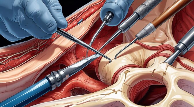
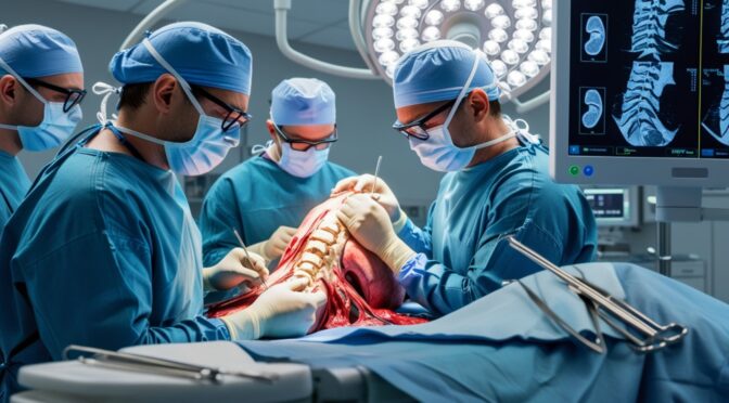
The Laminectomy Procedure Explained

The laminectomy procedure is typically performed under general anesthesia, ensuring that the patient is unconscious and free of pain during the surgery. The patient is positioned face-down on the operating table, allowing the surgeon access to the affected area of the spine. Once the patient is properly positioned, the surgical team prepares the site by cleaning it with antiseptic solutions to prevent infection.

The surgeon makes an incision over the affected vertebrae, carefully moving aside muscles and tissues to expose the spine. Using specialized instruments, the surgeon removes the lamina, creating more space within the spinal canal. Depending on the specific condition being treated, additional procedures may be performed, such as removing herniated disc material or excising bone spurs. In some cases, a spinal fusion may be recommended to stabilize the spine if significant bone is removed.

Once the decompression is complete, the surgeon closes the incision with sutures or staples and covers it with a sterile dressing. The entire procedure can take several hours, depending on the complexity and extent of the surgery. After the operation, patients are moved to a recovery room, where they are monitored as they awaken from anesthesia. Understanding each step of the laminectomy procedure can help alleviate concerns and prepare patients for what to expect on the day of surgery.

Recovery Process After Laminectomy

Recovery from a laminectomy can vary widely among patients, depending on factors such as age, overall health, and the complexity of the surgery. Initially, patients may experience some discomfort and swelling at the surgical site, which is a normal part of the healing process. Pain management is a crucial aspect of recovery, with medications prescribed to help control discomfort in the days following the surgery.

Patients are typically encouraged to begin moving soon after the procedure to promote healing and reduce the risk of complications such as blood clots. Gentle walking and physical therapy exercises can help improve circulation and prevent stiffness. While it may be tempting to rest extensively, early mobilization is essential for a smooth recovery and can expedite the return to normal activities.

The timeline for returning to work and other activities varies, with some patients able to resume light duties within a few weeks, while others may require a more extended period of recovery. Regular follow-up appointments with the surgeon are important to monitor progress and address any concerns that may arise. By adhering to post-operative instructions and actively participating in the recovery process, patients can optimize their outcomes and achieve the best possible results from their laminectomy.

Pain Management and Rehabilitation

Effective pain management is a cornerstone of successful recovery after laminectomy. In the immediate post-operative period, patients are likely to be prescribed a combination of medications to manage pain and inflammation. These may include opioids for severe pain, nonsteroidal anti-inflammatory drugs (NSAIDs), and muscle relaxants. It’s essential for patients to follow their prescribed medication regimen and communicate with their healthcare provider about any side effects or concerns.

Rehabilitation plays a critical role in restoring function and strength after laminectomy. Physical therapy often begins shortly after surgery and is tailored to the individual needs of the patient. A physical therapist will guide patients through exercises designed to improve flexibility, strength, and stability in the spine. As recovery progresses, the intensity and complexity of exercises are gradually increased to match the patient’s capabilities.

In addition to professional rehabilitation, patients are encouraged to incorporate gentle activities into their routines, such as walking, swimming, or yoga, as tolerated. These activities can enhance cardiovascular health, improve mood, and accelerate the healing process. By actively engaging in both formal rehabilitation and personal activities, patients can facilitate a smoother recovery and reduce the risk of future spinal issues.

Lifestyle Changes Post-Surgery

Adopting healthy lifestyle changes after laminectomy can significantly impact long-term outcomes and prevent recurrence of symptoms. One of the most important changes is maintaining a healthy weight, as excess body weight can place additional stress on the spine and hinder recovery. Patients should focus on a balanced diet rich in nutrients, including protein, vitamins, and minerals, to support tissue healing and overall health.

Incorporating regular exercise into daily life is another crucial lifestyle change. While the type and intensity of exercise should be tailored to individual abilities and recovery progress, staying active can help maintain spinal flexibility and strength, reducing the likelihood of future issues. Patients should consult with their healthcare providers or physical therapists to develop a safe and effective exercise plan that meets their needs.

Smoking cessation is particularly important for individuals recovering from laminectomy, as smoking can impair blood flow and delay healing. Quitting smoking not only benefits spinal health but also improves overall cardiovascular and respiratory health. By embracing these lifestyle changes, patients can enhance their recovery, support spinal health, and improve their quality of life long after surgery.

Frequently Asked Questions About Laminectomy

Patients considering laminectomy often have numerous questions about the procedure, recovery, and long-term outcomes. One common question is how long the effects of the surgery will last. While laminectomy can provide significant relief from symptoms, its longevity varies depending on the underlying condition and adherence to post-surgery recommendations. Some patients experience long-lasting relief, while others may require additional treatments in the future.

Another frequently asked question is whether laminectomy will limit physical activities. Post-surgery, patients are encouraged to gradually resume activities, with modifications as necessary. While some high-impact sports or activities may need to be adjusted, most patients can return to a full range of activities with appropriate precautions and conditioning. Consulting with healthcare providers about specific activities can ensure a safe and effective return to an active lifestyle.

Patients may also wonder about the potential for surgery to be unsuccessful. While laminectomy is generally effective in relieving symptoms, there is a possibility that some patients may not experience the desired outcome. Factors such as the extent of nerve damage or the presence of other spinal issues can influence results. Open communication with the surgical team and realistic expectations are essential components of the decision-making process.

Conclusion and Next Steps

Understanding laminectomy, from its indications and benefits to potential risks and recovery insights, is crucial for anyone considering this surgical option. By comprehensively exploring this procedure, patients can make informed decisions about their spinal health and confidently navigate their treatment journey. Laminectomy offers a promising path to relief for many individuals suffering from chronic back pain and neurological symptoms, enabling them to reclaim their quality of life.

For those considering laminectomy, the next steps involve a thorough discussion with healthcare providers, including surgeons and rehabilitation specialists, to evaluate the appropriateness of the procedure based on individual circumstances. Patients should feel empowered to ask questions, seek second opinions, and explore all available treatment options to ensure the best possible outcome.

Ultimately, laminectomy can be a transformative experience, providing significant relief and improved function for those struggling with debilitating spinal conditions. By approaching the decision with knowledge and preparation, patients can embark on the path to recovery with confidence, supported by a comprehensive understanding of what to expect before, during, and after surgery.

How to Tell If Your Neck Pain Is a Sign of Something Serious

Understanding Neck Pain: Common Causes

Neck pain is a widespread issue that affects millions of people globally, and its origins can be as varied as its sufferers. One of the most prevalent causes of neck discomfort is poor posture, often a result of our modern, sedentary lifestyles. Whether it’s hunching over a computer screen or spending long hours looking down at mobile devices, these habits strain the muscles and ligaments in the neck, leading to pain and stiffness. Additionally, sleeping in an awkward position or using an unsupportive pillow can also contribute to waking up with a sore neck.

Another common cause of neck pain is muscle strain or tension. Everyday activities, such as prolonged driving, carrying heavy bags on one shoulder, or sudden movements during sports, can cause the muscles in the neck to become overstretched or torn. This type of injury often leads to acute pain that can last for several days. Moreover, stress and anxiety can cause muscle tension in the neck and shoulders, exacerbating discomfort.

In some cases, neck pain is associated with underlying medical conditions. Arthritis, for example, can result in chronic neck pain due to inflammation of the joints in the cervical spine. Herniated discs, where the inner gel-like core of a spinal disc pushes out through a tear in the outer shell, can also press on nearby nerves, causing pain that radiates into the arm. While these causes are more severe, they are less common than the everyday strains and sprains most people experience.

When to Worry: Signs of Serious Neck Pain

While many instances of neck pain are benign and resolve on their own, certain symptoms should prompt further investigation. One critical sign to watch out for is pain that persists beyond a few days or becomes progressively worse. If your neck pain doesn’t improve with rest or over-the-counter medications, it might indicate an underlying condition that requires medical attention.

Another red flag is neck pain accompanied by neurological symptoms. If you experience numbness, tingling, or weakness in your arms or hands, it could suggest nerve compression or damage. This is often seen in conditions like cervical radiculopathy, where a nerve root in the neck is pinched. These symptoms should not be ignored, as prompt treatment can prevent permanent nerve damage.

Severe, sudden neck pain following an accident or injury is also a cause for concern. Trauma to the head or neck can result in fractures, dislocations, or even spinal cord injuries. In such cases, it’s crucial to seek immediate medical care to evaluate the extent of the injury and initiate appropriate treatment. Remember, it’s better to err on the side of caution and consult a healthcare professional if you have any doubts about the severity of your neck pain.

The Connection Between Neck Pain and Other Health Issues

Neck pain can sometimes be an indicator of more systemic health issues. For instance, certain infections can cause neck pain. Meningitis, an infection of the protective membranes covering the brain and spinal cord, often presents with a stiff neck, headache, and fever. This condition requires urgent medical treatment, as it can be life-threatening if not addressed promptly.

Heart-related problems can also manifest as neck pain. Angina, which is chest pain caused by reduced blood flow to the heart, can radiate to the neck, jaw, or shoulders. This type of pain is often accompanied by other symptoms like shortness of breath, sweating, or nausea. If you suspect your neck pain is related to a cardiac issue, it’s crucial to seek immediate medical attention.

Autoimmune diseases, such as rheumatoid arthritis or lupus, can cause chronic inflammation in various parts of the body, including the neck. These conditions often require long-term management with medications to control symptoms and prevent joint damage. Understanding the potential link between neck pain and other health issues is vital for accurate diagnosis and effective treatment.

How to Differentiate Between Minor and Severe Neck Pain

Differentiating between minor and severe neck pain can be challenging, but paying attention to specific characteristics can provide valuable clues. Minor neck pain typically resolves within a few days to a week with rest, gentle stretching, and over-the-counter pain relievers. It generally doesn’t interfere with daily activities and tends to improve with changes in posture or ergonomics.

In contrast, severe neck pain often persists for weeks or longer and may not respond to standard home remedies. It may be accompanied by other symptoms, such as headaches, dizziness, or difficulty swallowing, which can indicate a more serious underlying condition. Monitoring the duration and intensity of your pain, as well as any associated symptoms, can help determine whether professional evaluation is necessary.

Another key factor is the onset of pain. Sudden, intense pain following an accident or injury is more concerning than gradual discomfort that develops over time. Additionally, pain that radiates into the arms or causes weakness should be evaluated by a healthcare provider, as it may suggest nerve involvement. When in doubt, it’s always best to err on the side of caution and consult a medical professional for an accurate diagnosis.

Diagnostic Tests for Neck Pain: What to Expect

When neck pain doesn’t resolve on its own or is accompanied by concerning symptoms, your healthcare provider may recommend diagnostic tests to identify the underlying cause. One of the most common initial tests is an X-ray, which can reveal structural abnormalities in the spine, such as fractures or degenerative changes.

If nerve involvement is suspected, your doctor might order an MRI (magnetic resonance imaging) to get a detailed view of the soft tissues, including the spinal cord and nerve roots. This non-invasive test provides comprehensive images that can help identify herniated discs, tumors, or infections that might be causing your symptoms.

In some cases, a CT (computed tomography) scan or myelogram may be necessary to provide additional information about the bones and nerves in the neck. These tests can help pinpoint the cause of your pain and guide appropriate treatment. While the prospect of undergoing diagnostic tests can be daunting, they are valuable tools in understanding and managing your neck pain.

Treatment Options for Neck Pain: When to Seek Help

Treatment for neck pain varies depending on the underlying cause and severity of symptoms. For minor, acute pain, conservative measures such as rest, ice or heat application, and over-the-counter pain relievers are often sufficient. Gentle stretching and strengthening exercises can also help alleviate discomfort and prevent future episodes.

However, if your neck pain persists despite these measures or is accompanied by other concerning symptoms, it may be time to seek professional help. Physical therapy is a common treatment option that can improve range of motion, strengthen neck muscles, and reduce pain. A physical therapist can also provide guidance on posture and ergonomics to prevent future issues.

In more severe cases, interventions such as corticosteroid injections, nerve blocks, or even surgery may be necessary to address the underlying cause of your pain. It’s crucial to work closely with your healthcare provider to develop an individualized treatment plan that addresses both your symptoms and the root cause of your neck pain.

Lifestyle Changes to Prevent Neck Pain

Preventing neck pain often involves making lifestyle changes that reduce strain and promote overall neck health. One of the most effective strategies is improving posture, both at work and during leisure activities. Ensure your computer monitor is at eye level, use a chair with proper lumbar support, and take regular breaks to stretch and move around.

Incorporating regular exercise into your routine can also help prevent neck pain by strengthening the muscles that support your spine. Focus on exercises that improve flexibility and core strength, such as yoga or pilates, which can help maintain proper alignment and reduce the risk of injury.

Managing stress is another important aspect of preventing neck pain, as tension and anxiety can contribute to muscle tightness. Techniques such as deep breathing, meditation, or massage therapy can help reduce stress levels and promote relaxation. By adopting these healthy habits, you can significantly reduce the likelihood of experiencing neck pain.

The Importance of Posture in Neck Health

Good posture is fundamental to maintaining neck health and preventing pain. When your posture is aligned, the muscles and ligaments in your neck can function optimally, reducing the risk of strain and injury. Conversely, poor posture places excessive stress on your neck and shoulders, leading to discomfort and potential long-term issues.

To improve your posture, be mindful of your body position throughout the day. Keep your shoulders back, your head level, and avoid slouching or leaning forward. When sitting, ensure your feet are flat on the floor and your back is supported. Simple adjustments like these can have a significant impact on reducing neck pain and promoting overall well-being.

In addition to maintaining good posture while sitting, it’s important to be mindful of your posture during other activities, such as driving or using a smartphone. Use hands-free devices when possible, and hold your phone at eye level to avoid straining your neck. By making these small changes, you can protect your neck from unnecessary stress and discomfort.

When to Consult a Specialist: Chiropractors vs. Medical Doctors

Deciding whether to consult a chiropractor or a medical doctor for neck pain can depend on the nature and severity of your symptoms. Chiropractors specialize in the diagnosis and treatment of musculoskeletal disorders, often using manual adjustments to improve alignment and relieve pain. If your neck pain is related to posture or muscle tension, a chiropractor may provide effective relief.

However, if your neck pain is accompanied by neurological symptoms, such as numbness or weakness, or if it persists despite conservative treatment, consulting a medical doctor is advisable. A primary care physician or a specialist, such as a neurologist or orthopedist, can perform a comprehensive evaluation and recommend appropriate diagnostic tests and treatments.

Ultimately, the decision to see a chiropractor or medical doctor should be based on your specific symptoms and needs. In some cases, a combination of both chiropractic care and medical treatment may be beneficial. Collaborating with a healthcare team that understands your condition and goals can lead to the most effective management of your neck pain.

Conclusion: Taking Action for Your Neck Health

Understanding the potential causes and warning signs of neck pain is crucial for taking proactive steps to protect your health. By recognizing when neck pain may indicate a more serious issue, you can seek timely medical attention and avoid potential complications. Implementing lifestyle changes, such as improving posture and managing stress, can also play a significant role in preventing neck pain.

Whether you choose to consult a chiropractor, a medical doctor, or a combination of specialists, it’s important to advocate for your health and communicate openly about your symptoms and concerns. With the right support and treatment, most cases of neck pain can be effectively managed, allowing you to return to your normal activities and enjoy a pain-free life.

Don’t let neck pain disrupt your daily routine or diminish your quality of life. By staying informed and taking action, you can address neck pain head-on and maintain your overall well-being. Remember, your neck is an essential part of your body’s support system, and taking care of it is an investment in your health and future.

Understanding the Root Causes of Chronic Back Pain

What is Chronic Back Pain?

Chronic back pain is a persistent or recurring discomfort in the back that lasts for 12 weeks or more. Unlike acute back pain, which typically resolves with treatment, chronic back pain persists despite medical intervention. It can be a debilitating condition, leading to decreased quality of life, reduced mobility, and significant emotional distress. Chronic back pain does not discriminate; it affects people of all ages, backgrounds, and lifestyles, though certain populations may be more at risk due to occupational or genetic factors.

The nature of chronic back pain is complex, involving both physical and psychological components. It often manifests as a dull ache, sharp pain, or even a burning sensation that can radiate to other areas such as the legs or hips. This pain can vary in intensity, sometimes flaring up to become almost unbearable, while at other times, it may seem to fade into the background, only to return later. Understanding the patterns and triggers of your pain is crucial for effective management and treatment.

Recognizing chronic back pain as a multifaceted condition is the first step toward managing it effectively. It requires a comprehensive approach that addresses not just the physical symptoms but also the mental and emotional toll it can take. Effective management begins with understanding the underlying causes, which range from lifestyle factors to medical conditions, and involves a combination of treatments tailored to the individual needs of the sufferer.

Common Symptoms and Diagnosis

Common symptoms of chronic back pain include persistent pain in the lower, middle, or upper back that does not subside over time. This pain can lead to stiffness, reduced flexibility, and difficulty performing everyday activities. Sufferers may also experience muscle spasms, shooting pain down the legs, or numbness in the extremities. These symptoms can significantly affect one’s ability to work, engage in physical activities, and even perform simple tasks like sitting or standing for extended periods.

Accurate diagnosis of chronic back pain is essential for effective treatment. It typically begins with a thorough medical history and physical examination conducted by a healthcare professional. During this process, the doctor will assess the location, intensity, and duration of the pain, as well as any accompanying symptoms. They may also inquire about past injuries, lifestyle habits, and family history to identify potential contributing factors. Diagnostic imaging tests such as X-rays, MRI, or CT scans may be ordered to get a clearer picture of the spine and surrounding structures.

In some cases, further tests may be required to rule out specific conditions such as herniated discs, spinal stenosis, or degenerative disc disease. Blood tests may also be conducted to identify any underlying infections or inflammatory conditions. An accurate diagnosis is crucial as it forms the basis for developing a personalized treatment plan aimed at managing symptoms and addressing the root causes of the pain.

The Anatomy of the Back: Understanding the Structure

The human back is a complex structure composed of bones, muscles, ligaments, nerves, and other tissues that work together to support the body’s framework and facilitate movement. At the heart of this structure is the spine, a column of vertebrae that runs from the base of the skull to the pelvis. The spine is divided into three main sections: the cervical (neck), thoracic (upper back), and lumbar (lower back) regions. Each section plays a crucial role in maintaining posture and allowing flexibility and movement.

Intervertebral discs lie between each vertebra, acting as shock absorbers that cushion the bones during motion. These discs have a tough outer layer and a gel-like center, providing both support and flexibility to the spine. Ligaments and tendons connect the bones to each other and to muscles, enabling coordinated movement. The muscles in the back, including the erector spinae, latissimus dorsi, and trapezius, among others, contribute to the strength and stability necessary for a wide range of activities.

Nerves branching from the spinal cord pass through spaces between the vertebrae, transmitting signals between the brain and the rest of the body. Any disruption or damage to these structures can lead to pain, reduced mobility, and other symptoms associated with chronic back pain. Understanding the intricate anatomy of the back is essential for identifying the sources of pain and devising effective treatment strategies.

Lifestyle Factors Contributing to Chronic Back Pain

Several lifestyle factors can contribute to the development and persistence of chronic back pain. One of the most significant is sedentary behavior, which is increasingly common in modern society. Prolonged periods of sitting, particularly with poor posture, can put undue stress on the spine and supporting muscles, leading to pain and discomfort. Regular physical activity is crucial for maintaining spinal health, as it strengthens the muscles that support the back and enhances flexibility.

Obesity is another lifestyle factor that can exacerbate back pain. Excess body weight, particularly around the abdomen, increases the strain on the lower back, leading to discomfort and potential injury. A balanced diet and regular exercise are important for managing weight and reducing the risk of chronic back pain. Smoking is also a risk factor, as it reduces blood flow to the spine and can contribute to the degeneration of spinal discs.

Stress and poor sleep can further compound the problem. High stress levels can lead to muscle tension and exacerbate pain, while inadequate sleep can hinder the body’s ability to repair and heal itself. Adopting a healthy lifestyle that includes regular exercise, a nutritious diet, and stress management techniques can go a long way in preventing and alleviating chronic back pain.

Medical Conditions Linked to Chronic Back Pain

Several medical conditions are closely linked to chronic back pain. One of the most common is degenerative disc disease, a condition where the intervertebral discs lose hydration and elasticity over time, leading to pain and reduced mobility. It often occurs as a result of aging or injury and can be exacerbated by lifestyle factors such as smoking or obesity. The pain associated with degenerative disc disease can be chronic and may radiate to other areas of the body.

Another condition frequently associated with chronic back pain is spinal stenosis, which involves the narrowing of the spinal canal, placing pressure on the spinal cord and nerves. This can lead to symptoms such as numbness, tingling, or weakness in the legs. Herniated discs, where the inner gel of a disc protrudes through a tear in its outer layer, can also cause chronic back pain by compressing nearby nerves.

Arthritis, particularly osteoarthritis and rheumatoid arthritis, can affect the spine and lead to chronic back pain. These conditions cause inflammation of the joints, leading to pain, stiffness, and reduced range of motion. Fibromyalgia, a disorder characterized by widespread musculoskeletal pain, fatigue, and tenderness, can also contribute to chronic back pain. Identifying and addressing these underlying medical conditions is crucial for effective pain management and treatment.

The Role of Posture and Ergonomics

Posture plays a critical role in the onset and progression of chronic back pain. Poor posture, whether sitting, standing, or lying down, can place additional stress on the spine and supporting muscles, leading to discomfort and pain. Slouching, for example, can cause the spine to curve unnaturally, placing strain on the back muscles and ligaments. Over time, this can lead to chronic pain and other musculoskeletal issues.

Ergonomics, the study of people’s efficiency in their working environment, is closely related to posture. An ergonomic workspace is designed to support proper posture and reduce the risk of injury. This includes adjusting the height of chairs and desks, ensuring computer monitors are at eye level, and using supportive seating. Simple changes in the work environment can significantly reduce the strain on the back and help prevent chronic pain.

Being mindful of posture and making ergonomic adjustments can have a profound impact on back health. Regular breaks to stretch and move around during prolonged periods of sitting can also help alleviate tension and reduce the risk of developing chronic back pain. By prioritizing good posture and ergonomics, individuals can take proactive steps to protect their spine and maintain a healthy back.

Psychological Factors and Chronic Pain

The relationship between psychological factors and chronic back pain is complex and significant. Mental health issues such as depression, anxiety, and stress can exacerbate the perception of pain and contribute to its persistence. Chronic pain itself can lead to emotional distress, creating a vicious cycle that is difficult to break. Understanding the psychological components of pain is crucial for effective management and treatment.

Cognitive-behavioral therapy (CBT) is one approach that has been shown to be effective in managing chronic back pain. CBT helps individuals identify and change negative thought patterns and behaviors that may be contributing to their pain. By addressing the psychological aspects of pain, individuals can develop coping strategies that reduce the impact of pain on their daily lives.

Mindfulness and relaxation techniques can also play a role in managing chronic back pain. Practices such as meditation, deep breathing, and progressive muscle relaxation can help reduce stress and promote a sense of calm, which can alleviate pain. By incorporating psychological interventions into a comprehensive treatment plan, individuals can improve their overall well-being and reduce the impact of chronic back pain.

Treatment Options: From Medication to Therapy

There are numerous treatment options available for managing chronic back pain, ranging from medication to therapy. Over-the-counter pain relievers such as acetaminophen and nonsteroidal anti-inflammatory drugs (NSAIDs) are often the first line of treatment. These medications can help reduce pain and inflammation, providing temporary relief. In some cases, prescription medications such as muscle relaxants or opioids may be necessary, though these should be used with caution due to the risk of side effects and dependency.

Physical therapy is a highly effective treatment option for chronic back pain. A physical therapist can design a customized exercise program that strengthens the muscles supporting the spine, improves flexibility, and reduces pain. Techniques such as manual therapy, ultrasound, and electrical stimulation may also be used to enhance treatment outcomes. Consistent physical therapy can lead to significant improvements in pain and function.

Complementary therapies such as acupuncture, chiropractic care, and massage therapy can also provide relief for chronic back pain. These approaches focus on restoring balance to the body and promoting natural healing. In some cases, surgical intervention may be necessary, particularly if there is a structural issue that cannot be addressed through conservative treatments. A multidisciplinary approach that combines various treatments is often the most effective way to manage chronic back pain.

Prevention Strategies for Chronic Back Pain

Preventing chronic back pain involves adopting a proactive approach to spinal health. Regular exercise is one of the most effective ways to maintain a healthy back. Engaging in activities that strengthen the core muscles, improve flexibility, and promote cardiovascular fitness can help prevent injury and reduce the risk of chronic pain. Low-impact exercises such as swimming, walking, and yoga are particularly beneficial for back health.

Maintaining a healthy weight is also crucial for preventing chronic back pain. Excess weight can strain the spine and lead to pain and discomfort. A balanced diet that includes plenty of fruits, vegetables, lean proteins, and whole grains can support a healthy weight and provide the nutrients necessary for strong bones and muscles. Staying hydrated and avoiding smoking are additional lifestyle choices that can support back health.

Proper posture and ergonomics are important preventive measures. Being mindful of posture, particularly during activities such as sitting, lifting, and sleeping, can reduce the risk of chronic back pain. Ergonomic adjustments to the workspace, such as using supportive seating and adjusting the height of desks and monitors, can also help prevent strain on the spine. By taking these preventive steps, individuals can reduce their risk of developing chronic back pain and maintain a healthy, active lifestyle.

Conclusion: Taking Control of Your Back Health

Understanding the root causes of chronic back pain is an essential step in managing and preventing this debilitating condition. By recognizing the various factors that contribute to back pain, individuals can take proactive steps to address their symptoms and improve their overall well-being. From lifestyle changes and ergonomic adjustments to medical treatments and psychological interventions, there are numerous strategies available to manage chronic back pain effectively.

Taking control of your back health involves a comprehensive approach that addresses both the physical and psychological aspects of pain. Regular exercise, a balanced diet, and stress management techniques can support a healthy spine and reduce the risk of chronic pain. Seeking professional guidance from healthcare providers, physical therapists, and mental health professionals can provide valuable support and expertise in managing pain.

Empowering yourself with knowledge about chronic back pain can lead to a path of relief and recovery. By understanding the complexities of this condition and exploring various treatment options, individuals can take charge of their health and work towards a pain-free life. Remember, the journey to managing chronic back pain is personal and ongoing, but with determination and the right strategies, it is possible to achieve lasting relief and improved quality of life.

Understanding Foraminotomy: A Comprehensive Guide to Relief from Nerve Compression

What is Foraminotomy?

Foraminotomy is a surgical procedure designed to relieve nerve compression in the spine, specifically targeting the foramen. The foramen is a small opening between the bones in the spine through which nerve roots exit. When these openings become narrowed, either due to bone spurs, herniated discs, or other spinal conditions, it can compress the nerves, leading to pain, numbness, and weakness. Foraminotomy involves enlarging this passageway to alleviate the pressure on the affected nerves.

This minimally invasive surgery is often performed using advanced techniques that require only small incisions, reducing recovery time and minimizing risks compared to traditional open surgery. Surgeons typically use specialized instruments and imaging technology to guide the procedure with precision. The primary goal is to remove the source of compression while preserving as much of the natural anatomy as possible. This approach not only leads to pain relief but also helps maintain spinal stability.

Foraminotomy can be an effective solution for individuals who have not found relief through conservative treatments such as physical therapy, medication, or spinal injections. It offers a targeted approach to resolving nerve compression issues while minimizing collateral damage to surrounding tissues. Understanding the specifics of this procedure is crucial for patients considering it as a potential pathway to alleviate debilitating symptoms.

The Anatomy of Nerve Compression

Nerve compression occurs when excessive pressure is placed on a nerve by surrounding tissues, such as bones, cartilage, muscles, or tendons. In the context of spinal health, this pressure often arises from changes in the structure of the spine, including the narrowing of the foraminal canals. This narrowing, medically known as foraminal stenosis, can result from various degenerative conditions, impacting the normal functioning of spinal nerves.

The spinal column is a complex structure made up of vertebrae, intervertebral discs, ligaments, and nerve roots. The nerve roots exit the spinal column through the foramina, serving as conduits for nerve signals traveling between the spinal cord and the rest of the body. When these foramina are constricted, the nerve roots can become compressed, leading to a range of symptoms, from localized pain to radiating discomfort that follows the path of the nerve.

Understanding the anatomy involved in nerve compression is essential for recognizing the potential benefits of foraminotomy. By addressing the specific areas where compression occurs, foraminotomy aims to restore the normal function of the affected nerves. This targeted approach not only alleviates symptoms but also prevents further deterioration of the spinal structure, promoting long-term spinal health.

Common Causes of Nerve Compression

Several factors can contribute to the development of foraminal stenosis and subsequent nerve compression. One of the most common causes is degenerative disc disease, a condition wherein the intervertebral discs lose their cushioning ability over time. As the discs degenerate, they can bulge or herniate into the foraminal space, exerting pressure on the nerve roots and causing pain.

Another prevalent cause of foraminal narrowing is the formation of bone spurs, or osteophytes. These bony projections often develop as a natural response to degeneration or arthritis, as the body attempts to stabilize the spine. However, the growth of these spurs can encroach upon the foramina, leading to compression of the nerves. Other conditions, such as arthritis or scoliosis, can also contribute to foraminal stenosis by altering the structural integrity of the spine.

Traumatic injuries, such as fractures or dislocations, can further exacerbate nerve compression by altering the alignment of the spinal column. Additionally, lifestyle factors, including poor posture, obesity, and lack of physical activity, can increase the stress on the spine, potentially leading to or aggravating foraminal stenosis. Understanding these causes is vital for prevention and for determining when surgical intervention like foraminotomy may be necessary.

Symptoms Indicating the Need for Foraminotomy

Recognizing the symptoms of nerve compression is critical for determining the appropriate course of treatment. Common indicators that may suggest the need for foraminotomy include persistent pain in the back or neck that radiates into the arms or legs. This pain is often accompanied by numbness, tingling, or weakness in the extremities, which can significantly impact daily activities and overall quality of life.

Patients may also experience a loss of motor function or coordination, making it difficult to perform tasks that require fine motor skills. In severe cases, nerve compression can lead to muscle atrophy or changes in reflexes, further highlighting the need for medical evaluation. The intensity and location of these symptoms can vary depending on the specific nerves affected and the degree of compression.

It is essential for individuals experiencing these symptoms to seek medical consultation. A thorough evaluation, which may include imaging studies such as MRI or CT scans, can help identify the exact cause and location of nerve compression. This diagnostic process is crucial for determining whether foraminotomy is a viable option and ensuring the best possible outcome for the patient.

The Foraminotomy Procedure: What to Expect

Undergoing a foraminotomy involves several stages, beginning with a preoperative assessment to evaluate the patient’s overall health and suitability for surgery. This assessment typically includes a review of medical history, physical examination, and imaging studies to confirm the diagnosis of nerve compression. Once deemed a suitable candidate, the patient is scheduled for the procedure, which is usually performed on an outpatient basis.

During the operation, the surgeon makes a small incision near the affected area of the spine. Using specialized instruments and real-time imaging guidance, the surgeon carefully removes the bone or tissue causing the compression. The goal is to enlarge the foramina and relieve pressure on the nerve roots without compromising the stability of the spine. This precision minimizes trauma to the surrounding tissues and promotes faster healing.

Post-operatively, patients are monitored for a brief period before being discharged. Recovery instructions are provided, including guidelines for pain management, activity restrictions, and follow-up appointments. While the surgery itself is relatively quick, typically lasting one to two hours, the preparation and recovery processes are equally important in achieving successful outcomes.

Benefits of Foraminotomy for Nerve Compression Relief

Foraminotomy offers several significant benefits for individuals suffering from nerve compression. One of the primary advantages is the relief of pain and other debilitating symptoms, allowing patients to resume their normal activities with reduced discomfort. By directly addressing the source of compression, foraminotomy provides targeted relief that can significantly improve the patient’s quality of life.

The minimally invasive nature of the procedure means that patients often experience shorter recovery times and less postoperative pain compared to traditional open surgeries. The use of small incisions reduces the risk of infection, scarring, and other complications, making foraminotomy a safer option for many patients. Additionally, the preservation of spinal stability is a critical benefit, as it helps maintain the overall function and alignment of the spine.

Beyond physical improvements, foraminotomy can also have positive psychological effects. The reduction of chronic pain and restoration of mobility can lead to improved mental health and well-being. Patients often report increased energy levels and a more active lifestyle following successful foraminotomy, highlighting the comprehensive benefits of this surgical intervention.

Risks and Complications Associated with Foraminotomy

As with any surgical procedure, foraminotomy carries certain risks and potential complications. While the surgery is generally considered safe, it is important for patients to be aware of these risks to make informed decisions. Common complications include infection, bleeding, and adverse reactions to anesthesia, though these are relatively rare due to the minimally invasive nature of the procedure.

Nerve damage is another potential risk, albeit infrequent. Given the proximity of the surgical site to the spinal nerves, there is a small chance of injury during the procedure, which could lead to worsening of symptoms or new neurological deficits. Surgeons take great care to minimize this risk by using advanced imaging techniques and specialized instruments to guide their movements with precision.

Other complications may include cerebrospinal fluid leaks or recurrence of symptoms if the underlying cause is not fully addressed. It is crucial for patients to discuss these risks with their healthcare provider, weighing them against the potential benefits of the surgery. A thorough understanding of the procedure and its associated risks allows patients to approach foraminotomy with confidence and realistic expectations.

Recovery and Rehabilitation After Foraminotomy

The recovery process following foraminotomy is a critical component of achieving successful outcomes. Patients are typically encouraged to engage in a rehabilitation program that includes physical therapy and exercises designed to strengthen the spine and improve flexibility. These activities are tailored to the individual’s needs and progress, gradually increasing in intensity as the patient heals.

Pain management is an integral part of the recovery phase, with medications prescribed to alleviate discomfort and inflammation. Patients are advised to adhere to activity restrictions to prevent strain on the surgical site. This may include avoiding heavy lifting, twisting movements, and prolonged periods of sitting or standing during the initial weeks of recovery.

Regular follow-up appointments with the healthcare provider are essential to monitor the healing process and address any concerns that may arise. These visits provide an opportunity to assess the effectiveness of the surgery, adjust rehabilitation plans, and ensure that the patient is on track for a full recovery. With proper care and adherence to postoperative instructions, most patients can expect to return to their normal activities within a few months.

Alternative Treatments for Nerve Compression

While foraminotomy is an effective solution for many individuals with nerve compression, it is not the only treatment option available. Conservative approaches, such as physical therapy, chiropractic care, and pain management techniques, are often recommended as initial interventions. These methods aim to alleviate symptoms and improve function without the need for surgery.

For some patients, spinal injections, including epidural steroid injections, can provide temporary relief by reducing inflammation and swelling around the affected nerve. These injections are typically used in conjunction with other treatments to enhance their effectiveness and provide longer-lasting benefits. Additionally, lifestyle modifications, such as weight management, ergonomic adjustments, and exercise programs, can play a significant role in managing symptoms and preventing further deterioration.

In cases where conservative treatments fail to provide adequate relief, other surgical options may be considered. Procedures such as laminectomy, discectomy, or spinal fusion may be appropriate depending on the specific cause and location of the nerve compression. It is important for patients to explore all available options and work closely with their healthcare providers to determine the most suitable approach for their condition.

Conclusion: Is Foraminotomy Right for You?

Deciding whether foraminotomy is the right choice involves careful consideration of various factors, including the severity of symptoms, the underlying cause of nerve compression, and the patient’s overall health. For individuals who have not achieved relief through conservative treatments, foraminotomy offers a promising solution with its minimally invasive approach and targeted effectiveness.

Potential candidates should engage in open discussions with their healthcare providers, weighing the benefits and risks associated with the procedure. Understanding the specifics of the surgery, including the recovery process and long-term outcomes, is crucial for making informed decisions. Additionally, exploring alternative treatments and considering second opinions can provide a comprehensive view of available options.

Ultimately, the decision to undergo foraminotomy should be based on a thorough evaluation of individual needs and goals. With the right approach and support, foraminotomy can pave the way towards a pain-free future, restoring mobility and enhancing quality of life for those affected by nerve compression.

Common Causes of Shoulder Pain and How to Treat Them

Understanding Shoulder Anatomy

The shoulder is one of the most complex and flexible joints in the human body, allowing for a wide range of motion. This remarkable flexibility is due to its unique structure, comprising bones, muscles, tendons, and ligaments that work in harmony. The primary bones involved in the shoulder joint are the humerus (the upper arm bone), the scapula (shoulder blade), and the clavicle (collarbone). These bones form two main joints: the glenohumeral joint, which is the ball-and-socket joint allowing arm rotation, and the acromioclavicular joint, where the clavicle meets the scapula.

The rotator cuff, consisting of four key muscles and their tendons, envelops the shoulder joint, providing stability and facilitating movement. These muscles include the supraspinatus, infraspinatus, teres minor, and subscapularis. Each plays a crucial role in shoulder dynamics, enabling actions such as lifting, rotating, and reaching. Additionally, the bursa, a small sac filled with lubricating fluid, reduces friction and allows smooth gliding of the shoulder components during movement.

Understanding this intricate anatomy is essential as it lays the foundation for identifying the root causes of shoulder pain. The interdependence of muscles, tendons, and bones makes the shoulder susceptible to various injuries and conditions. When any of these components are compromised, it can lead to discomfort and hinder functionality, prompting the need for effective diagnosis and targeted treatment to restore health and mobility.

Common Causes of Shoulder Pain

Shoulder pain can arise from a multitude of sources, often stemming from overuse, injury, or underlying medical conditions. Identifying the specific cause is crucial for devising an appropriate treatment plan. Among the most prevalent causes are rotator cuff injuries, which account for a significant portion of shoulder-related complaints. These injuries can range from inflammation of the tendons to complete tears and are often the result of repetitive motions or acute trauma.

Another common culprit is shoulder impingement syndrome, which occurs when the rotator cuff tendons are compressed during shoulder movements. This condition is frequently seen in athletes and individuals who engage in activities that involve overhead motions. Frozen shoulder, or adhesive capsulitis, is another painful disorder characterized by stiffness and limited range of motion. It typically develops gradually and can severely impact daily activities.

Arthritis, particularly osteoarthritis and rheumatoid arthritis, also contributes significantly to shoulder pain. These conditions lead to the degeneration of cartilage, resulting in pain, swelling, and reduced mobility. Tendonitis and bursitis, involving inflammation of the tendons and bursa, respectively, are additional contributors to shoulder discomfort. Understanding these common causes is the first step toward addressing shoulder pain effectively.

Rotator Cuff Injuries

Rotator cuff injuries are among the most frequently diagnosed conditions affecting the shoulder, with symptoms ranging from mild discomfort to severe pain and restricted movement. The rotator cuff is essential for stabilizing the shoulder and facilitating various arm movements. Injuries can occur due to acute trauma, such as a fall, or through chronic wear and tear, often seen in individuals who perform repetitive overhead activities.

Tears in the rotator cuff can be partial or complete, with symptoms including pain during rest or activity, weakness in the shoulder, and difficulty performing tasks that require lifting or rotating the arm. Diagnosis typically involves a combination of physical examinations, imaging tests like MRI or ultrasound, and an assessment of medical history. Early diagnosis is crucial to prevent further damage and to start an effective treatment regimen.

Treatment for rotator cuff injuries ranges from conservative approaches such as rest, ice application, and physical therapy to more invasive procedures like corticosteroid injections or surgery. Physical therapy focuses on strengthening the shoulder muscles, improving flexibility, and restoring function. In cases where conservative treatments fail to alleviate symptoms, surgical intervention may be necessary to repair torn tendons and restore shoulder stability.

Shoulder Impingement Syndrome

Shoulder impingement syndrome is a common cause of shoulder pain, particularly in those who frequently engage in activities requiring repetitive overhead motions, such as swimming, tennis, or painting. This condition occurs when the shoulder’s rotator cuff tendons and bursa become compressed between the acromion (part of the scapula) and the humeral head during arm elevation. This compression leads to inflammation and irritation, manifesting as pain and reduced range of motion.

Symptoms of shoulder impingement include a persistent ache in the shoulder, pain when lifting the arm, and discomfort during activities that involve overhead movement. If left untreated, impingement can lead to more severe conditions, such as rotator cuff tears. Diagnosis is typically achieved through a combination of physical examinations, patient history, and imaging tests, such as X-rays or MRIs, to rule out other potential causes of the symptoms.

The primary goal of treatment is to reduce inflammation and restore normal function. Conservative treatments include rest, ice application, anti-inflammatory medications, and physical therapy to strengthen the shoulder muscles and improve flexibility. In cases where symptoms persist despite conservative measures, surgical intervention may be necessary to remove the impinging structures and create more space for the rotator cuff tendons to move freely.

Frozen Shoulder (Adhesive Capsulitis)

Frozen shoulder, or adhesive capsulitis, is a condition characterized by pain and stiffness in the shoulder joint, leading to a significant reduction in range of motion. This condition develops gradually and can last from several months to years, typically progressing through three stages: the freezing stage, the frozen stage, and the thawing stage. Each stage presents distinct symptoms and challenges, impacting the individual’s ability to perform daily activities.

The exact cause of frozen shoulder remains unclear, but it is often associated with prolonged immobilization, such as after surgery or injury, and certain medical conditions like diabetes. During the freezing stage, individuals experience increasing pain, particularly at night, and a gradual loss of shoulder movement. As the condition progresses into the frozen stage, pain may diminish, but stiffness persists, making it difficult to move the shoulder. The thawing stage is characterized by a gradual return of movement and decreased pain.

Treatment focuses on pain management and restoring mobility. Non-surgical options include physical therapy to improve range of motion, pain relief through medications, and, in some cases, corticosteroid injections to reduce inflammation. In severe cases, surgical intervention might be necessary to release the tight joint capsule. Early intervention and adherence to a rehabilitation program are crucial for recovery and preventing long-term disability.

Arthritis and Shoulder Pain

Arthritis is a prevalent cause of shoulder pain, with osteoarthritis and rheumatoid arthritis being the most common types affecting the shoulder joint. Osteoarthritis involves the degeneration of cartilage within the joint, leading to pain, stiffness, and reduced mobility. This type of arthritis is often age-related, but it can also occur due to previous injuries or repetitive stress on the shoulder.

Rheumatoid arthritis is an autoimmune disorder that causes inflammation of the joint lining, resulting in pain, swelling, and joint deformity. Unlike osteoarthritis, rheumatoid arthritis can affect individuals at any age and typically involves multiple joints. Symptoms include persistent pain, swelling, and a reduced range of motion, which can severely impact daily activities and quality of life.

Diagnosis of arthritis involves a combination of physical examinations, patient history, and imaging tests such as X-rays or MRI. Blood tests may also be conducted to identify markers of rheumatoid arthritis. Treatment aims to manage symptoms and slow disease progression. Conservative measures include pain management with medications, physical therapy to maintain joint function, and lifestyle modifications. In advanced cases, surgical options such as joint replacement may be considered to restore function and alleviate pain.

Tendonitis and Bursitis in the Shoulder

Tendonitis and bursitis are inflammatory conditions that commonly affect the shoulder, leading to pain and discomfort. Tendonitis involves inflammation of the tendons, often resulting from overuse or repetitive movements, while bursitis is the inflammation of the bursa, the small fluid-filled sacs that cushion the shoulder joint. Both conditions share similar symptoms, including pain, swelling, and reduced range of motion.

Tendonitis in the shoulder often occurs in individuals who engage in repetitive activities, such as athletes or manual laborers. The condition can cause sharp pain during movement and tenderness around the affected area. Bursitis, on the other hand, may present as a dull ache, particularly when moving the shoulder or applying pressure to the area. These conditions can significantly impact daily activities and lead to chronic pain if left untreated.

Treatment for tendonitis and bursitis focuses on reducing inflammation and restoring shoulder function. Initial management includes rest, ice application, and anti-inflammatory medications. Physical therapy plays a crucial role in rehabilitation, helping to strengthen the shoulder muscles and improve flexibility. In some cases, corticosteroid injections may be administered to provide relief. Early intervention is key to preventing further complications and ensuring a full recovery.

Diagnostic Methods for Shoulder Pain

Accurate diagnosis of shoulder pain is essential for effective treatment and management. The diagnostic process typically begins with a thorough physical examination and medical history review to identify potential causes and contributing factors. During the examination, a healthcare professional will assess the range of motion, strength, and any visible signs of injury or inflammation.

Imaging tests play a crucial role in diagnosing shoulder conditions. X-rays can reveal bone abnormalities, such as fractures or arthritis-related changes, while MRI and ultrasound provide detailed images of soft tissues, including muscles, tendons, and ligaments. These advanced imaging techniques help in identifying tears, inflammation, and other structural issues that may be causing pain.

In some cases, additional tests such as blood tests or joint aspiration may be conducted to rule out underlying medical conditions or infections. Once a diagnosis is established, a personalized treatment plan can be developed to address the specific cause of shoulder pain. Early diagnosis and intervention are vital to prevent long-term complications and ensure a successful recovery.

Treatment Options for Shoulder Pain

Treatment for shoulder pain varies depending on the underlying cause, severity, and individual patient needs. Conservative treatment options are typically the first line of defense and include rest, ice application, and pain management with medications. These measures aim to reduce inflammation and alleviate pain, allowing the shoulder to heal naturally.

Physical therapy is a cornerstone of shoulder pain treatment, focusing on strengthening the shoulder muscles, improving flexibility, and restoring normal function. A tailored exercise program can help prevent future injuries and enhance overall shoulder health. For persistent pain, corticosteroid injections may be administered to provide temporary relief by reducing inflammation.

In cases where conservative treatments fail to provide adequate relief, surgical intervention may be necessary. Common surgical procedures include arthroscopy to repair torn tissues, shoulder stabilization to address instability, and joint replacement for severe arthritis. Post-surgical rehabilitation is crucial for optimal recovery, involving a combination of physical therapy and gradual return to activities. Regardless of the treatment approach, early intervention and adherence to a rehabilitation program are key to achieving a successful outcome.

Preventive Measures and Exercises for Shoulder Health

Preventing shoulder pain involves a combination of lifestyle modifications, ergonomic adjustments, and targeted exercises to maintain shoulder strength and flexibility. Engaging in regular physical activity is essential for overall musculoskeletal health, but it’s important to incorporate exercises that specifically target the shoulder muscles. Strengthening exercises, such as shoulder presses and lateral raises, can help build muscle support and stability, reducing the risk of injury.

Stretching exercises are equally important to maintain flexibility and prevent stiffness. Incorporating stretches for the rotator cuff muscles and shoulder joint into a regular routine can enhance range of motion and reduce the likelihood of developing conditions such as frozen shoulder. Additionally, practicing good posture, especially during prolonged sitting or desk work, can alleviate strain on the shoulder and neck muscles.

Ergonomic adjustments in daily activities, such as ensuring proper desk height and chair support, can help minimize stress on the shoulders. For those engaged in repetitive activities or sports, using the correct techniques and equipment can prevent overuse injuries. Regular breaks and varying activities can also reduce the risk of developing shoulder pain. By adopting these preventive measures, individuals can maintain shoulder health and enjoy an active, pain-free lifestyle.