Understanding Laminectomy: Benefits, Risks, and Recovery Insights

What is a Laminectomy?

A laminectomy is a surgical procedure that involves the removal of the lamina, a part of the vertebra that covers the spinal canal. This surgery aims to relieve pressure on the spinal cord or nerves, which can be caused by conditions such as spinal stenosis or herniated discs. By removing this bony structure, the surgeon provides more space for the spinal cord and nerve roots, reducing pain and improving mobility.

In medical terms, the laminectomy is often referred to as a decompressive surgery. This procedure is particularly beneficial in cases where conservative treatments like medication or physical therapy have failed to provide relief. It’s important to note that while the term “laminectomy” may sound intimidating, it is a common and well-understood procedure in the field of spinal surgery.

Patients considering this surgery often have questions about what it entails, the expected outcomes, and the recovery process. Understanding these aspects can help individuals make informed decisions about their health and well-being.

Indications for Laminectomy Surgery

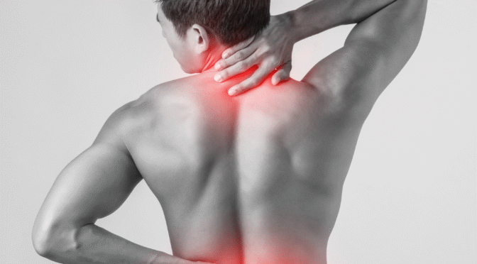
Laminectomy surgery is typically recommended for individuals experiencing significant spinal issues that interfere with daily life. These issues often include persistent pain, numbness, or weakness that affects the arms or legs. Such symptoms are usually caused by conditions like spinal stenosis, herniated discs, or degenerative disc disease.

For many patients, the decision to undergo a laminectomy comes after other less invasive treatments have been tried without success. These treatments may have included physical therapy, medications, or injections designed to reduce inflammation and relieve pain. When these options do not provide sufficient relief, a laminectomy may be the next step.

The decision to proceed with surgery is made in consultation with a medical professional, who will evaluate the patient’s overall health, the severity of symptoms, and the potential benefits and risks of the procedure. It’s a decision that requires careful consideration of all available information to ensure the best possible outcome.

Benefits of Laminectomy

The primary benefit of a laminectomy is relief from pain and symptoms caused by spinal cord or nerve compression. Many patients experience a significant reduction in pain, allowing them to return to activities they enjoy and improve their quality of life. The procedure can also enhance mobility and function, making everyday tasks easier and more manageable.

In addition to pain relief, a laminectomy can prevent further deterioration of the spine. By addressing the root cause of the symptoms, the surgery can stop or slow the progression of spinal conditions that, if left untreated, could lead to more severe complications. This preventive aspect is a crucial consideration for many patients and their healthcare providers.

Moreover, a successful laminectomy can reduce the need for ongoing pain medications, which often come with their own set of side effects and risks. By providing a long-term solution to chronic pain, the surgery can improve both physical and mental well-being, enhancing overall health and lifestyle.

Risks and Complications of Laminectomy

As with any surgical procedure, a laminectomy carries certain risks and potential complications. These can include infection, bleeding, or adverse reactions to anesthesia. While these risks are relatively rare, it’s important for patients to be aware of them and discuss any concerns with their surgical team before proceeding.

Another potential complication is the development of spinal instability, which may require additional surgery. This condition can occur when too much of the lamina or other supporting structures are removed, leading to increased movement in the spinal column. However, surgeons typically take measures to minimize this risk by preserving as much of the natural anatomy as possible.

Nerve damage is another concern, although it is uncommon. The delicate nature of spinal surgery means that extreme care is taken to avoid any harm to the spinal cord or nerves. A well-trained and experienced surgical team significantly reduces the likelihood of such complications, reinforcing the importance of choosing a reputable medical facility for the procedure.

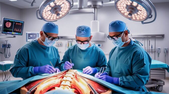
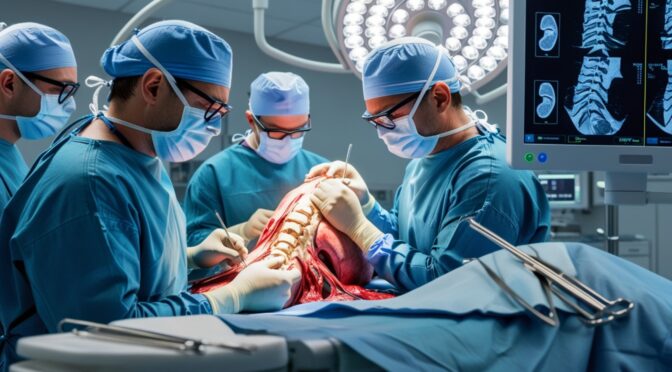
The Laminectomy Procedure: What to Expect

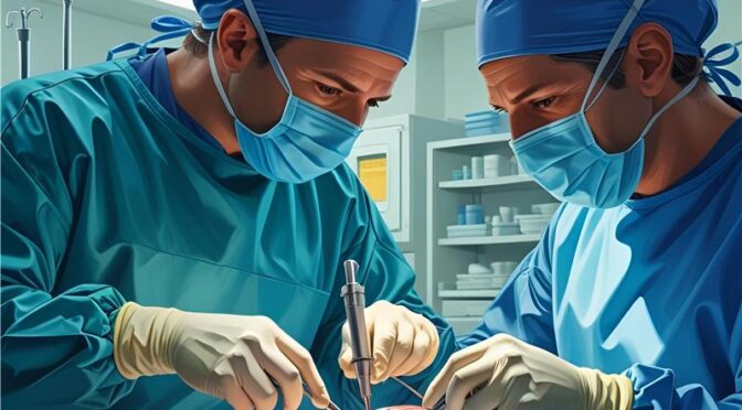
Understanding what happens during a laminectomy can help alleviate apprehension and prepare patients for the procedure. Typically performed under general anesthesia, the surgery involves making an incision in the back over the affected area of the spine. The surgeon then carefully removes the lamina to relieve pressure on the spinal cord or nerves.

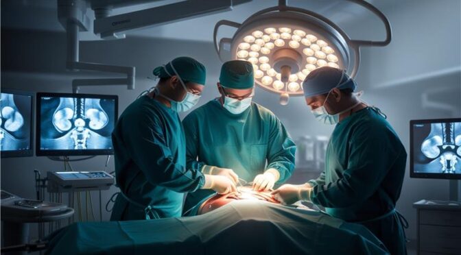
The duration of the procedure can vary depending on the complexity of the case and the number of vertebrae involved. On average, a laminectomy takes between one to three hours. Throughout the surgery, advanced imaging techniques like X-rays or MRI are used to guide the surgeon and ensure precision.

Post-surgery, patients are moved to a recovery area where they are closely monitored as the anesthesia wears off. Pain management is a priority during this phase, with medications provided to keep discomfort at a minimum. Recovery in the hospital typically lasts one to three days, after which most patients can return home to continue their recuperation.

Recovery After Laminectomy Surgery

Recovery from laminectomy surgery varies among individuals, but most patients can expect a gradual return to normal activities over a period of weeks to months. Initially, there may be some pain and stiffness, but these symptoms usually improve as the healing process progresses. Following post-operative care instructions is crucial to ensure a smooth recovery.

Physical therapy is often recommended to help strengthen the back muscles and improve flexibility. These exercises are tailored to the patient’s specific needs and are gradually increased in intensity as healing occurs. It’s important to adhere to the therapy regimen to maximize the benefits of the surgery and prevent future issues.

Patients are advised to avoid heavy lifting, twisting, or other activities that could strain the back during the initial recovery phase. Listening to one’s body and taking plenty of rest are key components of an effective recovery strategy. Regular follow-up appointments with the healthcare provider are essential to monitor progress and address any concerns.

Pain Management and Rehabilitation

Effective pain management is a critical aspect of recovery after a laminectomy. Initially, prescription pain medications may be necessary to control discomfort, but the goal is to transition to over-the-counter options as soon as possible. This minimizes the risk of dependency and side effects associated with stronger medications.

Rehabilitation plays a vital role in regaining strength and mobility. A physical therapist will design a program tailored to the patient’s needs, focusing on exercises that enhance flexibility, stability, and core strength. These exercises help prevent future spinal issues and support overall recovery.

In addition to physical therapy, incorporating practices such as yoga or Pilates can further aid in rehabilitation by promoting gentle stretching and strengthening of the back muscles. It’s important to consult with healthcare providers before starting any new exercise regimen to ensure it aligns with the recovery plan and does not exacerbate any conditions.

Lifestyle Changes Post-Laminectomy

Adopting certain lifestyle changes after a laminectomy can support the healing process and promote long-term spinal health. Maintaining a healthy weight is crucial, as excess body weight can place additional stress on the spine. A balanced diet rich in nutrients supports overall health and aids in recovery.

Regular exercise, beyond rehabilitation, is essential to keep the back strong and flexible. Low-impact activities such as walking, swimming, or cycling are excellent choices that provide cardiovascular benefits without putting undue stress on the back. Building a routine that incorporates these activities helps maintain the improvements achieved through surgery.

Posture plays a significant role in spinal health, so being mindful of body alignment during daily activities is important. Ergonomic adjustments at workstations and using supportive furniture can prevent unnecessary strain on the back. By making these lifestyle changes, individuals can protect the spine and enhance their quality of life after surgery.

Frequently Asked Questions About Laminectomy

  1. How long does it take to fully recover from a laminectomy?
    • Recovery can vary, but most patients see significant improvement within six weeks. Full recovery may take several months, depending on individual circumstances and adherence to post-surgery care and rehabilitation.Will I need a spinal fusion along with my laminectomy?
    • In some cases, a spinal fusion may be recommended to stabilize the spine. This decision is based on factors such as the extent of spinal instability and the patient’s overall condition.
  2. Can I return to work after a laminectomy?
    • Most patients can return to work within a few weeks, depending on the nature of their job and how physically demanding it is. Light-duty work may be possible earlier, while more strenuous occupations may require additional recovery time.
  3. Are there alternatives to laminectomy?
    • Alternatives may include physical therapy, medications, or minimally invasive procedures. The best course of action depends on the specific condition and severity of symptoms, as determined by a healthcare professional.
  4. How successful is laminectomy surgery?
    • Laminectomy is generally successful in relieving pain and improving mobility. Success rates are high, especially when patients follow post-operative care and rehabilitation guidelines.

Conclusion

Understanding the intricacies of laminectomy surgery, from benefits and risks to recovery and lifestyle changes, is crucial for anyone considering this procedure. By relieving pain and improving quality of life, laminectomy offers a path to enhanced mobility and well-being. If you or someone you know is contemplating this surgery, taking the time to research and consult with medical professionals is essential.

If you’re experiencing chronic back pain and considering laminectomy surgery, reach out to a healthcare provider to discuss your options. Knowledge and preparation are key to making informed decisions about your health, ensuring the best possible outcomes.

Understanding Foraminotomy – A Comprehensive Guide to Relief from Nerve Compression

What is Foraminotomy?

Foraminotomy is a surgical procedure designed to relieve nerve compression in the spine. This process involves enlarging the foramina, which are the openings between the vertebrae through which nerve roots exit the spinal column. By widening these passages, foraminotomy alleviates pressure on the nerves, potentially reducing pain and restoring function.

As someone considering this procedure, it’s crucial to understand its purpose and how it fits into the broader context of spinal health. Foraminotomy is typically an option when other treatments for nerve compression, such as physical therapy or medications, have not provided adequate relief. The procedure can be performed on any part of the spine, including the cervical, thoracic, and lumbar regions, depending on where the nerve compression is occurring.

In this guide, we’ll delve into the specifics of foraminotomy, exploring its indications, the surgical process, possible risks, recovery expectations, and alternatives. By comprehensively understanding foraminotomy, you’ll be better equipped to decide whether it’s the right step for your health journey.

Understanding Nerve Compression

Nerve compression occurs when a nerve is pressed or pinched, often resulting in pain, numbness, or weakness. This can happen due to various reasons, including herniated discs, bone spurs, or degenerative changes in the spine. The symptoms typically depend on the location and severity of the compression.

The spine is a complex structure comprising vertebrae, discs, nerves, and various supporting tissues. When any of these components become misaligned or degenerate, they can encroach upon the space within the foramina, leading to nerve compression. This condition can significantly affect your quality of life, causing discomfort that may radiate to other parts of the body, like the arms or legs.

Understanding the mechanics of nerve compression is essential for recognizing when surgical intervention, such as foraminotomy, might be necessary. It’s not uncommon for patients to initially seek non-surgical treatments. However, when these methods fail, addressing the problem at its source through surgery can provide lasting relief.

Indications for Foraminotomy Surgery

Foraminotomy surgery is indicated for individuals experiencing symptoms of nerve compression that have not resolved with conservative treatments. These symptoms can include chronic pain, tingling, numbness, or muscle weakness. If these occur in specific areas like the arms or legs, it may suggest that the nerve roots emanating from the spine are compressed.

Before recommending foraminotomy, healthcare providers usually exhaust non-surgical options such as physical therapy, anti-inflammatory medications, or steroid injections. When these treatments prove ineffective, and the patient’s quality of life is significantly impacted, foraminotomy may be considered a viable solution.

Selecting candidates for this surgery involves a thorough evaluation, including imaging studies like MRI or CT scans, to confirm the presence and location of nerve compression. By understanding these indications, patients can have informed discussions with their healthcare providers about whether foraminotomy is the most appropriate course of action.

The Foraminotomy Procedure: A Step-by-Step Overview

1. Preoperative Preparation: Before the procedure, patients typically undergo several evaluations, including a detailed medical history and physical examination. Pre-surgical imaging helps in planning the surgery with precision. Patients are advised to stop taking certain medications that could affect bleeding and are instructed on fasting prior to surgery.

2. Anesthesia and Incision: Foraminotomy is usually performed under general anesthesia, ensuring the patient is asleep and pain-free during the procedure. The surgeon makes a small incision over the affected area of the spine, carefully moving aside muscles and tissues to access the spine.

3. Enlarging the Foramen: Using specialized instruments, the surgeon removes bone or tissue obstructing the foramen, thus decompressing the nerve. This step requires precision and expertise to avoid damaging the nerve or spinal cord.

4. Closure and Recovery: Once the compression is relieved, the incision is closed with sutures or staples. The patient is then moved to a recovery area where they are monitored as the anesthesia wears off. Follow-up care includes instructions on activity restrictions and pain management.

Understanding this step-by-step process helps demystify the surgery, providing clarity on what to expect. This knowledge is empowering, allowing you to approach the procedure with confidence and peace of mind.

Risks and Complications Associated with Foraminotomy

As with any surgical procedure, foraminotomy carries certain risks and potential complications. While many patients experience significant relief, it’s important to be aware of these possibilities to make an informed decision.

1. Infection and Bleeding: Though rare, infections can occur at the surgical site. Proper post-operative care and hygiene are crucial to minimizing this risk. Bleeding is another possibility, though usually controlled during the procedure.

2. Nerve Damage: Although foraminotomy aims to relieve nerve impingement, there’s a slight risk of inadvertently damaging nerves during the surgery. Such damage could result in worsened symptoms or new neurological issues.

3. Recurrence of Symptoms: In some cases, symptoms may return if the underlying degenerative condition progresses or if new bone spurs develop. Regular follow-up and monitoring are essential to address any new issues promptly.

Understanding these risks allows you to weigh the benefits against potential complications, ensuring that your decision to undergo foraminotomy is well-considered and aligned with your health goals.

Recovery and Rehabilitation After Foraminotomy Surgery

Recovery from foraminotomy can vary depending on the individual’s overall health, the extent of the surgery, and adherence to post-operative care instructions. Typically, patients experience a gradual return to normal activities over several weeks.

1. Immediate Post-Operative Care: After surgery, you’ll be monitored in a recovery room until the effects of anesthesia diminish. Pain management is a priority, with medications provided to alleviate discomfort. You’ll receive specific instructions on wound care and physical activity limitations.

2. Physical Therapy and Rehabilitation: Engaging in a structured rehabilitation program is vital for a full recovery. Physical therapy focuses on strengthening and stabilizing the spine, improving flexibility, and gradually increasing activity levels to prevent future complications.

3. Long-Term Recovery: Full recovery can take several weeks to months. During this time, it’s important to follow your healthcare provider’s guidance, attend follow-up appointments, and report any concerns, such as persistent pain or neurological symptoms.

A comprehensive recovery plan not only ensures optimal healing but also maximizes the benefits of the surgery, helping you return to daily activities with renewed vigor and comfort.

Alternative Treatments for Nerve Compression

Before considering surgery, many patients explore alternative treatments for nerve compression. These non-surgical options can provide relief and may delay or eliminate the need for surgical intervention.

1. Physical Therapy: A customized physical therapy program can strengthen muscles, improve posture, and increase flexibility, potentially reducing nerve compression. Techniques such as stretching, strengthening exercises, and manual therapy are commonly employed.

2. Medications and Injections: Anti-inflammatory medications or corticosteroid injections can reduce swelling and alleviate pain. These treatments are often used in conjunction with physical therapy to provide short-term relief.

3. Lifestyle Modifications: Making changes to daily activities, such as improving ergonomics at work or adopting a regular exercise routine, can help manage symptoms. Weight management and smoking cessation are also beneficial in reducing spinal stress.

These alternatives offer valuable options for those not ready or eligible for surgery, providing pathways to manage symptoms and maintain a good quality of life.

Success Rates and Patient Outcomes of Foraminotomy

Foraminotomy has a high success rate, with many patients experiencing significant relief from symptoms and improved function. Success largely depends on the accuracy of the diagnosis and the skill of the surgical team.

1. Symptom Relief and Functionality: Most patients report a reduction in pain and a marked improvement in mobility and quality of life. Some studies indicate success rates as high as 80-90% for symptom relief post-surgery.

2. Long-Term Outcomes: Long-term benefits include sustained pain relief and a return to regular activities. However, ongoing management and lifestyle adjustments are crucial for maintaining these improvements.

3. Patient Satisfaction: Patient satisfaction is generally high, especially when realistic expectations are set and met. Open communication with your healthcare team ensures you understand the potential outcomes and are prepared for the recovery process.

Understanding these success metrics provides reassurance and hope, affirming foraminotomy as a viable solution for those suffering from nerve compression.

Frequently Asked Questions About Foraminotomy

1. Is foraminotomy a permanent solution? Foraminotomy can provide long-term relief, but underlying conditions like arthritis may continue to progress. Regular monitoring and lifestyle management are key to sustaining results.

2. How soon can I return to normal activities post-surgery? Recovery times vary, but most patients can resume light activities within a few weeks. Full recovery often takes several months, during which physical therapy plays a crucial role.

3. Are there any specific risks for older adults? While age can increase surgical risks, many older adults successfully undergo foraminotomy with positive outcomes. A thorough pre-surgical assessment helps mitigate age-related risks.

Conclusion: Is Foraminotomy the Right Choice for You?

Deciding whether foraminotomy is the right choice involves weighing the benefits of symptom relief against the potential risks and recovery time. This decision should be based on a comprehensive evaluation of your specific condition, symptoms, and overall health.

Consulting with a knowledgeable spine specialist is essential in this process. They can provide a personalized assessment, discuss alternatives, and help you understand what to expect from the surgery and recovery.

If you’re struggling with persistent nerve compression symptoms and other treatments have failed, foraminotomy could offer the relief you need. Embrace this opportunity to regain control of your life and restore your comfort and mobility.


Ready to take the next step? Contact us today to schedule a consultation with one of our spine specialists. Together, we’ll explore your options and find the best path to relief and recovery.

Understanding Discectomy: A Comprehensive Guide to Spine Surgery and Recovery

What is a Discectomy?

In the world of spine surgery, discectomy stands out as a common procedure aimed at relieving pain caused by herniated discs. But what exactly is a discectomy? At its core, a discectomy is a surgical intervention designed to remove part or all of a problematic spinal disc. This procedure primarily targets discs that have slipped or herniated, causing pressure on spinal nerves and leading to pain, numbness, or weakness.

When we talk about spinal discs, we’re referring to the cushion-like structures that sit between the vertebrae in our spine, acting as shock absorbers. A herniated disc occurs when the inner gel-like substance leaks out through a tear in the disc’s outer layer. This can result in significant discomfort, which is where a discectomy comes into play, offering much-needed relief and restoration of function.

Understanding the anatomy of the spine and the role of discs is crucial in appreciating the significance of a discectomy. The spinal column is a complex structure, and any disruption can impact the body significantly. Thus, when a discectomy is recommended, it’s often the result of careful consideration and diagnosis by a healthcare professional.

Indications for Discectomy

Deciding to undergo a discectomy is not a decision made lightly. There are specific indicators that suggest when this surgery might be necessary. Primarily, a discectomy is considered when conservative treatments, such as physical therapy, medication, or injections, fail to alleviate symptoms. This procedure is particularly beneficial for individuals experiencing severe back or leg pain, numbness, or weakness that interferes with daily activities.

One of the most common indications for a discectomy is sciatica, a condition characterized by pain that radiates along the path of the sciatic nerve. When a herniated disc compresses this nerve, a discectomy can provide significant relief. Additionally, if there is evidence of neurological deficits, such as loss of bladder or bowel control, immediate surgical intervention may be warranted.

It’s important to have a thorough discussion with a healthcare provider to understand if a discectomy is appropriate for your specific condition. Factors such as age, overall health, and the severity of symptoms will play a role in determining the best course of action.

The Discectomy Procedure Explained

The discectomy procedure itself is a meticulous process that requires precision and expertise. Typically performed under general anesthesia, the surgery involves making a small incision near the affected area of the spine. Using specialized instruments, the surgeon carefully removes the portion of the disc that is pressing on the nerve.

The goal of the procedure is to relieve nerve compression, which in turn alleviates pain and restores function. In some cases, the surgeon may opt for a microdiscectomy, a less invasive technique that uses a microscope to enhance visibility and precision. This approach often results in quicker recovery times and less postoperative discomfort.

Patients should be aware that while discectomy is generally safe, it is not without risks. However, the potential benefits, such as reduced pain and improved mobility, often outweigh the risks for those suffering from debilitating symptoms. Clear communication with the surgical team before and after the procedure is essential for a successful outcome.

Types of Discectomy Techniques

There are several discectomy techniques available, each tailored to address specific patient needs. The most common types include:

1.Open Discectomy: This traditional approach involves a larger incision and direct access to the spinal column. It provides the surgeon with a clear view of the affected area but may require a longer recovery period.

2.Microdiscectomy: As mentioned earlier, this minimally invasive technique uses a microscope to perform the surgery through a smaller incision. It is often preferred for its reduced recovery time and lower risk of complications.

3.Endoscopic Discectomy: This method involves using an endoscope to visualize and remove the herniated disc material. It’s one of the least invasive options and is typically associated with less postoperative pain.

    Choosing the right technique depends on various factors, including the location and severity of the herniated disc, the patient’s overall health, and the surgeon’s expertise. A thorough consultation with a spine specialist will help determine the most suitable approach for your condition.

    Risks and Potential Complications of Discectomy

    While discectomy is widely regarded as a safe and effective procedure, like any surgery, it carries certain risks and potential complications. Understanding these risks is crucial for making an informed decision.

    Common Risks:

    • Infection: Despite sterile techniques, there is always a risk of infection at the surgical site.
    • Bleeding: While minimal bleeding is expected, excessive bleeding can occur in rare cases.
    • Nerve Damage: Although uncommon, there is a risk of injury to the surrounding nerves during the procedure.

    Potential Complications:

    • Recurrent Herniation: In some cases, the disc may herniate again, necessitating additional treatment.
    • Chronic Pain: Some patients may continue to experience pain despite successful surgery.
    • Dural Tear: A tear in the dura mater (the outer membrane of the spinal cord) can lead to cerebrospinal fluid leakage.

    It’s essential to discuss these risks and complications with your surgeon to gauge the likelihood of occurrence based on your specific situation. Surgeons take every precaution to minimize these risks, ensuring the best possible outcome for their patients.

    Recovery Process After Discectomy

    Recovering from a discectomy is a gradual process that requires patience and adherence to postoperative guidelines. Most patients can expect to spend a short time in the hospital following the procedure, with discharge typically occurring within a day or two.

    Initial Recovery:

    • Pain Management: Pain is managed with medications prescribed by the healthcare team.
    • Mobility: Early mobilization is encouraged, often within hours of surgery, to promote healing and prevent complications.
    • Wound Care: Proper care of the surgical site is crucial to prevent infection.

    Long-term Recovery:

    • Physical Therapy: Engaging in a structured physical therapy program helps restore strength and flexibility.
    • Activity Restrictions: Lifting heavy objects and strenuous activities should be avoided for several weeks.
    • Follow-up Appointments: Regular follow-up visits with the surgeon ensure the recovery process is on track.

    Recovery timelines can vary based on individual factors, but most patients return to normal activities within 6 to 8 weeks. Adhering to medical advice and engaging in recommended exercises significantly contributes to a successful recovery.

    Post-operative Care and Rehabilitation

    Effective post-operative care and rehabilitation are pivotal to achieving optimal outcomes after discectomy. A well-structured rehabilitation program is tailored to address the individual’s specific needs and promote healing.

    Key Components of Rehabilitation:

    • Physical Therapy: A licensed therapist will guide exercises to improve strength, flexibility, and posture.
    • Pain Management: Continued use of prescribed medications or alternative therapies, such as massage, may be recommended.
    • Lifestyle Modifications: Incorporating ergonomic practices at work and home can prevent future strain on the spine.

    Tips for Successful Rehabilitation:

    • Consistency: Adhering to the rehabilitation plan is crucial for recovery.
    • Communication: Keep open lines of communication with the healthcare team regarding any concerns or setbacks.
    • Patience: Recovery is a journey, and progress may be gradual.

    Commitment to rehabilitation not only expedites recovery but also reduces the risk of future spinal issues. With the right support and dedication, patients can regain their quality of life and return to daily activities with confidence.

    Alternative Treatments to Discectomy

    For those who may not be ideal candidates for discectomy or prefer to explore non-surgical options first, several alternative treatments are available:

    1.Physical Therapy: A tailored exercise program can strengthen the muscles supporting the spine and alleviate pain.

    2.Chiropractic Care: Spinal manipulation by a qualified chiropractor may provide relief for some individuals.

    3.Pain Management: Medications, such as non-steroidal anti-inflammatory drugs (NSAIDs) or corticosteroid injections, can reduce inflammation and discomfort.

      Additionally, holistic approaches like acupuncture or yoga may benefit those seeking complementary therapies. It’s essential to discuss these alternatives with a healthcare provider to ensure they are suitable and safe for your specific condition.

      Frequently Asked Questions About Discectomy

      1. How long does the surgery take?

      Typically, a discectomy procedure lasts between 1 to 2 hours, depending on the complexity of the case.

      2. Will I need to wear a brace after surgery?

      In some cases, a brace may be recommended to support the spine during the initial recovery phase. This decision is made on a case-by-case basis.

      3. Can I drive after surgery?

      Driving is usually not advised immediately after surgery. Patients should wait until they are no longer taking narcotic pain medications and feel comfortable behind the wheel.

      4. Is the relief from pain immediate?

      Many patients experience relief from pain soon after surgery, but complete recovery may take several weeks to months.

      5. What are the chances of a successful outcome?

      The success rate of discectomy is generally high, with many patients experiencing significant improvement in symptoms.

      Conclusion: Is Discectomy the Right Choice for You?

      Deciding to undergo a discectomy is a significant decision that requires careful consideration of the potential benefits and risks. This procedure has helped countless individuals regain their quality of life by alleviating pain and restoring function. However, it’s crucial to engage in an open dialogue with your healthcare provider to determine if discectomy is the most appropriate option for your specific condition.

      In conclusion, understanding the intricacies of discectomy, from the procedure itself to the recovery process, empowers patients to make informed decisions about their health. Whether you choose surgery or explore alternative treatments, prioritizing your well-being and adhering to medical advice is paramount.

      For those contemplating discectomy, I encourage you to seek a consultation with a qualified spine specialist. They can provide a comprehensive evaluation and discuss the most suitable treatment plan tailored to your needs. Remember, the journey to recovery is personal, and taking the first step is often the most challenging yet rewarding decision.

      If you are experiencing persistent back pain or have been diagnosed with a herniated disc, don’t hesitate to reach out to a spine specialist today. Early intervention can make a significant difference in your quality of life. Schedule a consultation to explore your options and take the first step towards a pain-free future.

      Laminoplasty: A Comprehensive Guide to Understanding the Procedure and Its Benefits

      What is Laminoplasty?

      Laminoplasty is a surgical procedure that involves reshaping or repositioning the lamina, a part of the vertebra in the spine. This technique is primarily aimed at relieving pressure on the spinal cord or nerves caused by conditions such as spinal stenosis. By expanding the spinal canal, laminoplasty alleviates symptoms like pain, numbness, or weakness, thereby improving the patient’s quality of life.

      The concept of laminoplasty originated in Japan and has been adapted globally due to its effectiveness and minimally invasive nature compared to traditional laminectomy. Unlike laminectomy, where a part of the vertebra is removed, laminoplasty involves creating a “hinge” to open the spinal canal, offering structural stability and reducing post-surgical complications. This innovative method is particularly beneficial for patients requiring decompression but who wish to maintain the integrity of their spinal structure.

      In understanding laminoplasty, it’s crucial to recognize its role in modern spinal surgery. Its adaptability to different regions of the spine and the potential for fewer complications make it a preferred option for many surgeons and patients alike. As we explore further, the specific reasons for opting for laminoplasty will become clearer.

      The Purpose of Laminoplasty Surgery

      The primary purpose of laminoplasty surgery is to relieve pressure on the spinal cord and nerves. This pressure often occurs due to spinal stenosis, a condition where the spinal canal narrows, leading to discomfort and potential neurological issues. By expanding the spinal canal, laminoplasty effectively alleviates these symptoms, allowing patients to regain mobility and decrease pain.

      Laminoplasty also aims to preserve the stability and integrity of the spine. While traditional laminectomy involves removing parts of the vertebra, which can lead to instability, laminoplasty maintains the spinal column’s structure. This preservation minimizes the risk of post-operative complications and reduces the need for additional surgeries, making it a preferable option for those with specific spinal issues.

      The procedure is particularly beneficial for individuals who have not responded well to conservative treatments such as physical therapy or medications. When these treatments fail to provide relief, laminoplasty emerges as a viable and effective surgical solution, offering long-term benefits and improving the patient’s overall quality of life.

      Indications for Laminoplasty

      Laminoplasty is indicated for several conditions that lead to spinal canal narrowing. These include cervical myelopathy, ossification of the posterior longitudinal ligament (OPLL), and multilevel cervical spondylotic myelopathy. Patients experiencing persistent symptoms like neck pain, numbness, or coordination difficulties may find laminoplasty a suitable intervention.

      Furthermore, individuals with degenerative spine disorders that have not responded to non-surgical treatments may benefit from laminoplasty. Conditions such as herniated discs or bone spurs can contribute to nerve compression, and laminoplasty provides a method to alleviate these issues without compromising spinal stability. Surgeons often recommend this procedure when other surgical options pose a higher risk of complications or when preserving the spine’s structural integrity is crucial.

      It’s important for patients to undergo comprehensive diagnostic evaluations, including imaging studies like MRI or CT scans, to determine the appropriateness of laminoplasty. These evaluations help identify the specific areas of compression and guide surgeons in tailoring the procedure to the patient’s unique spinal anatomy. Ultimately, laminoplasty offers a targeted approach to addressing complex spinal conditions.

      The Laminoplasty Procedure: Step-by-Step

      The laminoplasty procedure is a meticulous and detail-oriented surgery designed to address specific spinal cord compression issues. Here’s a step-by-step overview of how the procedure is typically performed:

        1. Preparation: The patient is placed under general anesthesia to ensure comfort and immobility during the procedure. The surgical site is then sterilized to prevent infection.

        2. Incision and Exposure: A midline incision is made along the affected area of the spine. The muscles and soft tissues are carefully retracted to expose the lamina, the bony part of the vertebra that covers the spinal canal.

        3. Hinge Creation: Using specialized instruments, the surgeon creates a “hinge” on one side of the lamina. This involves making a partial cut that allows the lamina to be lifted, much like opening a door.

        4. Expansion: The lamina is gently lifted to expand the spinal canal. Small metal plates or grafts may be used to maintain the new position of the lamina, ensuring that the spinal cord has adequate space and reducing pressure.

        5. Closure: Once the desired decompression is achieved, the incision is closed with sutures or staples. A dressing is applied to protect the area and promote healing.

          This procedure typically lasts several hours, depending on the complexity and the number of levels being addressed. Throughout the surgery, precision is paramount to ensure the spinal cord and surrounding structures are not damaged, leading to a successful outcome.

          Benefits of Laminoplasty Compared to Other Treatments

          Laminoplasty offers several advantages over other surgical treatments for spinal conditions. One of the primary benefits is its ability to preserve the structural integrity of the spine. Unlike laminectomy, which involves removing parts of the vertebra, laminoplasty maintains the bone, reducing the risk of post-surgical spinal instability.

          Another significant benefit is the reduced likelihood of complications. By preserving the lamina and avoiding extensive bone removal, patients experience fewer issues related to spinal alignment and movement. This results in a more stable recovery process and a lower chance of needing additional surgeries, making laminoplasty a cost-effective option in the long term.

          Patients undergoing laminoplasty often experience quicker recovery times and improved outcomes compared to other procedures. The minimally invasive nature of the surgery means less disruption to surrounding tissues, leading to reduced pain and faster rehabilitation. Additionally, the preservation of spinal motion segments contributes to a more natural range of motion post-surgery, enhancing the overall quality of life for patients.

          Risks and Complications of Laminoplasty Surgery

          As with any surgical procedure, laminoplasty carries potential risks and complications. These can include infection, bleeding, or adverse reactions to anesthesia. However, with proper surgical techniques and post-operative care, these risks are minimized, ensuring a safe and effective procedure.

          Another potential complication is nerve damage or incomplete decompression, which can result in persistent symptoms or new neurological issues. Surgeons must exercise precision and skill to avoid causing harm to the spinal cord or surrounding nerves during the operation. Patients should discuss these risks with their healthcare provider to fully understand the potential outcomes and set realistic expectations for recovery.

          In rare cases, patients may experience complications related to the hardware used to secure the lamina in its new position. This can include hardware failure or displacement, necessitating revision surgery. Regular follow-ups with the surgical team can help identify and address any issues early on, ensuring optimal long-term results.

          Recovery and Rehabilitation After Laminoplasty

          Recovery from laminoplasty involves a comprehensive rehabilitation program tailored to the patient’s needs. Immediately after surgery, patients are typically monitored in a hospital setting to ensure there are no immediate complications. Pain management is a priority, with medications prescribed to alleviate discomfort and facilitate healing.

          Once discharged, patients begin a structured rehabilitation program that includes physical therapy. This therapy is designed to improve mobility, strengthen muscles, and promote spinal alignment. Exercises are gradually introduced to increase flexibility and endurance, aiding in the overall recovery process. Consistent participation in physical therapy is crucial for achieving the best outcomes.

          Patients are advised to avoid strenuous activities and heavy lifting during the initial recovery period. Adhering to the surgeon’s guidelines and attending follow-up appointments are essential for tracking progress and addressing any concerns. With diligence and commitment to rehabilitation, most patients can expect a significant improvement in symptoms and a return to normal activities within a few months.

          Patient Testimonials and Success Stories

          Many patients who undergo laminoplasty report significant improvements in their quality of life. One common theme in testimonials is the relief from chronic pain and the ability to return to daily activities. For instance, John, a 55-year-old man with cervical myelopathy, shared how laminoplasty enabled him to resume his passion for hiking without experiencing debilitating pain.

          Another success story comes from Lisa, a 62-year-old woman who struggled with numbness and weakness due to spinal stenosis. After undergoing laminoplasty, she regained full sensation in her limbs and expressed gratitude for the procedure’s life-changing impact. Her story highlights the potential for laminoplasty to restore function and enhance independence.

          These testimonials underscore the procedure’s effectiveness and the transformative experiences of those who have chosen laminoplasty. While individual results may vary, the positive outcomes shared by patients across different backgrounds offer hope and encouragement for those considering the surgery.

          Alternatives to Laminoplasty Surgery

          While laminoplasty is a highly effective treatment for certain spinal conditions, there are alternative options that may be suitable for some patients. These alternatives include:

          1. Laminectomy: This procedure involves removing part of the vertebra to relieve pressure on the spinal cord. Although it can be effective, it may result in spinal instability, making it less desirable for some patients.

          2. Spinal Fusion: Often performed in conjunction with other surgeries, spinal fusion involves permanently connecting two or more vertebrae. While it can stabilize the spine, it limits motion and may not be ideal for all patients.

          3. Non-Surgical Treatments: For patients with mild symptoms, conservative treatments such as physical therapy, medications, or epidural injections may be sufficient. These options aim to manage symptoms without surgery but may not provide long-term relief for severe cases.

            Patients should engage in thorough discussions with their healthcare providers to explore these alternatives and determine the best course of action based on their specific condition and lifestyle needs.

            Conclusion: Is Laminoplasty Right for You?

            Deciding whether laminoplasty is the right option involves careful consideration of various factors, including the severity of symptoms, overall health, and lifestyle goals. As we’ve explored, laminoplasty offers numerous benefits, from preserving spinal integrity to providing lasting relief from pain and neurological issues.

            If you’re experiencing persistent symptoms that have not responded to conservative treatments, laminoplasty may be a viable solution. Consulting with a skilled spinal surgeon is crucial for understanding the procedure’s potential impact and ensuring it aligns with your health objectives.

            Ultimately, the decision to undergo laminoplasty should be made collaboratively with your healthcare team, taking into account your unique circumstances and aspirations. If you’re ready to explore the possibilities offered by laminoplasty, we encourage you to reach out to a qualified specialist and embark on the journey towards improved spinal health and enhanced quality of life.


            Ready to take the next step? Contact us today to schedule a consultation and learn more about how laminoplasty can transform your life.

            Understanding Spondylolisthesis: A Comprehensive Guide to Its Meaning and Impact

            Understanding Spondylolisthesis: Definition and Overview

            Spondylolisthesis is a complex medical condition that involves the displacement of one vertebra over another. This term, derived from the Greek words “spondylo,” meaning spine, and “listhesis,” meaning slippage, is crucial for understanding how our spinal column can be affected by various factors. As we delve into the spondylolisthesis meaning, it becomes clear that this condition can significantly influence an individual’s spinal health.

            In the context of spinal disorders, spondylolisthesis is not just about physical discomfort but also about understanding the underlying mechanisms that cause vertebral slippage. The vertebrae, which are the bones forming the spine, should ideally be aligned perfectly. However, in spondylolisthesis, the misalignment can lead to significant issues, including nerve compression, which can manifest in various symptoms.

            For those seeking to comprehend the spondylolisthesis meaning, it is essential to grasp not only the definition but also the broader implications. Understanding this condition can help in recognizing early signs and symptoms, facilitating timely intervention and management. This overview sets the stage for a deeper exploration of causes, symptoms, and treatment options that can influence the quality of life of those affected.

            Causes of Spondylolisthesis

            Spondylolisthesis can result from numerous causes, each contributing differently to the condition’s onset. Among the most prevalent causes is congenital spondylolisthesis, where individuals are born with a defect in the vertebra that predisposes them to slippage. Understanding these congenital elements is vital for identifying individuals at risk from an early age.

            Another significant cause is acquired spondylolisthesis, often due to degenerative changes in the spine. As people age, the wear and tear on spinal structures can lead to conditions such as arthritis, which can weaken the vertebrae and their supporting structures. This degeneration increases the likelihood of vertebral slippage, particularly in the lower back, where stress and movement are most pronounced.

            Trauma and injury can also play a critical role in developing spondylolisthesis. High-impact activities or accidents can cause fractures or stress fractures in the vertebrae, leading to instability and eventual slippage. Recognizing these traumatic causes is crucial for implementing protective measures and reducing the risk of spondylolisthesis, especially in athletes or individuals engaged in physically demanding activities.

            Symptoms and Diagnosis of Spondylolisthesis

            Identifying the symptoms of spondylolisthesis is essential for timely diagnosis and management. Common signs include lower back pain, which can range from mild discomfort to severe pain that hinders daily activities. This pain often intensifies with movement and can extend to the thighs or buttocks, indicating nerve involvement due to vertebral slippage.

            In addition to pain, individuals with spondylolisthesis may experience symptoms such as muscle tightness, weakness, or numbness in the legs. These neurological symptoms arise from nerve compression, which occurs when the displaced vertebrae exert pressure on the spinal nerves. A comprehensive understanding of these symptoms can guide clinicians in distinguishing spondylolisthesis from other spinal disorders.

            Diagnosis typically involves a combination of physical examination and imaging studies. X-rays are crucial in visualizing the extent of vertebral slippage, while MRI or CT scans provide detailed views of the spinal structures and nerve involvement. Early and accurate diagnosis is vital for devising an effective treatment plan that addresses both the symptoms and the underlying cause of spondylolisthesis.

            Types of Spondylolisthesis

            Spondylolisthesis can be classified into several types, each with distinct characteristics and implications. The most common type is isthmic spondylolisthesis, which occurs when a defect in the pars interarticularis, a small segment of bone connecting the facet joints, leads to slippage. This type is often seen in younger individuals and can be linked to repetitive stress activities.

            Degenerative spondylolisthesis is another prevalent type, primarily affecting older adults. This type arises from age-related changes in the spine, such as disc degeneration and ligament laxity, which lead to vertebral instability. Recognizing the signs of degenerative spondylolisthesis is crucial for implementing appropriate interventions to manage symptoms and prevent progression.

            Less common types include traumatic and pathological spondylolisthesis. Traumatic spondylolisthesis results from acute injury or trauma that causes vertebral fractures, while pathological spondylolisthesis is associated with diseases such as tumors or infections that weaken the spinal structures. Understanding these types can aid in tailoring specific treatment strategies to address the unique aspects of each condition.

            Treatment Options for Spondylolisthesis

            Managing spondylolisthesis involves a range of treatment options tailored to the severity of the condition and the individual’s specific needs. Conservative treatments are often the first line of defense, focusing on relieving symptoms and improving function. These may include physical therapy to strengthen the core and back muscles, reducing stress on the spine and enhancing stability.

            Medications, such as nonsteroidal anti-inflammatory drugs (NSAIDs), can be effective in alleviating pain and inflammation associated with spondylolisthesis. In some cases, epidural steroid injections may be recommended to provide targeted relief from nerve irritation. Understanding these pharmacological options is crucial for achieving symptom control and improving quality of life.

            For individuals with severe spondylolisthesis or those who do not respond to conservative treatment, surgical intervention may be necessary. Surgical options, such as spinal fusion or decompression, aim to stabilize the spine and relieve nerve pressure. It is essential to weigh the benefits and risks of surgery carefully, as well as to consider the potential impact on long-term mobility and function.

            Impact of Spondylolisthesis on Daily Life

            Living with spondylolisthesis can significantly impact an individual’s daily life, affecting both physical and emotional well-being. The chronic pain and discomfort associated with this condition can limit mobility and hinder the ability to perform everyday tasks, leading to decreased independence and quality of life.

            In addition to physical limitations, the psychological impact of spondylolisthesis should not be underestimated. Chronic pain can lead to feelings of frustration, anxiety, and depression, further complicating the management of the condition. Understanding the emotional aspects is crucial for providing comprehensive care and support to individuals affected by spondylolisthesis.

            Despite these challenges, many individuals with spondylolisthesis can successfully manage their condition with appropriate treatment and lifestyle modifications. By adopting a proactive approach to care, including regular exercise, weight management, and adherence to medical recommendations, individuals can maintain their quality of life and continue to engage in activities they enjoy.

            Preventive Measures for Spondylolisthesis

            Prevention plays a vital role in mitigating the risk of developing spondylolisthesis, particularly for individuals predisposed to the condition. One effective preventive strategy is maintaining a healthy lifestyle that includes regular exercise and a balanced diet. Engaging in physical activities that strengthen the core and back muscles can enhance spinal stability and reduce the risk of vertebral slippage.

            Proper body mechanics and ergonomics are also crucial in preventing spondylolisthesis. Learning to lift objects safely, maintaining good posture, and using supportive equipment can minimize stress on the spine and prevent injury. For those involved in sports or physically demanding activities, using protective gear and practicing safe techniques can further reduce the risk.

            Regular medical check-ups and early intervention are essential components of a preventive strategy. By identifying potential risk factors and addressing them promptly, individuals can reduce the likelihood of developing spondylolisthesis or prevent its progression. Awareness and education about the condition are fundamental in encouraging proactive management and prevention.

            Living with Spondylolisthesis: Tips and Resources

            Living with spondylolisthesis requires a multifaceted approach to manage symptoms and maintain a fulfilling lifestyle. One effective strategy is developing a personalized exercise routine that focuses on strengthening the core and back muscles. Consulting with a physical therapist can provide valuable guidance in creating a safe and effective program tailored to individual needs.

            In addition to physical activity, adopting stress-management techniques can be beneficial in coping with the emotional aspects of spondylolisthesis. Practices such as yoga, meditation, and deep breathing exercises can help reduce stress and promote relaxation, contributing to overall well-being.

            Accessing resources and support networks can also be invaluable for individuals living with spondylolisthesis. Online forums, support groups, and educational materials can provide information, encouragement, and a sense of community. Sharing experiences and learning from others can empower individuals to take control of their condition and improve their quality of life.

            Frequently Asked Questions about Spondylolisthesis

            1. What is the main cause of spondylolisthesis?
            Spondylolisthesis can result from various causes, including congenital defects, degenerative changes, trauma, and pathological conditions. Each cause contributes differently to the condition’s onset and progression.

            2. How is spondylolisthesis diagnosed?
            Diagnosis typically involves a combination of physical examination and imaging studies, such as X-rays, MRI, or CT scans. These tools help visualize the extent of vertebral slippage and assess nerve involvement.

            3. Is surgery necessary for spondylolisthesis?
            Surgery is not always necessary and is typically considered for severe cases or when conservative treatments are ineffective. Surgical options aim to stabilize the spine and relieve nerve pressure.

            4. Can spondylolisthesis be prevented?
            While not all cases can be prevented, maintaining a healthy lifestyle, practicing proper body mechanics, and engaging in regular exercise can reduce the risk of developing spondylolisthesis.

            5. What are the long-term effects of living with spondylolisthesis?
            Long-term effects can vary but may include chronic pain, limited mobility, and potential emotional impacts. With appropriate management and lifestyle adjustments, individuals can maintain a good quality of life.

            Conclusion: Emphasizing Awareness and Management

            Understanding the meaning and impact of spondylolisthesis is crucial for effective management and improving quality of life. By recognizing the causes, symptoms, and treatment options, individuals can make informed decisions about their care and take proactive steps to manage the condition. Awareness and education are key to preventing spondylolisthesis and ensuring those affected receive the support and resources they need.

            As we conclude this comprehensive guide, I encourage you to take control of your spinal health by staying informed, seeking medical advice when necessary, and adopting preventive measures. If you or someone you know is affected by spondylolisthesis, reach out to healthcare professionals for guidance and support. Together, we can raise awareness and promote effective management of this condition

            Call to Action: If you suspect you may have symptoms of spondylolisthesis or are seeking more information, consult with a healthcare provider to explore your options. Early intervention can make a significant difference in managing your spinal health. Stay proactive and informed; your well-being is worth it.

            Understanding Thoracic Outlet Syndrome: Symptoms, Causes, and Treatment Options

            What is Thoracic Outlet Syndrome?

            Thoracic Outlet Syndrome (TOS) is a condition that arises when blood vessels or nerves in the thoracic outlet—the space between your collarbone and your first rib—become compressed. This compression can lead to a variety of symptoms, which can often be debilitating. Understanding the mechanics of TOS is crucial for identifying symptoms and seeking appropriate medical intervention.

            TOS is generally classified into three main types: neurogenic, venous, and arterial. Neurogenic TOS, which accounts for the majority of cases, involves compression of the brachial plexus, a network of nerves that sends signals from the spinal cord to the shoulder, arm, and hand. Venous TOS involves the subclavian vein, leading to symptoms such as swelling and discoloration, while arterial TOS affects the subclavian artery, potentially causing cold sensitivity and paleness in the arm.

            The diagnosis of TOS can be challenging due to its varied presentation. Many patients may not realize that their symptoms are connected to a singular underlying issue, as TOS can mimic other conditions. It’s essential for individuals experiencing unexplained symptoms in the upper body to consult healthcare professionals for a thorough evaluation.

            Common Symptoms of Thoracic Outlet Syndrome

            The symptoms of Thoracic Outlet Syndrome can vary significantly depending on the type, but they generally involve pain, discomfort, and impaired function in the affected areas. Common symptoms include:

            • Pain and Tingling: Many individuals report a tingling sensation or pain in the neck, shoulder, and arm. This discomfort can be exacerbated by certain positions or activities, such as lifting or reaching overhead.
            • Weakness: Muscle weakness is a prevalent symptom, particularly in the hands. This can lead to difficulty in performing tasks requiring fine motor skills.
            • Swelling and Discoloration: In cases of venous TOS, swelling and a blueish discoloration may occur in the arm, signaling impaired blood flow.

            These symptoms can have a significant impact on daily activities, making it crucial to recognize and address them promptly. Misdiagnosis or delayed treatment can lead to worsening symptoms and a decrease in quality of life.

            Causes and Risk Factors of Thoracic Outlet Syndrome

            The etiology of Thoracic Outlet Syndrome is multifaceted, involving both anatomical and lifestyle factors. Anatomical abnormalities, such as an extra rib or tight fibrous bands, can predispose individuals to TOS by increasing the likelihood of nerve or vessel compression. These structural variations can be congenital or develop over time due to repetitive stress.

            Lifestyle factors also play a significant role in the development of TOS. Individuals engaged in occupations or sports that require repetitive arm or shoulder movements are at a higher risk. For example, athletes such as swimmers or painters who frequently use overhead motions may experience symptoms due to the repetitive strain placed on the thoracic outlet.

            Moreover, poor posture can exacerbate the condition. Sitting or standing with hunched shoulders and a forward head posture can contribute to the compression of nerves and vessels. Awareness and modification of these risk factors can be an integral part of both prevention and management.

            How Thoracic Outlet Syndrome is Diagnosed

            Diagnosing Thoracic Outlet Syndrome can be complex due to the overlap of symptoms with other neurological or vascular conditions. A comprehensive evaluation by a healthcare professional is essential to differentiate TOS from other potential causes of pain and dysfunction.

            The diagnostic process typically begins with a detailed medical history and physical examination. During the examination, a healthcare provider may conduct several tests to assess nerve and blood vessel function in the affected area. These may include:

            • Adson’s Test: This test evaluates the compression of the subclavian artery by having the patient turn their head and take a deep breath, monitoring for changes in pulse.
            • Wright’s Test: This test involves moving the arm into specific positions to assess for changes in symptoms or pulse.

            Imaging studies such as X-rays, MRI, or CT scans may be utilized to identify anatomical abnormalities that could contribute to the condition. Additionally, nerve conduction studies or electromyography (EMG) may be performed to evaluate nerve function.

            Treatment Options for Thoracic Outlet Syndrome

            The treatment of Thoracic Outlet Syndrome typically involves a combination of conservative measures and, in some cases, surgical intervention. The primary goal of treatment is to relieve compression on the affected nerves or blood vessels, thereby alleviating symptoms and restoring function.

            Conservative treatment options often include physical therapy, which focuses on improving posture, strengthening muscles, and increasing flexibility. Specific exercises may be prescribed to target the muscles around the thoracic outlet, enhancing support and reducing strain.

            In addition to physical therapy, pain management strategies such as medication or injections may be recommended to control symptoms. Nonsteroidal anti-inflammatory drugs (NSAIDs) or muscle relaxants can help reduce pain and inflammation, while corticosteroid injections may provide temporary relief for severe symptoms.

            For individuals who do not respond to conservative treatment, surgical intervention may be considered. The decision to pursue surgery is typically based on the severity of symptoms and the presence of underlying anatomical abnormalities that are unlikely to improve with non-surgical measures.

            Physical Therapy and Rehabilitation for Thoracic Outlet Syndrome

            Physical therapy plays a pivotal role in the management of Thoracic Outlet Syndrome. A targeted rehabilitation program can help alleviate symptoms, improve function, and prevent recurrence. The focus of physical therapy is on correcting posture, strengthening muscles, and enhancing flexibility.

            Postural exercises are a cornerstone of physical therapy for TOS. These exercises aim to correct any imbalances and promote proper alignment of the shoulders and spine. Patients are taught to maintain a neutral shoulder position and avoid slouching, which can exacerbate compression of the thoracic outlet.

            Strengthening exercises target the muscles of the upper back, shoulders, and chest. By improving muscle strength and endurance, patients can better support the thoracic outlet and reduce the risk of compression. Stretching exercises are also important, as they enhance flexibility and alleviate tightness in the surrounding muscles.

            Regular sessions with a physical therapist, combined with a commitment to a home exercise program, can lead to significant improvements in symptoms and overall function. Patients are encouraged to actively participate in their rehabilitation to achieve optimal outcomes.

            Surgical Interventions for Thoracic Outlet Syndrome

            In cases where conservative treatment fails to provide relief, surgical intervention may be necessary to address Thoracic Outlet Syndrome. Surgery aims to remove the cause of compression, thereby alleviating symptoms and preventing long-term complications.

            Several surgical options are available, and the choice of procedure depends on the specific type of TOS and the underlying anatomical abnormalities. Common surgical interventions include:

            • First Rib Resection: This procedure involves the removal of the first rib to relieve pressure on the nerves and blood vessels.
            • Scalenectomy: This involves the removal of one or more of the scalene muscles, which may be contributing to compression.
            • Clavicle Repositioning: In some cases, repositioning or removing part of the clavicle can alleviate symptoms.

            Surgery for TOS is generally considered when symptoms are severe and significantly impact quality of life. It is important for patients to discuss the potential risks and benefits of surgery with their healthcare provider to make an informed decision. Recovery from surgery typically involves a period of rehabilitation to restore strength and function.

            Coping Strategies and Lifestyle Changes

            Living with Thoracic Outlet Syndrome can be challenging, but adopting certain coping strategies and lifestyle changes can help manage symptoms and improve quality of life. Awareness and adaptation are key components of successful management.

            Ergonomic adjustments in the workplace or home environment can minimize strain on the thoracic outlet. For instance, ensuring that workstations are set up to promote good posture, with computer screens at eye level and chairs that provide adequate support, can reduce the risk of exacerbating symptoms.

            Incorporating regular breaks and stretching exercises into daily routines can also be beneficial. Simple activities such as shoulder rolls, neck stretches, and arm circles can help alleviate tension and promote circulation. Additionally, maintaining a healthy lifestyle, including regular exercise, a balanced diet, and stress management techniques, can contribute to overall well-being.

            Support from healthcare providers, family, and friends is invaluable. Joining support groups or connecting with others who have TOS can provide emotional support and practical advice for managing the condition.

            Preventive Measures for Thoracic Outlet Syndrome

            Preventing Thoracic Outlet Syndrome involves minimizing risk factors and adopting habits that promote musculoskeletal health. While some risk factors, such as anatomical abnormalities, cannot be changed, many lifestyle modifications can reduce the likelihood of developing TOS.

            Key preventive measures include:

            • Maintaining Good Posture: Proper posture reduces strain on the neck and shoulder area, decreasing the likelihood of compression. Awareness of posture during daily activities is essential.
            • Regular Exercise: Engaging in exercises that strengthen the back, shoulders, and chest can enhance muscle support and flexibility. This helps prevent the development of muscular imbalances.
            • Avoiding Repetitive Strain: Limiting repetitive overhead activities and ensuring proper technique when lifting or carrying objects can prevent undue stress on the thoracic outlet.

            Education and awareness are crucial components of prevention. Individuals should be informed about the signs and symptoms of TOS, as early detection can lead to more effective management and reduce the risk of chronic issues.

            Conclusion: Understanding and Managing Thoracic Outlet Syndrome

            Thoracic Outlet Syndrome is a complex condition that requires a comprehensive approach to diagnosis and treatment. By understanding the symptoms, causes, and available treatment options, individuals are better equipped to seek appropriate care and make informed decisions about their health.

            Management of TOS often involves a combination of lifestyle modifications, physical therapy, and, in some cases, surgical intervention. With proper treatment and coping strategies, individuals can reduce symptoms and improve their quality of life.

            If you suspect you may be experiencing symptoms of Thoracic Outlet Syndrome, I encourage you to consult with a healthcare professional for a thorough evaluation. Early intervention can make a significant difference in managing this condition effectively. Remember, you don’t have to navigate this journey alone—support and resources are available to help you every step of the way. Are you or someone you know struggling with symptoms of Thoracic Outlet Syndrome? Don’t wait for symptoms to worsen. Reach out to a healthcare professional today to start your journey toward relief and recovery. Stay informed, stay proactive, and take charge of your health.

            Understanding Spinal Stenosis: Causes, Symptoms, and Treatment Options

            What is Spinal Stenosis?

            Spinal stenosis is a condition characterized by the narrowing of spaces within the spine, which can exert pressure on the nerves traveling through the spine. This condition can occur in any part of the spine, but it is most commonly found in the lower back and the neck. As someone living with spinal stenosis, understanding its nature is crucial for managing its effects on daily life.

            The spine is a complex structure composed of bones, muscles, ligaments, and nerves. When the spinal canal narrows, it can lead to a range of symptoms, from mild discomfort to severe pain. Often, this narrowing is gradual and occurs over time, making early detection and management essential for preventing further complications.

            For us, knowing the intricacies of spinal stenosis helps in identifying symptoms and seeking timely medical intervention. Equipped with this knowledge, we can make informed decisions regarding treatment options and lifestyle adjustments that may alleviate symptoms and improve quality of life.

            Causes of Spinal Stenosis

            Several factors contribute to the development of spinal stenosis. One of the primary causes is degenerative changes due to aging. As we age, our bones and ligaments can thicken or enlarge, leading to a narrowing of the spinal canal. This natural progression can be exacerbated by conditions such as arthritis, which can further impact the spine’s structural integrity.

            Herniated discs are another common cause. Discs that act as cushions between our vertebrae may dry out and crack with age, allowing the inner material to escape and press on the spinal cord or nerves. Additionally, the formation of bone spurs, often due to osteoarthritis, can lead to further narrowing of the spinal canal.

            In some cases, spinal stenosis may result from congenital spinal deformities or conditions such as scoliosis. Understanding these various causes allows us to identify risk factors and adopt preventive measures, such as maintaining a healthy lifestyle and seeking regular medical evaluations.

            Common Symptoms of Spinal Stenosis

            Recognizing the symptoms of spinal stenosis is vital for early diagnosis and treatment. Common symptoms include pain, numbness, tingling, and muscle weakness. These symptoms generally occur in the back, neck, arms, or legs, depending on the affected area of the spine.

            Pain associated with spinal stenosis can vary from mild to severe and may worsen with activities such as walking or standing for extended periods. In more advanced cases, individuals might experience balance issues, making it challenging to maintain stability while walking or performing daily tasks.

            We should be vigilant about these symptoms and seek medical advice if they persist or worsen. Early intervention can significantly improve outcomes and prevent further complications, enhancing our overall well-being and mobility.

            Diagnosing Spinal Stenosis

            Diagnosing spinal stenosis involves a comprehensive approach that includes a medical history review, physical examination, and diagnostic imaging. During a medical history review, healthcare providers gather information about symptoms, duration, and intensity, as well as any previous spinal issues or treatments.

            Physical examinations assess mobility, reflexes, and muscle strength, helping to pinpoint areas of concern. These evaluations are often supplemented by imaging tests such as X-rays, MRIs, or CT scans, which provide detailed views of the spine’s structure and can identify areas of narrowing.

            These diagnostic tools are invaluable for confirming the presence of spinal stenosis and determining its severity. By collaborating with healthcare professionals, we can develop a tailored treatment plan that addresses the specific needs of our condition.

            Treatment Options for Spinal Stenosis

            A variety of treatment options are available for managing spinal stenosis, ranging from conservative measures to surgical interventions. Conservative treatments typically include physical therapy, medications, and lifestyle modifications aimed at reducing pain and improving function.

            Physical therapy focuses on strengthening the muscles supporting the spine, enhancing flexibility, and improving balance. Medications, such as non-steroidal anti-inflammatory drugs (NSAIDs) or corticosteroid injections, can help alleviate pain and inflammation. Lifestyle modifications, including weight management and posture improvement, can also play a significant role in symptom management.

            In cases where conservative treatments are insufficient, surgical options may be considered. Procedures like laminectomy or spinal fusion can relieve pressure on the spinal cord or nerves, providing long-term relief. By exploring these options with our healthcare team, we can select the most appropriate course of action to enhance our quality of life.

            Lifestyle Changes to Manage Spinal Stenosis

            Adopting lifestyle changes can significantly impact the management of spinal stenosis. Regular exercise tailored to individual abilities and limitations is beneficial for maintaining flexibility, strength, and endurance. Activities such as swimming, walking, or yoga are generally gentle on the spine while promoting overall health.

            Maintaining a healthy weight is crucial, as excess body weight can place additional stress on the spine, exacerbating symptoms. A balanced diet rich in nutrients supports bone health and can aid in weight management, contributing to overall well-being.

            Furthermore, ergonomic adjustments in everyday activities, such as using supportive chairs or practicing proper lifting techniques, can minimize strain on the spine. By integrating these lifestyle changes, we can manage symptoms more effectively and improve our daily functioning.

            Surgical vs. Non-Surgical Treatment Approaches

            The decision between surgical and non-surgical treatment approaches for spinal stenosis depends on various factors, including the severity of symptoms and the effectiveness of conservative treatments. Non-surgical approaches are often preferred initially, given their lower risk and potential for symptom management without invasive procedures.

            Surgical interventions are typically considered when non-surgical methods fail to provide relief or if there is a risk of significant nerve damage. Surgical procedures aim to decompress the spinal cord or nerves, alleviating pain and improving function. While surgery carries inherent risks, advancements in techniques have improved outcomes and recovery times.

            For us, weighing the pros and cons of each approach in consultation with healthcare professionals will guide us toward the best decision for our individual circumstances. By understanding both options, we can make informed choices that prioritize our health and quality of life.

            Recovery and Rehabilitation from Spinal Stenosis

            Recovery and rehabilitation from spinal stenosis vary depending on the treatment approach and individual factors. Post-treatment, whether surgical or non-surgical, rehabilitation focuses on restoring function, reducing pain, and preventing future complications.

            Physical therapy plays a pivotal role in rehabilitation, offering exercises tailored to our specific needs and abilities. These exercises aim to strengthen muscles, improve flexibility, and enhance balance, contributing to overall recovery. Additionally, pain management strategies, such as medications or alternative therapies, may be incorporated to alleviate discomfort.

            Adhering to a structured rehabilitation plan is essential for achieving optimal outcomes. Regular follow-up appointments with healthcare providers allow for monitoring progress and making necessary adjustments to the rehabilitation program. By actively participating in recovery, we can maximize our potential for a successful outcome and return to our daily activities.

            Frequently Asked Questions about Spinal Stenosis

            1. Can spinal stenosis be prevented?

            While spinal stenosis cannot always be prevented, adopting a healthy lifestyle, including regular exercise and maintaining a healthy weight, can reduce the risk and slow its progression.

            2. Is spinal stenosis a progressive condition?

            Spinal stenosis can be progressive, particularly if left untreated. Early intervention and management can help mitigate its progression and alleviate symptoms.

            3. Are there alternative therapies for spinal stenosis?

            Yes, alternative therapies such as acupuncture, chiropractic care, or massage may provide symptom relief for some individuals. However, these should complement, not replace, conventional treatments.

            Conclusion

            Understanding spinal stenosis is crucial for effectively managing its symptoms and improving our quality of life. By recognizing the causes, symptoms, and available treatment options, we can take proactive steps toward maintaining spinal health. Whether through lifestyle changes, conservative treatments, or surgical interventions, a comprehensive approach tailored to our unique needs will empower us to live life to the fullest.

            If you or a loved one is experiencing symptoms of spinal stenosis, I encourage you to seek medical advice promptly. Early diagnosis and intervention can make a significant difference in managing this condition effectively. Take the first step towards better spinal health today and consult with a healthcare professional to explore your options.