Bulging Disc vs. Herniated Disc: How Doctors Differentiate Using Modern Imaging
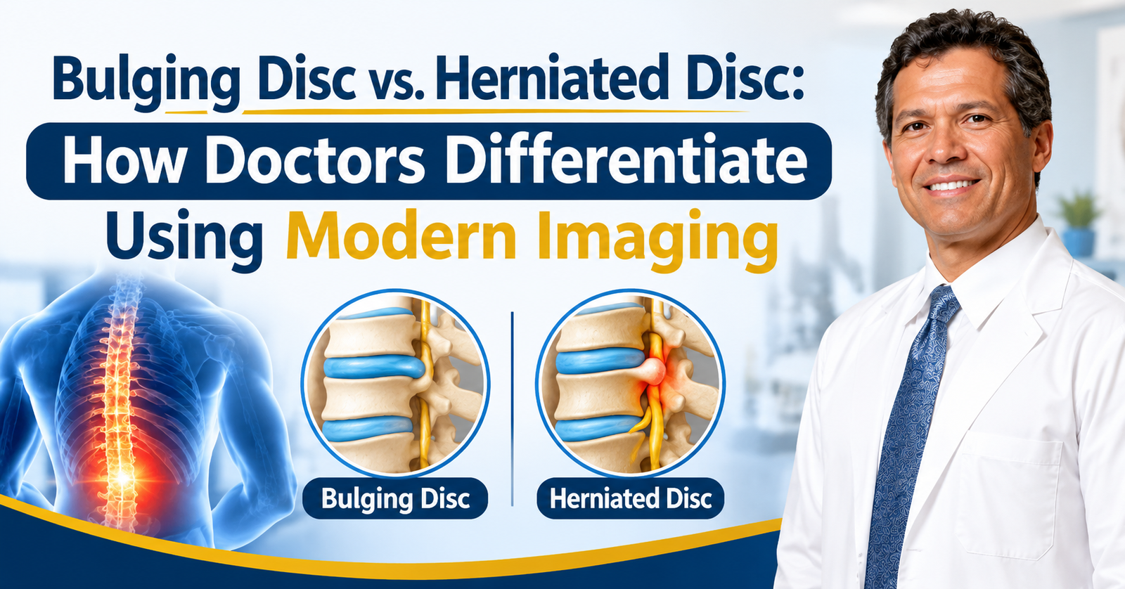
Bulging disc vs. herniated disc? These are terms that are used quite interchangeably in an MRI report and they are, in fact, two very different things. If you’re over 40, you very likely have some degree of disc degeneration (that’s the condition that affects pretty much all of us over 40 – Arthritis Foundation). The term itself doesn’t dictate your treatment. The diagnosis of spinal disc pain in the USA is very much based on degenerative disc disease imaging. Back pain diagnostic imaging (i.e., an MRI, CT or X-ray) can reliably differentiate between a bulging disc and a herniated disc. That imaging will allow your doctor to establish the cause and then they can go on to recommend the next step – whether that be physio, injections or a surgical referral.
Before the Scan: How Doctors Assess Disc Symptoms
How Doctors Examine Disc Symptoms
Your doctor’s assessment starts long before they order a scan. The physical examination is key and they’ll check your reflexes, muscle strength and sensation (pinprick, vibration, etc.). They’ll want to know when your symptoms started, whether lifting or twisting makes the pain worse, and what makes it better.
Straight Leg Raise Test for Disc Pain
The straight leg raise is a common test. You lie down and the doctor lifts your leg up in a straight line. If you feel pain radiating down the leg with that movement, it may indicate a ruptured disc. The test has a sensitivity of 73%-98%, but much lower specificity (11%-61%). The crossed straight leg raise (i.e., testing the other leg) is more specific (88%-98%).
Red Flags Doctors Watch For
Your provider also watches for red flags, such as:
- Loss of bladder or bowel control
- Progressive leg weakness or numbness
- Fever or a history of cancer
- Difficulty walking or balance problems
When Nerve Problems Need an EMG or Nerve Conduction Study
Most low back pain resolves on its own over 4–6 weeks, so imaging is generally deferred unless there are red flags or the condition is getting worse despite conservative management. If there is still doubt about nerve involvement / nerve problems, an EMG or nerve conduction study can tell you which nerves are involved before you even consider imaging.
Reading the Scan: How Doctors Identify a Bulge or Herniation
Once an imaging study is ordered, the radiologist will look at the shape and position of the disc material, not just its size. MRI is still the modality of choice for degenerative disc disease with a sensitivity of circa 75%. Pooled study data show a sensitivity of about 80.9% and a specificity of about 81% for lumbar disc herniation. X-rays won’t show a herniation on their own, but they can show disc height loss and/or vertebral shifts that can indicate a herniation.
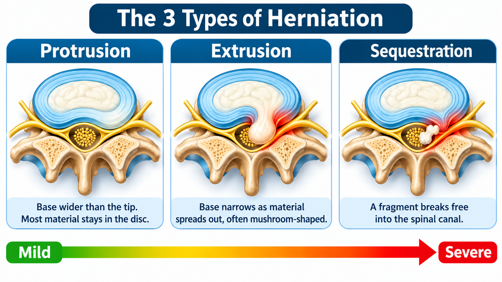
A bulging disc is what you see on the scan. The outer wall is still there; it’s just bulging out by about 25%- 50% of the disc’s circumference. A herniated disc is then further classified according to its shape:
- Protrusion: the base attached to the disc is wider than the tip, so most of the material remains within the disc.
- Extrusion: the base tapers as the material is displaced and spreads out, frequently resulting in a mushroom shape.
- Sequestration: a fragment moves freely within the spinal canal.
Radiologists also grade nerve root compression as mild, moderate or severe, depending on the amount of canal narrowing. They’ll note whether the herniation is central, paracentral or in the foramen and L5-S1 is by far the most common level.
After the Report: What the Findings Mean for Your Care
Your report can be completely at odds with the language you’d expect in that context, but it’s pivotal in determining your treatment pathway. A diagnosis of a herniated disc doesn’t in any way mean that you need surgery. It’s up to your doctor to decide whether that is in fact the cause of your pain. The evidence supports this. 20% of people under 60% and 36% of people over 60 have a herniated disc and are completely asymptomatic. Disc bulges are also seen in around 30% of pain-free adults under 40.
Scans are often completely asymptomatic, so your team will compare the scan findings with your clinical examination before deciding on treatment. Most people with a herniated disc will improve without surgery. One study showed that around 70% of patients on conservative management will have disc resorption within 6 months. Surgery is generally only indicated if the non-operative treatments (meds, muscle pain medication, physio, injections etc.) have failed to help after 6 weeks. It may also be indicated if you go on to develop a progressive deterioration of your weakness, numbness or loss of bladder control.
Conclusion
The difference between a bulging disc and a herniated disc is just a matter of what the imaging shows and whether that correlates with your symptoms. The label isn’t that important. What matters is the full clinical picture your doctor has from their examination and the scan.
Many imaging findings are completely painless, so don’t jump to conclusions. Dr. Rodolfo Gari provides fully individualized non-surgical first-line care (muscle pain relief and pain management) based on your individual diagnosis.
Frequently Asked Questions (FAQs)
What is the difference between a bulging disc and a herniated disc?
A bulging disc is one where the outer wall of the disc is still present, but it bulges out (usually across 25%-50% of the circumference). A herniated disc is one where the disc material pushes through the outer wall and is described as a protrusion, an extrusion or a sequestration depending on the shape. Both terms can appear in the same MRI report, and neither one alone determines your treatment.
What are the common symptoms of a bulging or herniated disc?
Common symptoms are back/neck pain, pain radiating down the leg or arm, numbness/tingling and muscle weakness. A lot of the bulges and herniations will be completely asymptomatic, though and only be picked up on a scan. Loss of bladder or bowel control, progressive leg weakness, fever and difficulty walking are all ‘red flags’ and require urgent medical attention.
Does a herniated disc always require surgery?
No. Most people with a herniated disc will in fact, improve without surgery and around 70% of those on conservative management will have resorption of the disc within 6 months. Surgery is only really considered in the instance where the non-operative options (meds, physio, injections etc.) have failed to make any difference after 6 weeks or where there’s a deterioration in the symptoms of weakness, numbness or loss of bladder control.
Is an MRI or CT scan better for diagnosing a herniated disc?
MRI is still the modality of choice for degenerative disc disease (sensitivity circa 75%) as opposed to CT. Pooled data gives MRI a sensitivity of circa 80.9% and a specificity of circa 81% for lumbar disc herniation. X-ray won’t show the herniation itself but can show disc height loss and/or vertebral shifts, which can indicate the problem.